



Oxychlororaphine

Based on 1 Customer Validation
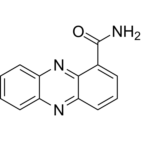
Oxychloroaphine could be isolated from the bacterium Pantoea agglomerans naturally present in soil. Oxychloroaphine has broad-spectrum antifungal activity. Oxychloroaphine has cytotoxicity in a dose-dependent manner and induces apoptosis. Oxychloroaphine can be used in research of cancer.
For research use only. We do not sell to patients.
- Purity : 98.63%
- CAS No.: 550-89-0
- Formula: C13H9N3O
- Molecular Weight:223.23
-
Storage:Powder -20°C, 3 years , 4°C, 2 years ; In solvent -80°C, 6 months , -20°C, 1 month

To place orders, for customer services and technical support, please contact: MedChemExpress USA
Tel: 609-228-6898 E-mail: [email protected] [email protected]


-
 Biological Activity
-
Chemical Information
-
Solvent & Solubility
- Protocols
- Purity & Documentation
- References
-
 Help & FAQs


-
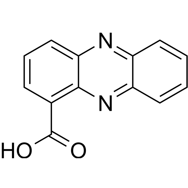
HY-33037
Phenazine-1-carboxylic acidFungal Inhibitor -

HY-W042191R
Oxychlororaphine (Standard)Antifungal Agent -
HY-113345
ZymostenolRORγ Agonist -
HY-17021
EsomeprazoleH+, K+-ATPase Inhibitor -

HY-17021R
Esomeprazole (Standard)H+, K+-ATPase Inhibitor -

HY-P2092
Cyclo(L-leucyl-L-tryptophyl)Antibacterial/Antifungal Agent -
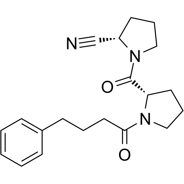
HY-100475
KYP-2047POP Inhibitor -
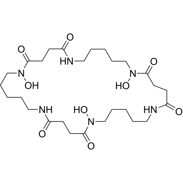
HY-124452
NocardamineSiderophore -

HY-113345R
Zymostenol (Standard)RORγ Agonist -
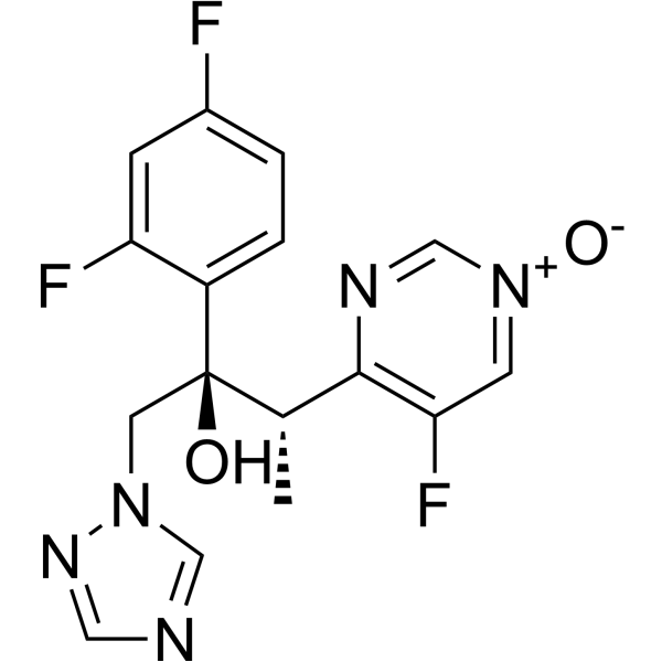
HY-137989
Voriconazole N-oxideAntifungal Agent -

HY-N12230
Penicolinate BPicolinic Acid Derivative -
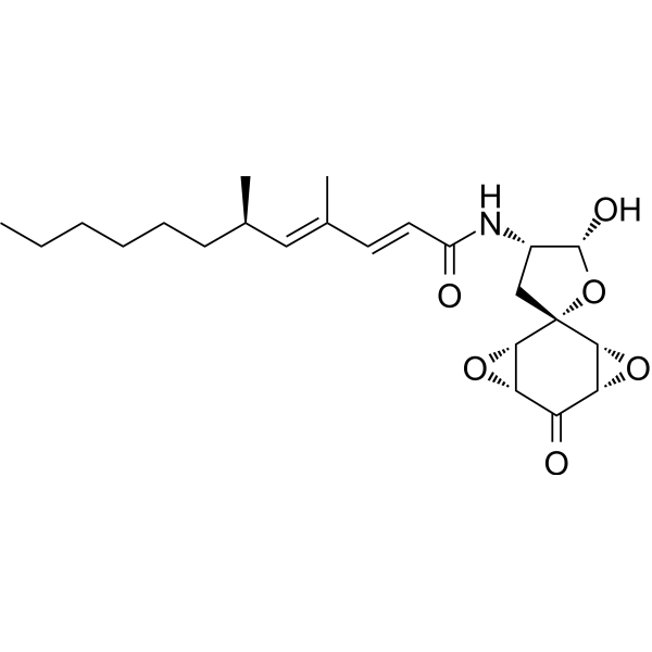
HY-121780
AranorosinAntifungal Antibiotic
All Caspase Isoforms
More

Biological Activity
Description
In Vitro
Oxychloroaphine (1-256 μM; 24 h) has cytotoxicity with IC50 values for A549, HeLa, and SW480 cancer cell lines between 32 and 40 μM[2].
Oxychloroaphine (1-150 μM; A549, HeLa, and SW480 cancer cell lines) causes cell membrane damage, leading to increase apoptosis and leakage of lactate dehydrogenase, and increases production of cytochrome c protein[2].
Oxychloroaphine (32 μM; A549 and SW480 cells) induces cycle arrest at G1 phase and induction of sub-G phase[2].
Oxychloroaphine (48 h; A549 cells) induces downregulation of antiapoptotic Bcl-2 protein and the activation of proapoptotic protein caspase-3 led to the cleavage of PARP[2].
MedChemExpress (MCE) has not independently confirmed the accuracy of these methods. They are for reference only. Further protocols information, click here.
-
Cell Line:A549, HeLa, and SW480 cancer cell lines
-
Concentration:1, 2, 4, 8, 16, 32, 64, 128, and 256 μM
-
Incubation Time:24 hours
-
Result:Inhibited cell proliferative in a dose-dependent manner.


Chemical Information
-
CAS No. 550-89-0
-
Appearance Solid
-
Molecular Weight 223.23
-
Formula C13H9N3O
-
Color Light yellow to brown
-
SMILES
O=C(C1=CC=CC2=NC3=CC=CC=C3N=C21)N
-
Structure Classification
-
Initial Source
Phytopathogenic bacteria
-
Shipping
Room temperature in continental US; may vary elsewhere.
-
Storage
Powder -20°C 3 years 4°C 2 years In solvent -80°C 6 months -20°C 1 month


Solvent & Solubility
In Vitro:
DMSO : 10 mg/mL (44.80 mM; ultrasonic and warming and heat to 60°C; Hygroscopic DMSO has a significant impact on the solubility of product, please use newly opened DMSO)
Please refer to the solubility information to select the appropriate solvent. Once prepared, please aliquot and store the solution to prevent product inactivation from repeated freeze-thaw cycles.
Storage method and period of stock solution: -80°C, 6 months; -20°C, 1 month. When stored at -80°C, please use it within 6 months. When stored at -20°C, please use it within 1 month.
Please refer to the solubility information to select the appropriate solvent. Once prepared, please aliquot and store the solution to prevent product inactivation from repeated freeze-thaw cycles.
Storage method and period of stock solution: -80°C, 6 months; -20°C, 1 month. When stored at -80°C, please use it within 6 months. When stored at -20°C, please use it within 1 month.
Concentration (start) × Volume (start) = Concentration (final) × Volume (final)

Protocols
-
Apoptosis
Apoptosis, also called programmed cell death, is generally characterized by distinct morphological characteristics.
-
TUNEL staining for apoptotic DNA fragmentation
TUNEL staining detects DNA strand breaks by using terminal deoxynucleotidyl transferase to add labeled nucleotides to exposed 3′-OH DNA termini, generating either microscopic staining in fixed cells or tissue sections, or fluorescence/cytometric signal in cell suspensions. TUNEL positivity reflects DNA fragmentation but should not be interpreted alone as definitive apoptosis, because TUNEL can also label necrotic, autolytic, mechanically damaged, or DNA-repair-associated DNA breaks.
-
Cell Cytotoxicity Assay
Cytotoxicity assays are usually based on the assessment of cell membrane damage, which can also be indirectly detected by measuring cell viability. Detection methods include MTT assay, CKK-8 assay, LDH assay and ATP assay, etc.
-
Mammalian live/dead viability and cytotoxicity staining
Live/dead viability and cytotoxicity staining assays are based on the simultaneous detection of intracellular esterase activity in metabolically active (viable) cells and membrane integrity loss in non-viable cells. In commonly used dual-staining approaches, membrane-permeant fluorogenic substrates are converted by intracellular esterases into fluorescent products in live cells, while impermeant DNA-binding dyes selectively enter cells with compromised plasma membranes and label nucleic acids in dead or dying cells, enabling discrimination between viable and non-viable populations by fluorescence microscopy or flow cytometry.
-
Annexin V plus membrane-impermeant dye apoptosis staining
Annexin V-based apoptosis assays rely on the detection of phosphatidylserine (PS) externalization from the inner leaflet of the plasma membrane to the outer leaflet, an early biochemical hallmark of apoptosis. Fluorescently labeled Annexin V binds PS in a calcium-dependent manner, enabling identification of early apoptotic cells by flow cytometry or fluorescence microscopy. When combined with a membrane-impermeant DNA-binding dye (e. g. , propidium iodide), this approach allows discrimination between viable (Annexin V−/dye−), early apoptotic (Annexin V+/dye−), and late apoptotic or necrotic (Annexin V+/dye+) cell populations by assessing membrane integrity and PS exposure.
-
Apoptosis Solutions
Apoptosis is a regulated, generally non-lytic cell-death pathway that removes unwanted, damaged, infected, or abnormal cells through coordinated morphological changes, caspase activation, DNA fragmentation, and membrane remodeling. The intrinsic apoptosis pathway is controlled mainly by mitochondrial outer membrane permeabilization, BCL-2 family proteins, cytochrome c release, apoptosome formation, caspase-9 activation, and downstream executioner caspase-3/7 activation. The extrinsic apoptosis pathway is initiated by death receptors such as Fas, TNFR, and TRAIL receptors, which recruit adaptor proteins and activate caspase-8 before engaging executioner caspases or mitochondrial amplification through BID cleavage. Apoptosis is linked to many phenotypes, including cancer cell killing, tissue homeostasis, immune regulation, neurodegeneration, infection response, and treatment-induced cytotoxicity; unresolved questions include how apoptosis interacts with necroptosis, pyroptosis, ferroptos
-
Cell Viability Determination by MTT Colorimetric Assay
The following protocol uses the MTT colorimetric assay as a classic literature-established method for assessing cell viability/metabolic activity in cultured mammalian cells. MTT[3-(4,5-dimethylthiazol-2-yl)-2,5-diphenyltetrazolium bromide] is reduced by metabolically active cells to a colored formazan product; the amount of formazan is quantified spectrophotometrically and provides an indirect measure of metabolically active viable cells. Importantly, MTT reduction reflects cellular oxidoreductase/metabolic activity rather than an absolute direct count of living cells, so changes in cellular metabolism can alter the signal independently of cell number.


Purity & Documentation
-
Data Sheet (276 KB)
-
SDS (396 KB)
- English - EN (396 KB)
- Français - FR (396 KB)
- Deutsch - DE (396 KB)
- Norwegian - NO (396 KB)
- Español - ES (396 KB)
- Swedish - SV (396 KB)
- Italian - IT (396 KB)
- Korean - KR (396 KB)
- Portuguese - PT (396 KB)
-
Handling Instructions (2659 KB)
References
[1]. Li S, et, al. Comparative metabolomics and transcriptomics analyses provide insights into the high-yield mechanism of phenazines biosynthesis in Pseudomonas chlororaphis GP72. J Appl Microbiol. 2022 Nov;133(5):2790-2801. [Content Brief]
[2]. Ali HM, et, al. Isolation of Bioactive Phenazine-1-Carboxamide from the Soil Bacterium Pantoea agglomerans and Study of Its Anticancer Potency on Different Cancer Cell Lines. J AOAC Int. 2016 Sep;99(5):1233-9. [Content Brief]


Complete Stock Solution Preparation Table
Please refer to the solubility information to select the appropriate solvent. Once prepared, please aliquot and store the solution to prevent product inactivation from repeated freeze-thaw cycles.
Storage method and period of stock solution: -80°C, 6 months; -20°C, 1 month. When stored at -80°C, please use it within 6 months. When stored at -20°C, please use it within 1 month.
| Optional Solvent | Concentration Solvent Mass | 1 mg | 5 mg | 10 mg | 25 mg |
|---|---|---|---|---|---|
| DMSO | 1 mM | 4.4797 mL | 22.3984 mL | 44.7968 mL | 111.9921 mL |
| 5 mM | 0.8959 mL | 4.4797 mL | 8.9594 mL | 22.3984 mL | |
| 10 mM | 0.4480 mL | 2.2398 mL | 4.4797 mL | 11.1992 mL | |
| 15 mM | 0.2986 mL | 1.4932 mL | 2.9865 mL | 7.4661 mL | |
| 20 mM | 0.2240 mL | 1.1199 mL | 2.2398 mL | 5.5996 mL | |
| 25 mM | 0.1792 mL | 0.8959 mL | 1.7919 mL | 4.4797 mL | |
| 30 mM | 0.1493 mL | 0.7466 mL | 1.4932 mL | 3.7331 mL | |
| 40 mM | 0.1120 mL | 0.5600 mL | 1.1199 mL | 2.7998 mL |


Oxychlororaphine Related Classifications
HY-W042191 Related Classifications
 Powered by Bioz