



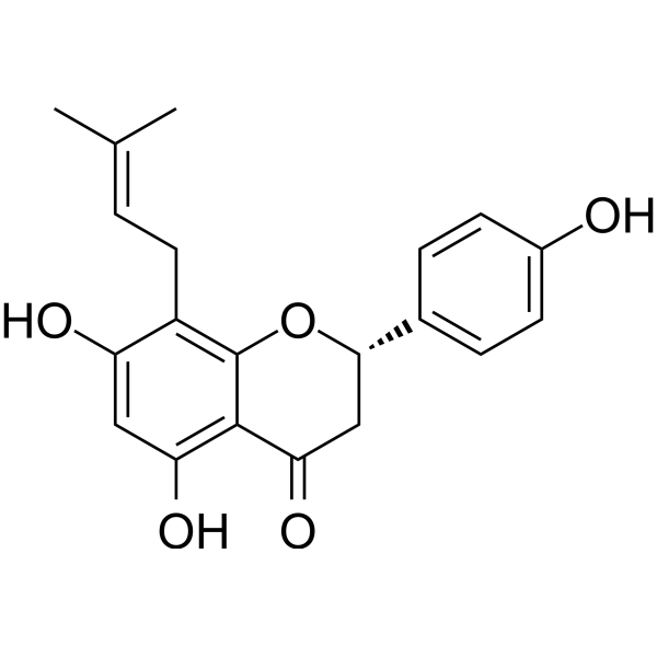
8-Prenylnaringenin
Based on 1 publication(s) in Google Scholar
8-Prenylnaringenin is an orally active prenyl flavonoid. 8-Prenylnaringenin can be isolated from the hop spike Humulus lupulus. 8-Prenylnaringenin activates the PI3K/Akt pathway and the AMPK pathway, upregulates OXPHOS complexes (II, III, and V) and Sirt1, and reduces ROS production and SOD activity. 8-Prenylnaringenin improves muscle atrophy and obesity and inhibits angiogenesis. 8-Prenylnaringenin exhibits anticancer activity against glioblastoma and colon cancer. 8-Prenylnaringenin also has LH/FSH regulatory activity. 8-prenylnaringenin may be used in bone health research.
Nur für Forschungszwecke. Wir verkaufen nicht an Patienten.
- Reinheit : 99.63%
- CAS. Nr.: 53846-50-7
- Formel: C20H20O5
- Molecular Weight:340.37
-
Speicherung:
4°C, protect from light
* In solvent : -80°C, 6 months; -20°C, 1 month (protect from light)

To place orders, for customer services and technical support, please contact: MedChemExpress USA
Tel: 609-228-6898 E-mail: [email protected] [email protected]


-
 Biologische Aktivität
-
Chemical Information
-
Lösungsmittel & Löslichkeit
- Protokoll
- Reinheit & Dokumentation
- Verweise
-
 Help & FAQs


-
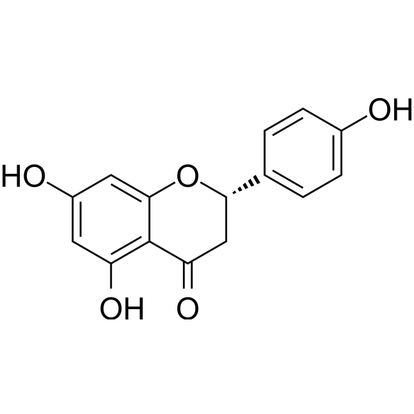
HY-N0100
NaringeninFlavonoid -
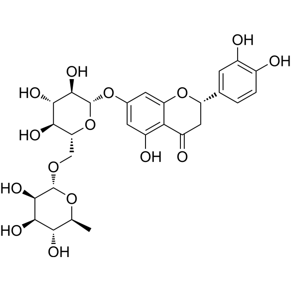
HY-N0636
EriocitrinAntioxidant Agent -
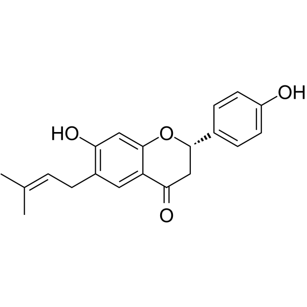
HY-N0233
BavachinPhytoestrogen -
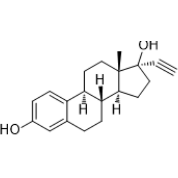
HY-B0216
EthinylestradiolEndogenous Metabolite -
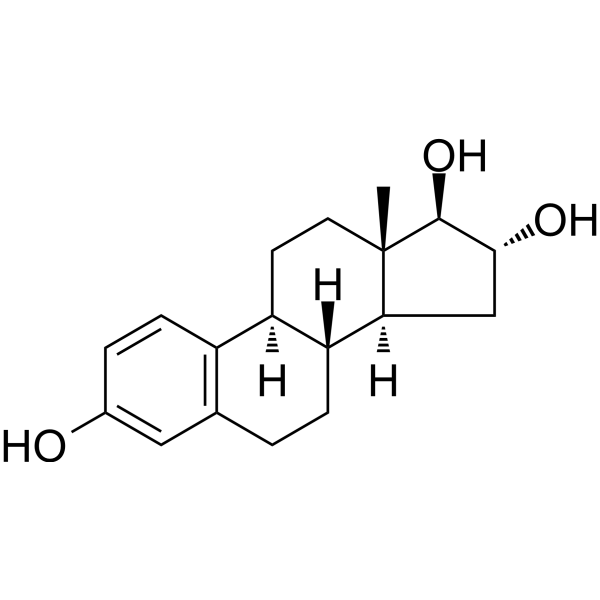
HY-B0412
EstriolEstrogen -
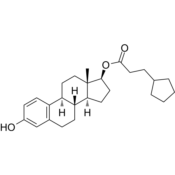
HY-B1100
Estradiol cypionateEstrogen Receptor/ERR Inhibitor -
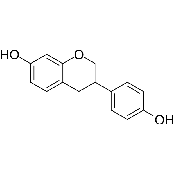
HY-100583A
(±)-EquolNonsteroidal Estrogen -
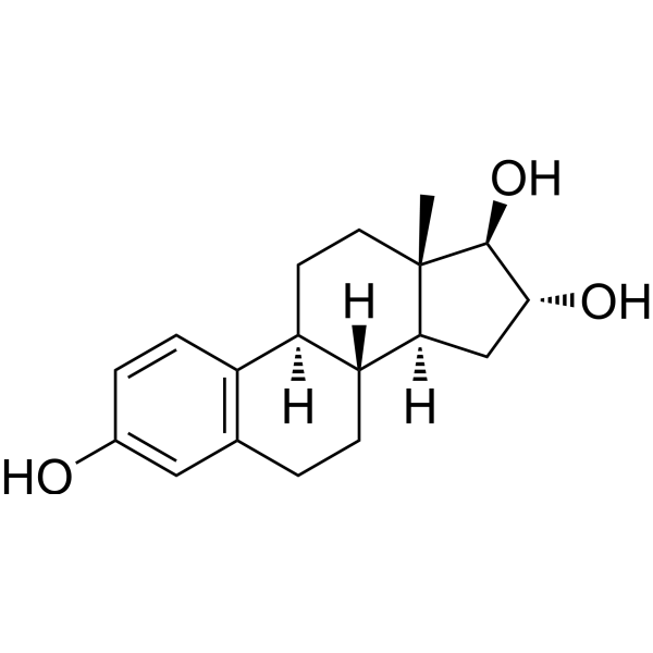
HY-B0412R
Estriol (Standard) -
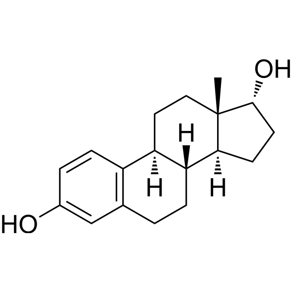
HY-B0141A
Alpha-Estradiol5α-reductase Inhibitor -
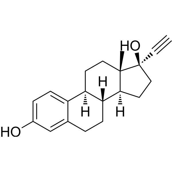
HY-B0216R
Ethinylestradiol (Standard) -
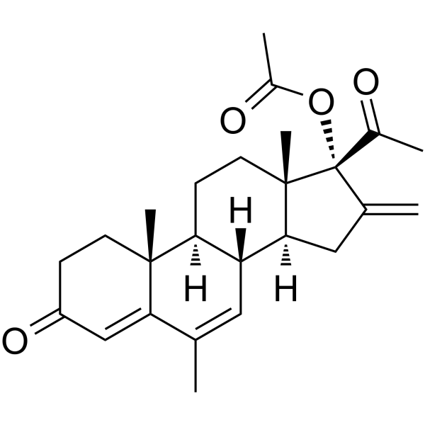
HY-111614
Melengestrol acetateContraceptive Agent -
HY-B0521
AltrenogestProgesterone Receptor Agonist
Publications Citing Use of MedChemExpress (MCE) 8-Prenylnaringenin
MoreAlle AMPK Isoform-spezifische Produkte anzeigen
More

Biologische Aktivität
Beschreibung
|
SIRT1 |
Cellular Effect
|
Cell Line

|
Type | Value | Description | References |
|---|---|---|---|---|
| Ishikawa | IC50 |
19.1 μM
Compound: 8-PN
|
Cytotoxicity against human Ishikawa cells after 96 hrs by SRB assay
Cytotoxicity against human Ishikawa cells after 96 hrs by SRB assay
|
[PMID: 28812892] |
| NCI-H460 | IC50 |
3.1 μM
Compound: 7
|
Inhibition of ABCG2 expressed in human NCI-H460 cells assessed as inhibition of PhA accumulation after 2 to 20 hrs relative to fumitremorgin C
Inhibition of ABCG2 expressed in human NCI-H460 cells assessed as inhibition of PhA accumulation after 2 to 20 hrs relative to fumitremorgin C
|
[PMID: 21275386] |
In Vitro
8-Prenylnaringenin (48 h) exhibits strong inhibitory effect against human colon cancer HCT-116 cells with an IC50 value of 23.83 μg/mL[1].
8-Prenylnaringenin (0.1-10 μM; 0.25-4 h) activates the PI3K/Akt pathway in mouse C2C12 myotubes[2].
8-Prenylnaringenin (10–500 μM; 24 h) shows higher cytotoxicity against human glioblastoma U-118 MG cells (IC50 ≈ 138 μM) than normal BJ fibroblasts (IC50 = 172 μM)[3].
8-Prenylnaringenin (0.001-20 μM; 48 h) reduces reactive oxygen species (ROS) production, SOD activity in MCF-7 breast cancer cells[4].
8-Prenylnaringenin inhibits BME cell invasion with IC50 values ranging from 3 to 10 μM[5].
8-Prenylnaringenin (1-30 μM) inhibits angiogenesis induced by bFGF, VEGF, or a combination of both cytokines in BME cells in a dose-dependent manner[5].
MedChemExpress (MCE) has not independently confirmed the accuracy of these methods. They are for reference only. Further protocols information, click here.
-
Cell Line:MCF-7 (human breast cancer cell line)
-
Concentration:0.01 μM, 1 μM
-
Incubation Time:48 h
-
Result:Upregulated expression of OXPHOS complexes (II, III, V), Sirt1 (sixfold increase at 1 μM).
In Vivo
8-Prenylnaringenin (0.0005%-0.005% wt/wt in high-fat diet; p.o. via mixed diet; 8 weeks) prevents high-fat diet-induced obesity in C57BL/6J male mice by promoting adiponectin secretion and activating the AMPK pathway[6].
8-Prenylnaringenin (126-1260 ppm; p.o. via soy-free diet; 3 months) suppresses serum LH/FSH levels and stimulates uterine weight in ovariectomized Sprague-Dawley rats[7].
8-Prenylnaringenin (1.77 mg/kg; s.c.; daily; 10 weeks) does not improve biomechanical properties or bone mineral density of lumbar vertebrae and femora in ovariectomized Sprague-Dawley rats[8].
MedChemExpress (MCE) has not independently confirmed the accuracy of these methods. They are for reference only.
-
Animal Model:C57BL/6J mice (male, 230-290 g initial weight, 5-week-old) fed with high-fat diet[6].
-
Dosage:0.0005% wt/wt, 0.005% wt/wt 8-PN in high-fat diet
-
Administration:Oral administration via mixed high-fat diet; 8 weeks
-
Result:Prevented high-fat diet-induced body weight gain and fat accumulation in WAT.
Reduced liver total lipid and triglyceride levels in the liver.
Inhibitd ALT activity.
Promoted AMPK phosphorylation.
Regulated SREBP-1 and PGC-1α expression via AMPK activation.


Chemical Information
-
CAS. Nr. 53846-50-7
-
Appearance Solid
-
Molecular Weight 340.37
-
Formel C20H20O5
-
Color White to off-white
-
SMILES
O=C1C[C@@H](C2=CC=C(O)C=C2)OC3=C(C/C=C(C)\C)C(O)=CC(O)=C13
-
Structure Classification
-
Initial Source
-
Versand
Room temperature in continental US; may vary elsewhere.
-
Speicherung
4°C, protect from light
* In solvent : -80°C, 6 months; -20°C, 1 month (protect from light)


Publications (1)
-
Journal Impact Factor
-
Most Recent


Lösungsmittel & Löslichkeit
In Vitro:
DMSO : 100 mg/mL (293.80 mM; Need ultrasonic; Hygroscopic DMSO has a significant impact on the solubility of product, please use newly opened DMSO)
Please refer to the solubility information to select the appropriate solvent. Once prepared, please aliquot and store the solution to prevent product inactivation from repeated freeze-thaw cycles.
Storage method and period of stock solution: -80°C, 6 months; -20°C, 1 month (protect from light). When stored at -80°C, please use it within 6 months. When stored at -20°C, please use it within 1 month.
Please refer to the solubility information to select the appropriate solvent. Once prepared, please aliquot and store the solution to prevent product inactivation from repeated freeze-thaw cycles.
Storage method and period of stock solution: -80°C, 6 months; -20°C, 1 month (protect from light). When stored at -80°C, please use it within 6 months. When stored at -20°C, please use it within 1 month.
Konzentration (Stammlösung) × Volumen (Stammlösung) = Konzentration (Ziellösung) × Volumen (Ziellösung)
In Vivo:
Select the appropriate dissolution method based on your experimental animal and administration route.
- For the following dissolution methods, please ensure to first prepare a clear stock solution using an In Vitro approach and then sequentially add co-solvents:
- To ensure reliable experimental results, the clarified stock solution can be appropriately stored based on storage conditions. As for the working solution for In Vivo experiments, it is recommended to prepare freshly and use it on the same day.
- The percentages shown for the solvents indicate their volumetric ratio in the final prepared solution. If precipitation or phase separation occurs during preparation, heat and/or sonication can be used to aid dissolution.
Add each solvent one by one: 10% DMSO 40% PEG300 5% Tween-80 45% Saline
Solubility: ≥ 2.08 mg/mL (6.11 mM); Clear solution
This protocol yields a clear solution of ≥ 2.08 mg/mL (saturation unknown).
Taking 1 mL working solution as an example, add 100 μL DMSO stock solution (20.8 mg/mL) to 400 μL PEG300, and mix evenly; then add 50 μL Tween-80 and mix evenly; then add 450 μL Saline to adjust the volume to 1 mL.
Preparation of Saline: Dissolve 0.9 g sodium chloride in ddH₂O and dilute to 100 mL to obtain a clear Saline solution.
Add each solvent one by one: 10% DMSO 90% (20% SBE-β-CD in Saline)
Solubility: ≥ 2.08 mg/mL (6.11 mM); Clear solution
This protocol yields a clear solution of ≥ 2.08 mg/mL (saturation unknown).
Taking 1 mL working solution as an example, add 100 μL DMSO stock solution (20.8 mg/mL) to 900 μL 20% SBE-β-CD in Saline, and mix evenly.
Preparation of 20% SBE-β-CD in Saline (4°C, storage for one week): 2 g SBE-β-CD powder is dissolved in 10 mL Saline, completely dissolve until clear.
In Vivo Dissolution Calculator
Please enter the basic information of animal experiments:
-
-
-
-
Recommended: Prepare an additional quantity of animals to account for potential losses during experiments.
Please enter your animal formula composition:
-
%DMSO +
Recommended: Keep the proportion of DMSO in working solution below 2% if your animal is weak.
-
%+
-
+%Tween-80 + +
-
%Saline +
The co-solvents required include: DMSO, . All of co-solvents are available by MedChemExpress (MCE). , Tween 80. All of co-solvents are available by MedChemExpress (MCE).
Working solution concentration: 0.22 mg/mL
Method for preparing stock solution: mg drug dissolved in μL DMSO. Stock solution concentration: mg/mL. * In solvent : -80°C, 6 months; -20°C, 1 month (protect from light)
1. Take μL DMSO stock solution;
2. Add μL .
μL , mix evenly;
3. Then add μL Tween 80, mix evenly;
4. Then add μL
Please ensure that the stock solution in the first step is dissolved to a clear state, and add co-solvents in sequence. You can use ultrasonic heating (ultrasonic cleaner, recommended frequency 20-40 kHz), vortexing, etc. to assist dissolution.

Protokoll
-
Human pluripotent stem cell endothelial-cell differentiation
Human pluripotent stem cell endothelial differentiation is based on stepwise developmental patterning: early activation of WNT/GSK3β inhibition promotes mesodermal or vascular progenitor entry, followed by endothelial specification using VEGF-related signaling, BMP4, FGF2, Notch modulation, or cAMP depending on the published protocol. Endothelial differentiation is read out by acquisition of CD31, CD34, VE-cadherin/CD144, KDR/VEGFR2, vWF, Tie2, NOS3, acetylated LDL uptake, tube/network formation, barrier function, and in vivo vessel-forming capacity where tested.
-
Vascular/Branching Fractal Analysis
Vascular/branching fractal analysis quantifies the geometric complexity of vessel trees or vascular networks from segmented 2D images, commonly by converting vessels into binary and/or skeletonized maps and estimating fractal dimension using box-counting or related approaches. Fractal dimension is interpreted as an image-derived readout of vascular branching complexity, space filling, or density, and has been applied to retinal photographs, fluorescein angiography, OCT angiography, capillary perfusion maps, and in vitro Matrigel angiogenesis networks. The assay readout is generated from vessel-positive pixels after image preprocessing, vessel segmentation, binarization, and optional skeletonization; reported outputs include fractal dimension, vessel density, branchpoint density, endpoint density, vessel length density, tortuosity, and generation-based branching metrics when VESGEN-style analysis is used. The biological interpretation is limited to quantitative vascular patterning and s
-
ROS/oxidative-stress fluorescent staining
ROS/oxidative-stress fluorescent staining uses cell-permeant fluorogenic probes that become fluorescent after oxidation inside cells or tissues; commonly used examples include DCFH-DA/DCFDA for broad cellular oxidant detection, DHE for superoxide-related signal detection, MitoSOX for mitochondrial superoxide-related signal detection, and CellROX probes for oxidative-stress-associated fluorescence readouts. The assay detects probe oxidation rather than a single ROS species unless the probe and analysis method have been chemically validated for that species. DCFH-DA enters cells, is deacetylated by intracellular esterases to DCFH, and produces fluorescent DCF after oxidation, so the readout is used as an operational measure of total cellular oxidative stress rather than a species-specific ROS measurement. DHE and MitoSOX can report superoxide-related oxidation, but red fluorescence alone can include non-specific ethidium-like oxidation products; HPLC or optimized spectral approaches are
-
Endothelial Tube Formation Assay
Endothelial tube formation assay evaluates the ability of endothelial cells to attach, migrate, align, and organize into capillary-like networks when cultured on gelled basement membrane extract or Matrigel; the readout is the morphology and quantity of tube-like networks, which reflects an in vitro endothelial morphogenesis step related to angiogenesis. Basement membrane extract/Matrigel provides laminin-rich extracellular matrix cues that support endothelial differentiation into capillary-like structures, but it can contain biologically active growth factors, so growth-factor-reduced matrix is preferred when testing defined angiogenic stimulators or inhibitors.
-
Research Protocol for Metabolic Diseases
AMP-activated protein kinase, AMPK, is a conserved cellular energy sensor that responds to reduced cellular energy status and coordinates metabolism by increasing ATP-generating catabolic pathways while suppressing ATP-consuming anabolic processes. In metabolic disease research, the AMPK pathway is experimentally relevant because it regulates hepatic lipid synthesis, fatty acid oxidation, glucose production, skeletal-muscle glucose disposal, mTORC1-linked biosynthesis, autophagy, mitochondrial homeostasis, and whole-body energy balance. The central pathway logic is that energy stress, metformin, exercise-like stimulation, or direct AMPK activators increase AMPKα Thr172 phosphorylation and downstream substrate phosphorylation, including ACC and RAPTOR. Phosphorylation of ACC suppresses lipogenesis and supports fatty acid oxidation, whereas phosphorylation of RAPTOR suppresses mTORC1 signaling and links cellular energy status to growth and protein synthesis control. The pathway is linked
-
Endothelial Cell Migration/Angiogenic Sprouting Assay
Endothelial cell migration and angiogenic sprouting assays are in vitro (and partially ex vivo-adapted) functional models that quantify the ability of endothelial cells to undergo coordinated migration, extracellular matrix invasion, and multicellular organization into capillary-like sprouts in response to pro-angiogenic stimuli such as VEGF, bFGF, or conditioned microenvironments. These assays are used to model early angiogenic events including tip-cell formation, directional migration, and lumen-like sprout extension, which collectively reflect angiogenic activation and vascular morphogenesis processes observed in vivo.


Reinheit & Dokumentation
-
Data Sheet (282 KB)
-
SDS (252 KB)
- English - EN (252 KB)
- Français - FR (252 KB)
- Deutsch - DE (252 KB)
- Norwegian - NO (252 KB)
- Español - ES (252 KB)
- Swedish - SV (252 KB)
- Italian - IT (252 KB)
- Korean - KR (252 KB)
- Portuguese - PT (252 KB)
-
Handling Instructions (2659 KB)
Verweise
[1]. Koosha S, et al. Antiproliferative and apoptotic activities of 8-prenylnaringenin against human colon cancer cells. Life Sci. 2019 Jul 3:116633. [Content Brief]
[2]. Mukai R, et al. 8-Prenylnaringenin promotes recovery from immobilization-induced disuse muscle atrophy through activation of the Akt phosphorylation pathway in mice. Am J Physiol Regul Integr Comp Physiol. 2016 Dec 1;311(6):R1022-R1031. [Content Brief]
[3]. Stompor M, et al. In Vitro Effect of 8-Prenylnaringenin and Naringenin on Fibroblasts and Glioblastoma Cells-Cellular Accumulation and Cytotoxicity. Molecules. 2017 Jun 30;22(7). [Content Brief]
[4]. Blanquer-Rosselló MM, et al. Effect of xanthohumol and 8-prenylnaringenin on MCF-7 breast cancer cells oxidative stress and mitochondrial complexes expression. J Cell Biochem. 2013 Dec;114(12):2785-94. [Content Brief]
[5]. Pepper MS, et al. 8-prenylnaringenin, a novel phytoestrogen, inhibits angiogenesis in vitro and in vivo. J Cell Physiol. 2004 Apr;199(1):98-107. [Content Brief]
[6]. Okada F, et al. 8-Prenylnaringenin suppresses obesity in high-fat diet-fed C57BL/6J mice via adiponectin secretion. J Clin Biochem Nutr. 2025 Jul;77(1):64-73. [Content Brief]
[7]. Christoffel J, et al. Effects of 8-prenylnaringenin on the hypothalamo-pituitary-uterine axis in rats after 3-month treatment. J Endocrinol. 2006 Mar;188(3):397-405. [Content Brief]
[8]. Hoffmann DB, et al. Effects of 8-Prenylnaringenin and Whole-Body Vibration Therapy on a Rat Model of Osteopenia. J Nutr Metab. 2016;2016:6893137. [Content Brief]


Complete Stock Solution Preparation Table
Please refer to the solubility information to select the appropriate solvent. Once prepared, please aliquot and store the solution to prevent product inactivation from repeated freeze-thaw cycles.
Storage method and period of stock solution: -80°C, 6 months; -20°C, 1 month (protect from light). When stored at -80°C, please use it within 6 months. When stored at -20°C, please use it within 1 month.
| Optional Solvent | Concentration Solvent Mass | 1 mg | 5 mg | 10 mg | 25 mg |
|---|---|---|---|---|---|
| DMSO | 1 mM | 2.9380 mL | 14.6899 mL | 29.3798 mL | 73.4495 mL |
| 5 mM | 0.5876 mL | 2.9380 mL | 5.8760 mL | 14.6899 mL | |
| 10 mM | 0.2938 mL | 1.4690 mL | 2.9380 mL | 7.3449 mL | |
| 15 mM | 0.1959 mL | 0.9793 mL | 1.9587 mL | 4.8966 mL | |
| 20 mM | 0.1469 mL | 0.7345 mL | 1.4690 mL | 3.6725 mL | |
| 25 mM | 0.1175 mL | 0.5876 mL | 1.1752 mL | 2.9380 mL | |
| 30 mM | 0.0979 mL | 0.4897 mL | 0.9793 mL | 2.4483 mL | |
| 40 mM | 0.0734 mL | 0.3672 mL | 0.7345 mL | 1.8362 mL | |
| 50 mM | 0.0588 mL | 0.2938 mL | 0.5876 mL | 1.4690 mL | |
| 60 mM | 0.0490 mL | 0.2448 mL | 0.4897 mL | 1.2242 mL | |
| 80 mM | 0.0367 mL | 0.1836 mL | 0.3672 mL | 0.9181 mL | |
| 100 mM | 0.0294 mL | 0.1469 mL | 0.2938 mL | 0.7345 mL |


8-Prenylnaringenin Related Classifications
HY-N2787 Related Classifications
 Powered by Bioz