



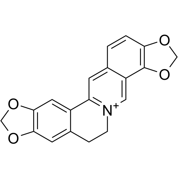
Coptisine
Based on 13 publication(s) in Google Scholar
Coptisine is an orally active and brain-penetrant alkaloid found in Coptis chinensis. Coptisine is a reversible, uncompetitive IDO inhibitor with a Ki of 5.8 μM and an IC50 of 6.3 μM. Coptisine suppresses neuroinflammation, reduces Aβ plaque burden and shows neuroprotective activity. Coptisine shows anti-inflammation activity by blocking NF-κB, MAPK, and PI3K/Akt activation. Coptisine inhibits cancer cells proliferation, induces DNA damage, G2/M phase cell cycle arrest, apoptosis, ROS production and mitochondrial dysfunction. Coptisine inhibits Rho/ROCK pathway activation, reduces arrhythmia, limits cardiac injury marker release, reduces infarct size, and preserves cardiac function in rat myocardial ischemia/reperfusion models. Coptisine downregulates HMGCR and upregulates LDLR and CYP7A1 to modulate cholesterol metabolism, reduces abnormal serum lipid levels, and promotes fecal bile acid excretion. Coptisine can be used for the research of cancer, hypercholesterolemia, Alzheimer’s disease, inflammatory disorders and cardiovascular disease.
Nur für Forschungszwecke. Wir verkaufen nicht an Patienten.
- CAS. Nr.: 3486-66-6
- Formel: C19H14NO4
- Molecular Weight:320.32
-
Speicherung:
Please store the product under the recommended conditions in the Certificate of Analysis.

To place orders, for customer services and technical support, please contact: MedChemExpress USA
Tel: 609-228-6898 E-mail: [email protected] [email protected]


-
 Biologische Aktivität
-
Chemical Information
- Protokoll
- Reinheit & Dokumentation
- Verweise
-
 Help & FAQs


-
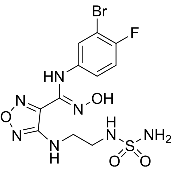
HY-15689
EpacadostatIDO1 Inhibitor -
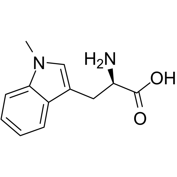
HY-16724
IndoximodIDO Inhibitor -
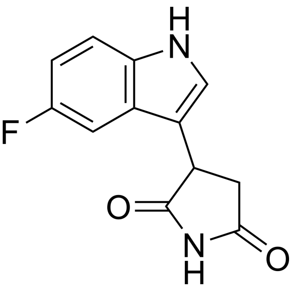
HY-101111
PF-06840003IDO-1 Inhibitor -
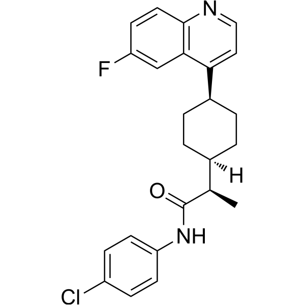
HY-101560
LinrodostatIDO1 Inhibitor -
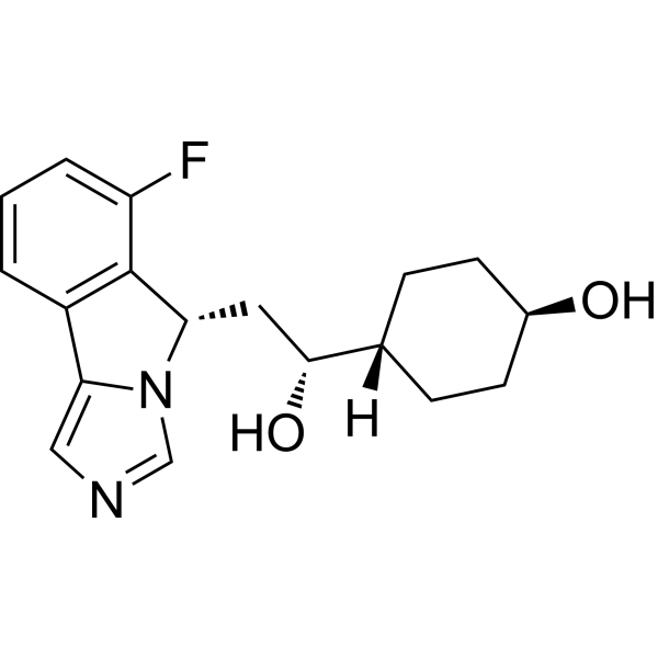
HY-18770B
NavoximodIDO Inhibitor -
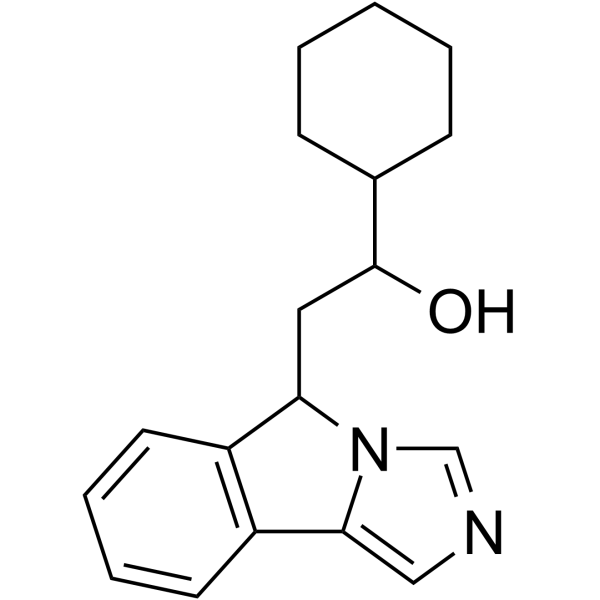
HY-13983
IDO-IN-7 -

HY-15689R
Epacadostat (Standard)IDO1 Inhibitor -
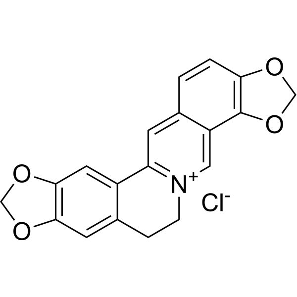
HY-N0736
Coptisine chlorideBacterial Inhibitor -
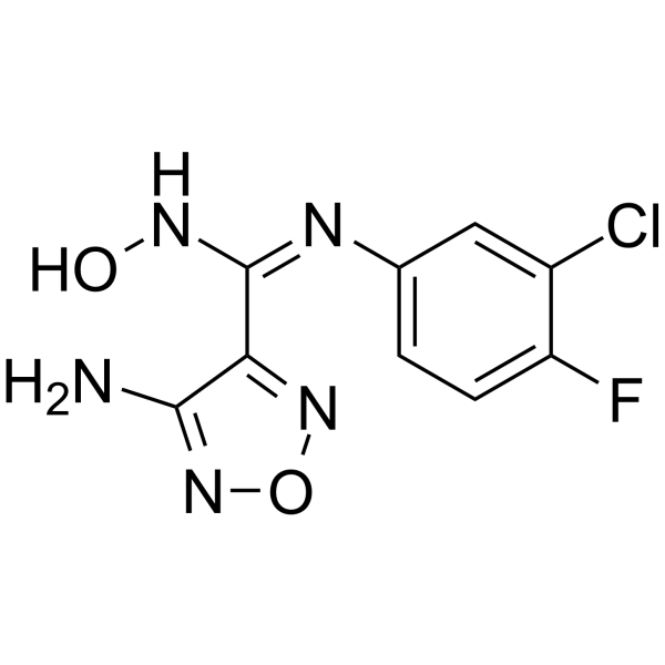
HY-15683
IDO5LIDO Inhibitor -
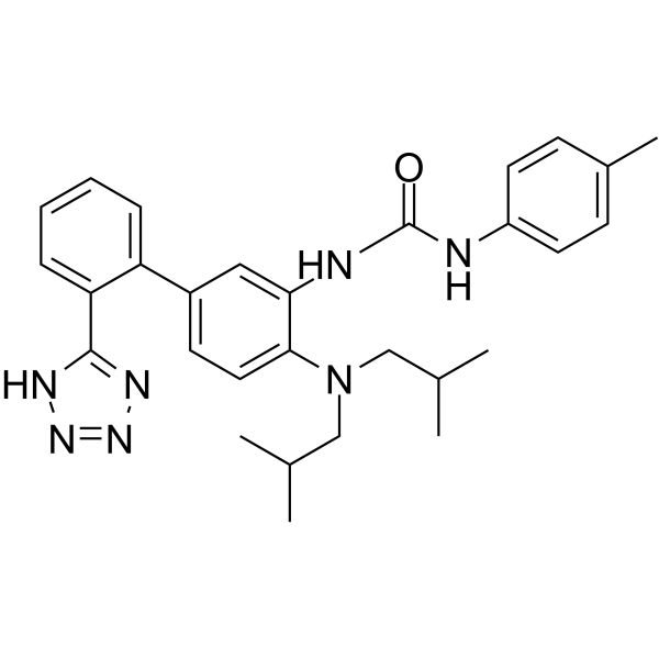
HY-100771
PCC0208009IDO Inhibitor -
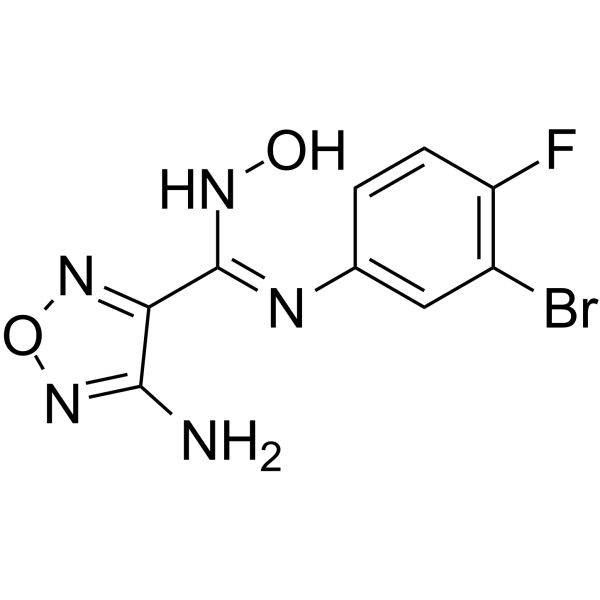
HY-79531
IDO-IN-1IDO Inhibitor -
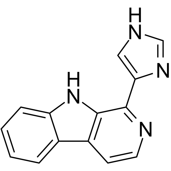
HY-151108
IDO1/TDO-IN-4IDO1/TDO Inhibitor
Publications Citing Use of MedChemExpress (MCE) Coptisine
More- Phytomedicine. 2025 Nov 25:148:157446. [Abstract]
- J Med Chem. 2026 Aug 13;69(15):19338-19354.
- Int Immunopharmacol. 2024 Feb 15:128:111433. [Abstract]
- Molecules. 2024 May 14;29(10):2304. [Abstract]
- Food Sci Nutr. 2026 Jul 15;14(7):e72107.
- Mater Technol (N Y N Y). 2026 Jan 27;41.
- Naunyn Schmiedebergs Arch Pharmacol. 2025 May;398(5):5465-5474. [Abstract]
- Vet Microbiol. 2026 May:316:110992. [Abstract]
- DNA Cell Biol. 2020 Oct 2. [Abstract]
- Biochem Biophys Res Commun. 2026 Mar 26:806:153415. [Abstract]
- Planta Med. 2024 Jun;90(7-08):523-533. [Abstract]
- SSRN. 2024 Apr 1.
- J Oncol. 2022 Jun 26:2022:9864411. [Abstract]
 Customer Validation & Images

-
WB

-
IF

-
WB

-
Others

Alle DNA/RNA Synthesis Isoform-spezifische Produkte anzeigen
More

Biologische Aktivität
Beschreibung
IC50 & Target
[4]|
IDO 6.3 μM (IC50) |
IDO 5.8 μM (Ki) |
Cellular Effect
|
Cell Line

|
Type | Value | Description | References |
|---|---|---|---|---|
| A2780/Taxol | IC50 |
>=24.33 μM
Compound: 3
|
Antiproliferative activity against human A2780T cells assessed as reduction in cell viability incubated for 48 hrs by CCK-8 assay
Antiproliferative activity against human A2780T cells assessed as reduction in cell viability incubated for 48 hrs by CCK-8 assay
|
[PMID: 39213483] |
| A549/TR | IC50 |
>=24.33 μM
Compound: 3
|
Antiproliferative activity against human A549/Taxol cells assessed as reduction in cell viability incubated for 48 hrs by CCK-8 assay
Antiproliferative activity against human A549/Taxol cells assessed as reduction in cell viability incubated for 48 hrs by CCK-8 assay
|
[PMID: 39213483] |
| LoVo | IC50 |
>=24.33 μM
Compound: 3
|
Antiproliferative activity against human LoVo cells assessed as reduction in cell viability incubated for 48 hrs by CCK-8 assay
Antiproliferative activity against human LoVo cells assessed as reduction in cell viability incubated for 48 hrs by CCK-8 assay
|
[PMID: 39213483] |
| Panel NCI-60 (60 carcinoma cell lines) | GI50 |
260 nM
Compound: Coptisine
|
Growth inhibitory activity against human cancer cell line in the NCI's anticancer drug screening program
Growth inhibitory activity against human cancer cell line in the NCI's anticancer drug screening program
|
[PMID: 15743190] |
| PC-3 | IC50 |
>=24.33 μM
Compound: 3
|
Antiproliferative activity against human PC-3 cells assessed as reduction in cell viability incubated for 24 hrs by CCK-8 assay
Antiproliferative activity against human PC-3 cells assessed as reduction in cell viability incubated for 24 hrs by CCK-8 assay
|
[PMID: 39213483] |
In Vitro
Coptisine (0.1-100 μM; 48 h) potently inhibits proliferation of human lung adenocarcinoma A5499, H460,
and H2170 cells (IC50 = 18.09, 29.50, and 21.60 μM) and other tested human cancer cell lines, with moderate selectivity relative to normal human umbilical vein endothelial cells[1].
Coptisine (12.5-50 μM; 48 h) induces concentration-dependent DNA damage in human lung adenocarcinoma A549 cells, as shown by increased γH2AX expression[1].
Coptisine (12.5-50 μM; 48 h) induces concentration-dependent G2/M phase cell cycle arrest in human lung adenocarcinoma A549 cells, mediated by downregulated cyclin B1, cdc2, and cdc25C expression and upregulated p21 expression[1].
Coptisine (12.5-50 μM; 48 h) induces concentration-dependent apoptosis in human lung adenocarcinoma A549 cells, inducing concentration-dependent activation of caspase 3/7, caspase 8, caspase 9, and cleavage of PARP[1].
Coptisine (12.5-50 μM; 0.5-24 h) induces time- and concentration-dependent reactive oxygen species generation in human lung adenocarcinoma A549 cells[1].
Coptisine (12.5-50 μM; 24 h) induces concentration-dependent mitochondrial dysfunction in human lung adenocarcinoma A549 cells, including loss of mitochondrial membrane potential and altered Bax, Bcl-2, and cytochrome c expression[1].
Coptisine potently inhibits recombinant human IDO as a reversible, uncompetitive inhibitor with a Ki of 5.8 μM and an IC50 of 6.3 μM[4].
Coptisine inhibits IDO activity in HEK 293 cells with an IC50 of 7.1 μM[4].
Coptisine (10 μM; 5 h pre-incubation) reverses amyloid-β peptide 1-42 and interferon-γ-induced IDO activation and restores cell viability in PC12 cells[4].
Coptisine inhibits LPS (HY-D1056)-stimulated inflammation by blocking NF-κB, MAPK, and PI3K/Akt activation in
macrophages[5].
MedChemExpress (MCE) has not independently confirmed the accuracy of these methods. They are for reference only.
-
Cell Line:human lung adenocarcinoma A549
-
Concentration:12.5, 25, 50 μM
-
Incubation Time:48 h
-
Result:Caused a concentration-dependent upregulation of pH2AX, a marker of DNA double-strand breaks, relative to untreated control cells.
-
Cell Line:human lung adenocarcinoma A549
-
Concentration:12.5, 25, 50 μM
-
Incubation Time:48 h
-
Result:Caused concentration-dependent G2/M phase arrest, with 26.5%, 29.9%, and 36.8% of cells in G2/M phase after treatment with 12.5, 25, and 50 μM, respectively, compared to 16.95% in control cells.
Accompanied this arrest by concentration-dependent downregulation of cyclin B1, cdc2, and cdc25C, and upregulation of p21.
-
Cell Line:human lung adenocarcinoma A549
-
Concentration:12.5, 25, 50 μM
-
Incubation Time:48 h
-
Result:Caused concentration-dependent induction of apoptosis: treatment with 50 μM resulted in 58.5% early apoptotic cells and 24.2% late apoptotic cells; treatment with 25 μM resulted in 26.4% early apoptotic cells; treatment with 12.5 μM resulted in 10.8% early apoptotic cells, compared to 8.6% early and 4.6% late apoptotic cells in control cultures.
Caused concentration-dependent activation of caspase 3/7, with a 19-fold increase in activity at 50 μM relative to control.
Induced concentration-dependent upregulation of active caspase 8, active caspase 9, and cleaved PARP.
-
Cell Line:PC12
-
Concentration:10 μM (pre-incubation)
-
Incubation Time:5 h (pre-incubation); 24 h (amyloid-β peptide 1-42 treatment); 24 h (interferon-γ treatment)
-
Result:Down-regulated the enhanced IDO activity induced by combined 25 μM amyloid-β peptide 1-42 and 1000 U/mL interferon-γ treatment.
Restored the reduced cell viability caused by these stimuli to levels comparable to untreated control cells.
In Vivo
Coptisine (23.35-70.05 mg/kg/day; i.g.; daily; 4 weeks) dose-dependently improves hypercholesterolemia in HFHC-fed Syrian golden hamsters[3].
Coptisine (482.5-1728 mg/kg; p.o.; single dose) has low acute toxicity in Kunming mice, with an LD50 of 880.18 mg/kg following a single oral dose[3].
Coptisine (154 mg/kg/day; p.o.; daily; 90 days) is well-tolerated in SD rats with no observable toxicity via daily oral administration for 90 days[3].
Coptisine (50 mg/kg; p.o.; once daily; 1 month) normalizes serum IDO activity, suppresses neuroinflammation, reduces Aβ plaque burden, restores neuronal integrity, and completely reverses cognitive impairment in A-PPswe/PS1ΔE9 transgenic Alzheimer's disease mice[4].
Coptisine attenuates obesity-related inflammation through LPS/TLR-4-mediated signaling pathway in Syrian golden hamsters[5].
MedChemExpress (MCE) has not independently confirmed the accuracy of these methods. They are for reference only.
-
Animal Model:Sprague-Dawley (male, 280±20 g, left anterior descending coronary artery occlusion for 30 min followed by reperfusion)[2]
-
Dosage:3 mg/kg; 10 mg/kg; 30 mg/kg
-
Administration:p.o.; single dose 10 min before ischemia, plus an additional dose 4 h after reperfusion (24 h reperfusion studies); single dose 10 min before ischemia (3 h reperfusion studies)
-
Result:Significantly decreased I/R-induced arrhythmia score and reduced incidence of premature ventricular complexes and ventricular tachycardia.
Significantly reduced infarct size compared with I/R controls.
Significantly reduced serum levels of aspartate transaminase, lactate dehydrogenase, and creatine kinase-MB compared with I/R controls.
Significantly attenuated I/R-induced reductions in left ventricular ejection fraction and fractional shortening at 24 h post-reperfusion.
Significantly reduced the percentage of TUNEL-positive apoptotic cardiomyocytes, dose-dependently decreased cleaved caspase-3 expression, and upregulated Bcl-2 protein expression compared with I/R controls.
Reduced DNA fragmentation indicative of apoptosis.
Significantly decreased heart tissue levels of proinflammatory cytokines IL-1β, IL-6, and TNF-α, inhibited NF-κB p65 translocation from cytoplasm to nucleus, reduced Rho, ROCK1, and ROCK2 protein expression, and attenuated phosphorylation of myosin phosphatase targeting subunit-1 compared with I/R controls.
-
Animal Model:Syrian golden hamsters (male, 4-week-old, 100±5 g, HFHC diet-induced hypercholesterolemia)[3]
-
Dosage:23.35 mg/kg/day; 46.7 mg/kg/day; 70.05 mg/kg/day
-
Administration:I.g.; daily; 4 weeks
-
Result:Reduced serum total cholesterol (TC) by 10.9% and increased fecal cholesterol by 29% at 23.35 mg/kg.
Reduced serum TC by 24.3% and low-density lipoprotein cholesterol (LDL-c) by 20.0%, increased high-density lipoprotein cholesterol (HDL-c) by 24.8%, increased fecal cholesterol by 44% and fecal total bile acids (TBA) by 31.6% at 46.7 mg/kg.
Reduced serum TC by 26.7%, LDL-c by 22.2%, and triglycerides (TG) by 15.4%, increased HDL-c by 41.7%, increased fecal cholesterol by 51.5% and fecal TBA by 61.4% at 70.05 mg/kg.
Reduced body weight by 9.2% at 70.05 mg/kg/day after 33 days of treatment versus HFHC group.
Suppressed liver Hmgcr mRNA expression, increased liver Ldlr mRNA expression by 8.5-fold, 9.78-fold, and 11-fold, increased liver Cyp7a1 mRNA expression (21% increase at 70.05 mg/kg), and increased liver Srebp-2 mRNA expression across all doses versus HFHC group.
Reduced liver HMGCR protein expression by 16.4%, increased liver SREBP-2, LDLR, and CYP7A1 protein expression by 46.0%, 51.9%, and 107% at 70.05 mg/kg versus HFHC group.
Increased liver LDLR protein expression by 30.7% and CYP7A1 protein expression at 46.7 mg/kg versus HFHC group.
-
Animal Model:B6C3-Tg (APPswe, PSEN1dE9)85Dbo/J (A-PPswe/PS1ΔE9) (8-month-old male)[4]
-
Dosage:50 mg/kg
-
Administration:p.o.; once daily; 1 month
-
Result:Reduced escape latency to levels indistinguishable from wild-type mice.
Showed significantly shorter latency to first target crossing, increased number of target platform crossings, and increased time spent in the target quadrant, with performance matching wild-type mice.
Normalized serum IDO activity (measured by kynurenine/tryptophan ratio) to wild-type levels, without altering IDO mRNA or protein expression in brain tissue.
Reduced brain expression of GFAP (astrocytic activation marker) and CD11b (microglial activation marker) to wild-type levels.
Restored hippocampal MAP2 (neuronal marker) immunoreactivity in the CA1 region, which was reduced in AD control mice.
Significantly reduced hippocampal Aβ1-42 plaque burden to near wild-type levels.
- Annexin V plus membrane-impermeant dye apoptosis staining
- BrdU Incorporation Assay
- ROS/oxidative-stress fluorescent staining
- Protocol for Cell Cycle
- Research Protocol for Cardiovascular Diseases
- Research Protocol for Inflammation-related Diseases
- Research Protocol for Neurological Diseases
- Alzheimer’s Disease Modeling
- Cell Viability Determination by MTT Colorimetric Assay
- Detection of Apoptosis
- TUNEL staining for apoptotic DNA fragmentation
- Flow cytometric DNA-content cell-cycle staining
- Cardiac voltage-sensitive optical mapping
- Mitochondrial membrane-potential and mitochondrial mass staining
- Apoptosis Solutions
- Genotoxicity/Mutagenicity Study


Chemical Information
-
CAS. Nr. 3486-66-6
-
Molecular Weight 320.32
-
Formel C19H14NO4
-
SMILES
C1(C(CC[N+]2=C1C=C(C=C3)C(C4=C3OCO4)=C2)=C5)=CC6=C5OCO6
-
Synonyms
Coptisin
-
Structure Classification
-
Initial Source
-
Versand
Room temperature in continental US; may vary elsewhere.
-
Speicherung
Please store the product under the recommended conditions in the Certificate of Analysis.


Publications (13)
-
Journal Impact Factor
-
Most Recent
-
Phytomedicine
Jiao-tai-wan and its component coptisine attenuate PCOS by regulating mitochondrial cholesterol import through suppression of SIRT1 ubiquitination. [Abstract]2025 Nov 25:148:157446. PMID: 41145090
Coptisine purchased from MedChemExpress. Usage Cited in: Phytomedicine. 2025 Nov 25:148:157446. [Abstract]
Coptisine chloride (10 μM). Western blot analysis of StAR in the cytoplasm and mitochondria of theca cells.
Coptisine purchased from MedChemExpress. Usage Cited in: Phytomedicine. 2025 Nov 25:148:157446. [Abstract]
Coptisine chloride (10 μM). Confocal imaging of COXIV/Filipin III/PI and colocalization analysis of COXIV and Filipin III in ovarian theca cells.
Coptisine purchased from MedChemExpress. Usage Cited in: Phytomedicine. 2025 Nov 25:148:157446. [Abstract]
Western blot analysis of SIRT1 in theca cells treated with coptisine chloride (10 μM), MG132, and bafilomycin A1.
Coptisine purchased from MedChemExpress. Usage Cited in: Phytomedicine. 2025 Nov 25:148:157446. [Abstract]
SPR results for coptisine chloride and the SIRT1 protein.
-
-
Int Immunopharmacol
Coptisine inhibits aggressive and proliferative actions of fibroblast like synoviocytes and exerts a therapeutic potential for rheumatoid arthritis. [Abstract]2024 Feb 15:128:111433. PMID: 38181676 -
Molecules
Discovery of Nine Dipeptidyl Peptidase-4 Inhibitors from Coptis chinensis Using Virtual Screening, Bioactivity Evaluation, and Binding Studies. [Abstract]2024 May 14;29(10):2304. PMID: 38792165 -
-
-
Naunyn Schmiedebergs Arch Pharmacol
Coptisine inhibits lipid accumulation in high glucose- and palmitic acid-induced HK-2 cells by regulating the AMPK/ACC/CPT-1 signaling pathway. [Abstract]2025 May;398(5):5465-5474. PMID: 39560754 -
Vet Microbiol
The Chinese medicine monomer Schisandrin C inhibits PRRSV infection by regulating the OGT-PI3K/AKT/mTOR signaling pathway. [Abstract]2026 May:316:110992. PMID: 41865607 -
DNA Cell Biol
Coptisine Blocks Secretion of Exosomal circCCT3 from Cancer-Associated Fibroblasts to Reprogram Glucose Metabolism in Hepatocellular Carcinoma. [Abstract]2020 Oct 2. PMID: 33001706 -
Biochem Biophys Res Commun
2026 Mar 26:806:153415. PMID: 41662796 -
Planta Med
Novel Approaches for the Analysis and Isolation of Benzylisoquinoline Alkaloids in Chelidonium majus. [Abstract]2024 Jun;90(7-08):523-533. PMID: 38843792 -
-
J Oncol
Pseudogene MSTO2P Interacts with miR-128-3p to Regulate Coptisine Sensitivity of Non-Small-Cell Lung Cancer (NSCLC) through TGF- β Signaling and VEGFC. [Abstract]2022 Jun 26:2022:9864411. PMID: 35794983


Reinheit & Dokumentation
Verweise
[1]. Rao PC, et al. Coptisine-induced cell cycle arrest at G2/M phase and reactive oxygen species-dependent mitochondria-mediated apoptosis in non-small-cell lung cancer A549 cells. Tumour Biol. 2017;39(3):1010428317694565. [Content Brief]
[2]. Guo J, et al. Coptisine protects rat heart against myocardial ischemia/reperfusion injury by suppressing myocardial apoptosis and inflammation. Atherosclerosis. 2013;231(2):384-391. [Content Brief]
[3]. He K, et al. The safety and anti-hypercholesterolemic effect of coptisine in Syrian golden hamsters. Lipids. 2015;50(2):185-194. [Content Brief]
[4]. Yu D, et al. The IDO inhibitor coptisine ameliorates cognitive impairment in a mouse model of Alzheimer's disease. J Alzheimers Dis. 2015;43(1):291-302. [Content Brief]
[5]. Chen HB, et al. Anti-inflammatory activity of coptisine free base in mice through inhibition of NF-κB and MAPK signaling pathways. Eur J Pharmacol. 2017;811:222-231. [Content Brief]

Calculators
Konzentration (Stammlösung) × Volumen (Stammlösung) = Konzentration (Ziellösung) × Volumen (Ziellösung)


Coptisine Related Classifications
HY-N0430 Related Classifications
 Powered by Bioz
Keywords
- Coptisine
- 3486-66-6
- Coptisin
- Indoleamine 2,3-Dioxygenase (IDO)
- NF-κB
- p38 MAPK
- PI3K
- Akt
- Apoptosis
- Reactive Oxygen Species (ROS)
- Mitochondrial Metabolism
- DNA/RNA Synthesis
- ROCK
- LDLR
- A549 cells
- non-small-cell lung cancer cells
- SD rats
- Kunming mice
- PC12 cells
- rat myocardial ischemia/reperfusion models
- Syrian golden hamsters
- IDO
- APP/PS1 transgenic mice
- HEK 293 cells
- Inhibitor
- inhibitor
- inhibit