



Bisindolylmaleimide X hydrochloride
Based on 2 publication(s) in Google Scholar
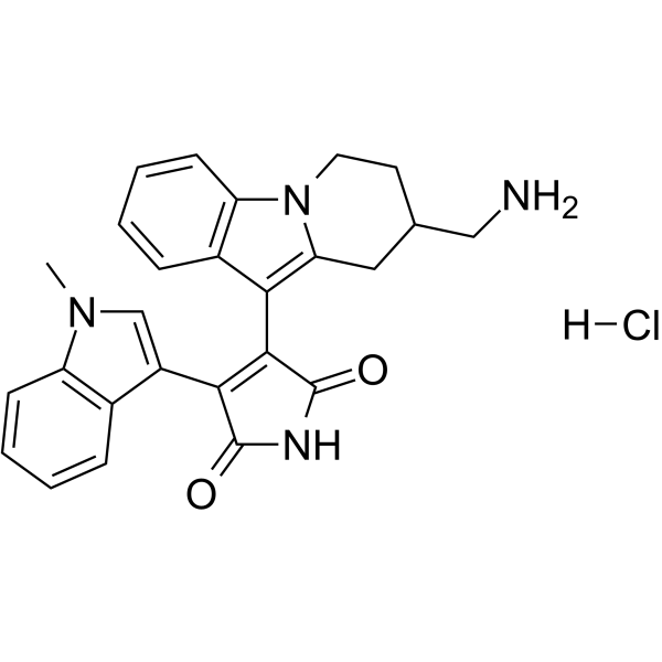
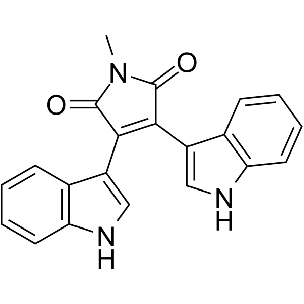
Bisindolylmaleimide X hydrochloride(Ro 31-8425 hydrochloride, BIM-X hydrochloride) is a cell-penetrating PKC inhibitor. Bisindolylmaleimide X hydrochloride is a potent cyclin-dependent kinase 2 (CDK2) antagonist with an IC50 of 200 nM. Bisindolylmaleimide X hydrochloride inhibits the proliferation of CD4 T cells in vitro. Bisindolylmaleimide X hydrochloride inhibits eNOS-Ser1177 phosphorylation in human embryonic vein endothelial cells. Bisindolylmaleimide X hydrochloride can be used for research on the immune system and cardiovascular diseases.
For research use only. We do not sell to patients.
- Purity : 98.90%
- CAS No.: 145317-11-9
- Formula: C26H25ClN4O2
- Molecular Weight:460.96
-
Storage:
4°C, sealed storage, away from moisture and light
* In solvent : -80°C, 6 months; -20°C, 1 month (sealed storage, away from moisture and light)

To place orders, for customer services and technical support, please contact: MedChemExpress USA
Tel: 609-228-6898 E-mail: [email protected] [email protected]


-
 Biological Activity
-
Chemical Information
-
Solvent & Solubility
- Protocols
- Purity & Documentation
- References
-
 Help & FAQs


-
HY-13689
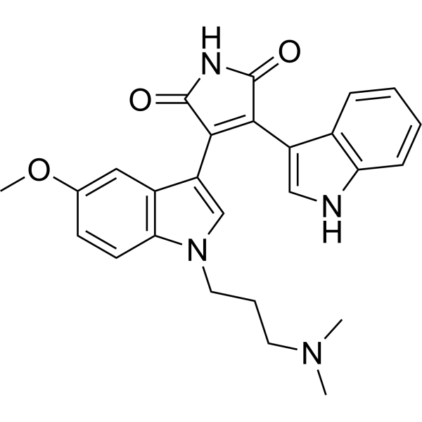
Go 6983PKC Inhibitor -
HY-129624A
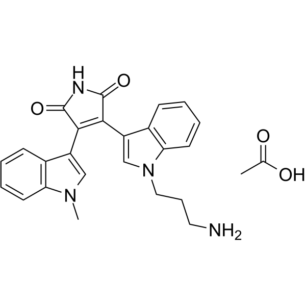
Bisindolylmaleimide VIII acetatePKC Inhibitor -
HY-10342
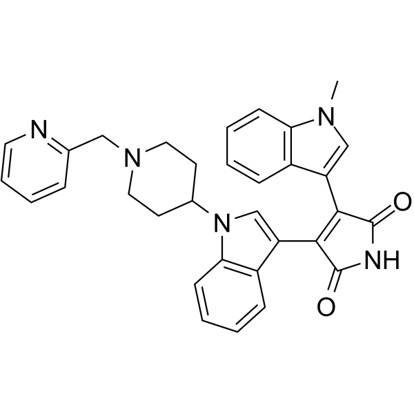
EnzastaurinPKCβ Inhibitor -
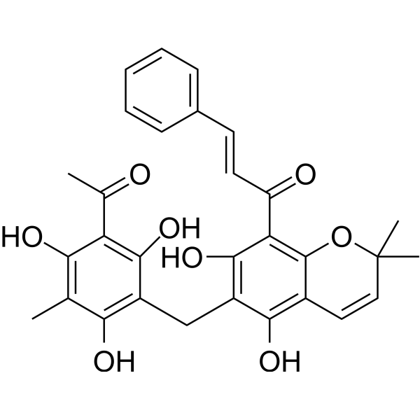
HY-18980
RottlerinPKC Inhibitor -
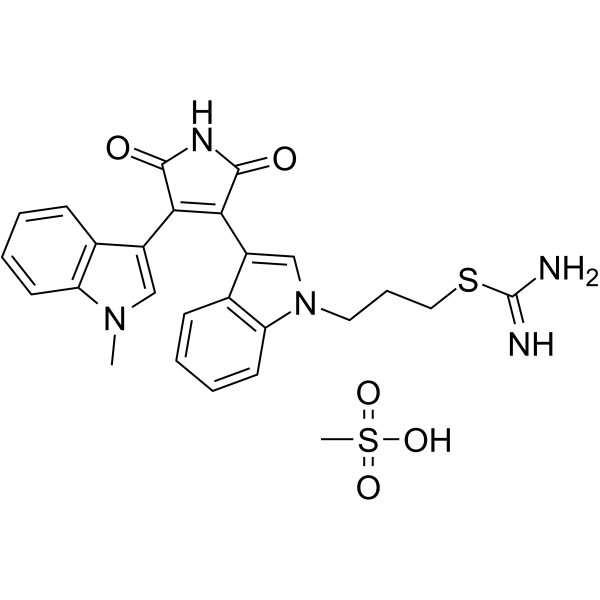
HY-13866
Ro 31-8220 mesylatePCK Inhibitor -
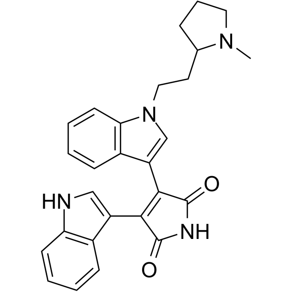
HY-108604
Bisindolylmaleimide II -
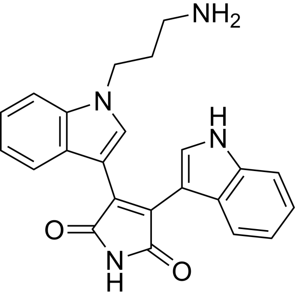
HY-117393
Bisindolylmaleimide IIIPKC Inhibitor -
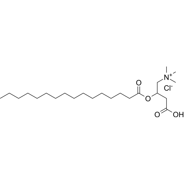
HY-101017
Palmitoylcarnitine chlorideAcylcarnitine -
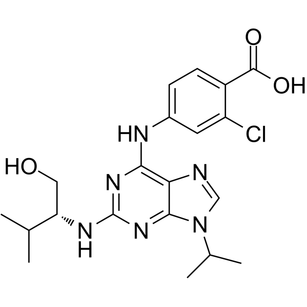
HY-18299
Purvalanol BCDK Inhibitor -
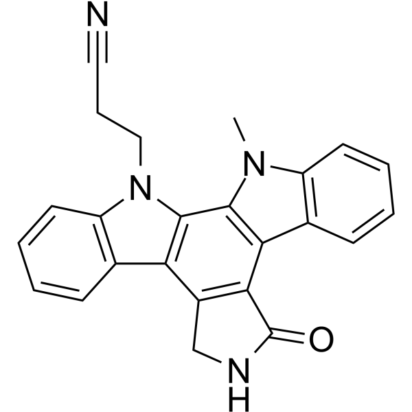
HY-10183
Go6976PKC Inhibitor -
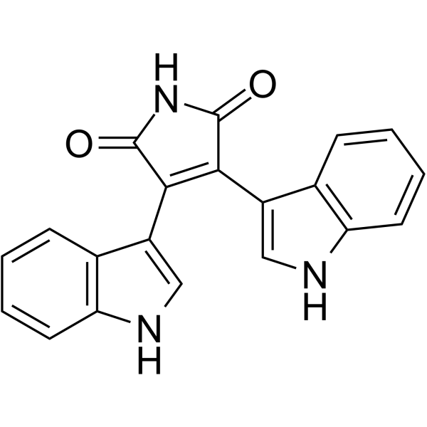
HY-108254
Bisindolylmaleimide IVPKC/HCMV Inhibitor -
HY-112400
Bisindolylmaleimide VS6K Inhibitor
Publications Citing Use of MedChemExpress (MCE) Bisindolylmaleimide X hydrochloride
More

Biological Activity
Description
IC50 & Target
[1]|
PKC |
CDK2 200 nM (IC50) |
In Vitro
Bisindolylmaleimide X hydrochloride (BIM-X hydrochloride; Ro31-8425 hydrochloride) derivatives acts as an useful tool to compare inhibitor potencies and selectivities for a target of interest[1].
MedChemExpress (MCE) has not independently confirmed the accuracy of these methods. They are for reference only. Further protocols information, click here.


Chemical Information
-
CAS No. 145317-11-9
-
Appearance Solid
-
Molecular Weight 460.96
-
Formula C26H25ClN4O2
-
Color Light yellow to orange
-
SMILES
O=C(C(C1=C(CC(CN)CC2)N2C3=C1C=CC=C3)=C4C5=CN(C)C6=C5C=CC=C6)NC4=O.[H]Cl
-
Synonyms
BIM-X hydrochloride; Ro31-8425 hydrochloride
-
Shipping
Room temperature in continental US; may vary elsewhere.
-
Storage
4°C, sealed storage, away from moisture and light
* In solvent : -80°C, 6 months; -20°C, 1 month (sealed storage, away from moisture and light)


Publications (2)
-
Journal Impact Factor
-
Most Recent
-
Cell Chem Biol
Identifying enhancers of innate immune signaling as broad-spectrum antivirals active against emerging viruses. [Abstract]2022 Jul 21;29(7):1113-1125.e6. PMID: 35728599 -
PeerJ
Icariside II induces rapid phosphorylation of endothelial nitric oxide synthase via multiple signaling pathways. [Abstract]2022 Oct 25:10:e14192. PMID: 36312762


Solvent & Solubility
In Vitro:
DMSO : 20.83 mg/mL (45.19 mM; Need ultrasonic; Hygroscopic DMSO has a significant impact on the solubility of product, please use newly opened DMSO)
H2O : < 0.1 mg/mL (insoluble)
Please refer to the solubility information to select the appropriate solvent. Once prepared, please aliquot and store the solution to prevent product inactivation from repeated freeze-thaw cycles.
Storage method and period of stock solution: -80°C, 6 months; -20°C, 1 month (sealed storage, away from moisture and light). When stored at -80°C, please use it within 6 months. When stored at -20°C, please use it within 1 month.
Please refer to the solubility information to select the appropriate solvent. Once prepared, please aliquot and store the solution to prevent product inactivation from repeated freeze-thaw cycles.
Storage method and period of stock solution: -80°C, 6 months; -20°C, 1 month (sealed storage, away from moisture and light). When stored at -80°C, please use it within 6 months. When stored at -20°C, please use it within 1 month.
Concentration (start) × Volume (start) = Concentration (final) × Volume (final)
In Vivo:
Select the appropriate dissolution method based on your experimental animal and administration route.
- For the following dissolution methods, please ensure to first prepare a clear stock solution using an In Vitro approach and then sequentially add co-solvents:
- To ensure reliable experimental results, the clarified stock solution can be appropriately stored based on storage conditions. As for the working solution for In Vivo experiments, it is recommended to prepare freshly and use it on the same day.
- The percentages shown for the solvents indicate their volumetric ratio in the final prepared solution. If precipitation or phase separation occurs during preparation, heat and/or sonication can be used to aid dissolution.
Add each solvent one by one: 10% DMSO 40% PEG300 5% Tween-80 45% Saline
Solubility: ≥ 2.08 mg/mL (4.51 mM); Clear solution
This protocol yields a clear solution of ≥ 2.08 mg/mL (saturation unknown).
Taking 1 mL working solution as an example, add 100 μL DMSO stock solution (20.8 mg/mL) to 400 μL PEG300, and mix evenly; then add 50 μL Tween-80 and mix evenly; then add 450 μL Saline to adjust the volume to 1 mL.
Preparation of Saline: Dissolve 0.9 g sodium chloride in ddH₂O and dilute to 100 mL to obtain a clear Saline solution.
In Vivo Dissolution Calculator
Please enter the basic information of animal experiments:
-
-
-
-
Recommended: Prepare an additional quantity of animals to account for potential losses during experiments.
Please enter your animal formula composition:
-
%DMSO +
Recommended: Keep the proportion of DMSO in working solution below 2% if your animal is weak.
-
%+
-
+%Tween-80 + +
-
%Saline +
The co-solvents required include: DMSO, . All of co-solvents are available by MedChemExpress (MCE). , Tween 80. All of co-solvents are available by MedChemExpress (MCE).
Working solution concentration: 0.22 mg/mL
Method for preparing stock solution: mg drug dissolved in μL DMSO. Stock solution concentration: mg/mL. * In solvent : -80°C, 6 months; -20°C, 1 month (sealed storage, away from moisture and light)
1. Take μL DMSO stock solution;
2. Add μL .
μL , mix evenly;
3. Then add μL Tween 80, mix evenly;
4. Then add μL
Please ensure that the stock solution in the first step is dissolved to a clear state, and add co-solvents in sequence. You can use ultrasonic heating (ultrasonic cleaner, recommended frequency 20-40 kHz), vortexing, etc. to assist dissolution.

Protocols
-
Kinase activity and phosphorylation assays
Kinase activity assays measure the ability of kinases to transfer phosphate groups from ATP to specific substrates, while phosphorylation assays detect the presence and levels of phosphorylated proteins. Common methods include radiolabeled ATP incorporation (e. g. ,), ADP release detection via bioluminescence (e. g. ,[3]), enzyme-linked immunosorbent assays (ELISA) for phospho-specific epitopes (e. g. ,[6]), and microtiter-based formats for high-throughput screening (e. g. ,[8]). The ADP-Glo assay quantifies kinase activity by measuring ADP produced during phosphorylation using a luciferase-based system. Radiometric assays involve autoradiography or scintillation counting after incorporation of 32P-labeled ATP into substrate proteins. ELISA-based approaches rely on phospho-specific antibodies to detect activated kinases in cell lysates or purified samples.
-
Western Blot
Western blotting (WB) is a commonly used experimental method in molecular biology, biochemistry, and immunogenetics for identifying and quantifying target proteins. It combines gel electrophoresis with immunoassay, enabling researchers to analyze protein expression, post-translational modifications, and molecular weight.
-
Cell Cytotoxicity Assay
Cytotoxicity assays are usually based on the assessment of cell membrane damage, which can also be indirectly detected by measuring cell viability. Detection methods include MTT assay, CKK-8 assay, LDH assay and ATP assay, etc.
-
Endothelial Tube Formation Assay
Endothelial tube formation assay evaluates the ability of endothelial cells to attach, migrate, align, and organize into capillary-like networks when cultured on gelled basement membrane extract or Matrigel; the readout is the morphology and quantity of tube-like networks, which reflects an in vitro endothelial morphogenesis step related to angiogenesis. Basement membrane extract/Matrigel provides laminin-rich extracellular matrix cues that support endothelial differentiation into capillary-like structures, but it can contain biologically active growth factors, so growth-factor-reduced matrix is preferred when testing defined angiogenic stimulators or inhibitors.
-
Protocol for Kinase activity and phosphorylation assays
Kinase activity assays measure transfer of phosphate from ATP to a protein or peptide substrate, generating phosphorylated substrate, ADP, or incorporated radiolabeled phosphate as the readout; phosphorylation assays measure site-specific phosphorylation in cells or tissues as a proxy for kinase-pathway activation, inhibition, or substrate regulation. Phosphorylation can be detected by phospho-specific Western blot, immunoprecipitation kinase assay, phospho-immunofluorescence, phospho-flow cytometry, luminescent ADP detection, radiolabeled ATP incorporation, or reporter-based pathway assays, and these readouts can be applied to cancer cells, primary neurons, mouse tumors, organoids, inflammatory macrophages, ferroptosis studies, and mitophagy studies when the kinase target is biologically relevant.
-
Cell Viability Determination by MTT Colorimetric Assay
The following protocol uses the MTT colorimetric assay as a classic literature-established method for assessing cell viability/metabolic activity in cultured mammalian cells. MTT[3-(4,5-dimethylthiazol-2-yl)-2,5-diphenyltetrazolium bromide] is reduced by metabolically active cells to a colored formazan product; the amount of formazan is quantified spectrophotometrically and provides an indirect measure of metabolically active viable cells. Importantly, MTT reduction reflects cellular oxidoreductase/metabolic activity rather than an absolute direct count of living cells, so changes in cellular metabolism can alter the signal independently of cell number.


Purity & Documentation
-
Data Sheet (278 KB)
-
SDS (393 KB)
- English - EN (393 KB)
- Français - FR (393 KB)
- Deutsch - DE (393 KB)
- Norwegian - NO (393 KB)
- Español - ES (393 KB)
- Swedish - SV (393 KB)
- Italian - IT (393 KB)
- Korean - KR (393 KB)
- Portuguese - PT (393 KB)
-
Handling Instructions (2659 KB)
References
[1]. Levy O, et al. A cell-based drug delivery platform for treating central nervous system inflammation. J Mol Med (Berl). 2021 May;99(5):663-671. [Content Brief]
[2]. Song W, et al. Icariside II induces rapid phosphorylation of endothelial nitric oxide synthase via multiple signaling pathways. PeerJ. 2022 Oct 25;10:e14192. [Content Brief]
[3]. Brehmer D, et al. Proteome-wide identification of cellular targets affected by bisindolylmaleimide-type protein kinase C inhibitors. Mol Cell Proteomics. 2004 May;3(5):490-500. [Content Brief]


Complete Stock Solution Preparation Table
Please refer to the solubility information to select the appropriate solvent. Once prepared, please aliquot and store the solution to prevent product inactivation from repeated freeze-thaw cycles.
Storage method and period of stock solution: -80°C, 6 months; -20°C, 1 month (sealed storage, away from moisture and light). When stored at -80°C, please use it within 6 months. When stored at -20°C, please use it within 1 month.
| Optional Solvent | Concentration Solvent Mass | 1 mg | 5 mg | 10 mg | 25 mg |
|---|---|---|---|---|---|
| DMSO | 1 mM | 2.1694 mL | 10.8469 mL | 21.6939 mL | 54.2346 mL |
| 5 mM | 0.4339 mL | 2.1694 mL | 4.3388 mL | 10.8469 mL | |
| 10 mM | 0.2169 mL | 1.0847 mL | 2.1694 mL | 5.4235 mL | |
| 15 mM | 0.1446 mL | 0.7231 mL | 1.4463 mL | 3.6156 mL | |
| 20 mM | 0.1085 mL | 0.5423 mL | 1.0847 mL | 2.7117 mL | |
| 25 mM | 0.0868 mL | 0.4339 mL | 0.8678 mL | 2.1694 mL | |
| 30 mM | 0.0723 mL | 0.3616 mL | 0.7231 mL | 1.8078 mL | |
| 40 mM | 0.0542 mL | 0.2712 mL | 0.5423 mL | 1.3559 mL |


Bisindolylmaleimide X hydrochloride Related Classifications
HY-108136A Related Classifications
 Powered by Bioz