



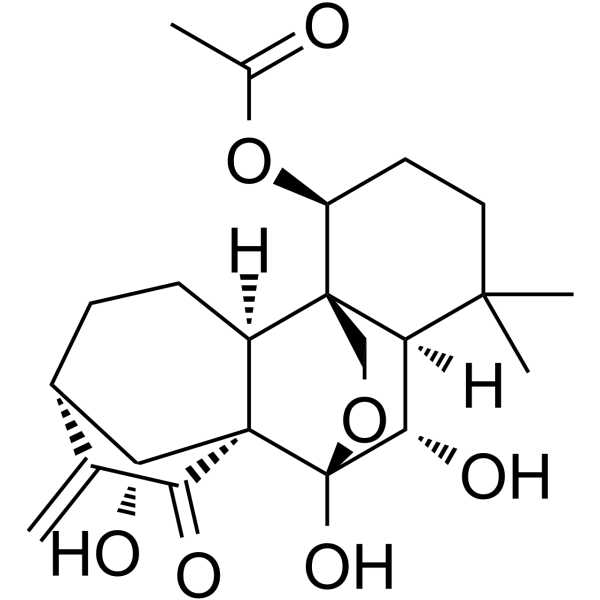
Lasiokaurin
Based on 1 publication(s) in Google Scholar
Lasiokaurin is an anticancer agent. Lasiokaurin can be isolated from Rabdosia rubescens (Hemsl.) H. Hara. Lasiokaurin inhibits the function of the PDPK1-AKT axis, with a Kd value of 75.93 μM for PDPK1. Lasiokaurin inhibits mTOR, STAT3, MAPK and NF-κB. Lasiokaurin reduces the mRNA and protein expression levels of PLK1. Lasiokaurin decreases DNA synthesis levels, induces cell Apoptosis, and regulates Autophagy processes. Lasiokaurin inhibits tumor growth in xenograft models. Lasiokaurin can be used in research related to nasopharyngeal carcinoma, triple-negative breast cancer.
Nur für Forschungszwecke. Wir verkaufen nicht an Patienten.
- Reinheit : 99.68%
- CAS. Nr.: 28957-08-6
- Formel: C22H30O7
- Molecular Weight:406.47
-
Speicherung:
-20°C, protect from light
* In solvent : -80°C, 6 months; -20°C, 1 month (protect from light)

To place orders, for customer services and technical support, please contact: MedChemExpress USA
Tel: 609-228-6898 E-mail: [email protected] [email protected]


-
 Biologische Aktivität
-
Chemical Information
-
Lösungsmittel & Löslichkeit
- Protokoll
- Reinheit & Dokumentation
- Verweise
-
 Help & FAQs


Publications Citing Use of MedChemExpress (MCE) Lasiokaurin
MoreAlle DNA/RNA Synthesis Isoform-spezifische Produkte anzeigen
More

Biologische Aktivität
Beschreibung
IC50 & Target
[1]|
PLK1 |
Cellular Effect
|
Cell Line

|
Type | Value | Description | References |
|---|---|---|---|---|
| A549 | IC50 |
10.9 μM
Compound: 8
|
Antiproliferative activity against human A549 cells incubated for 72 hrs by CCK8 cells
Antiproliferative activity against human A549 cells incubated for 72 hrs by CCK8 cells
|
[PMID: 31200238] |
| A549 | IC50 |
13.47 μM
Compound: 6
|
Antiproliferative activity against human A549 cells assessed as reduction in cell viability after 72 hrs by MTT assay
Antiproliferative activity against human A549 cells assessed as reduction in cell viability after 72 hrs by MTT assay
|
[PMID: 31202992] |
| A549 | IC50 |
6.7 μM
Compound: 19
|
Cytotoxicity against human A549 cells after 48 hrs by MTT assay
Cytotoxicity against human A549 cells after 48 hrs by MTT assay
|
[PMID: 23819871] |
| A549 | IC50 |
7.6 μM
Compound: 13
|
Cytotoxicity against human A549 cells after 48 hrs by MTT method
Cytotoxicity against human A549 cells after 48 hrs by MTT method
|
[PMID: 21534539] |
| B16 | IC50 |
32.57 μM
Compound: 5
|
Antiproliferative activity against mouse B16 cells assessed as reduction in cell viability after 48 hrs by MTT assay
Antiproliferative activity against mouse B16 cells assessed as reduction in cell viability after 48 hrs by MTT assay
|
[PMID: 31877536] |
| Bel-7402 | IC50 |
3.59 μM
Compound: 6
|
Antiproliferative activity against human Bel7402 cells assessed as reduction in cell viability after 72 hrs by MTT assay
Antiproliferative activity against human Bel7402 cells assessed as reduction in cell viability after 72 hrs by MTT assay
|
[PMID: 31202992] |
| HCT-116 | IC50 |
13.48 μM
Compound: 5
|
Antiproliferative activity against human HCT-116 cells assessed as reduction in cell viability after 48 hrs by MTT assay
Antiproliferative activity against human HCT-116 cells assessed as reduction in cell viability after 48 hrs by MTT assay
|
[PMID: 31877536] |
| HepG2 | IC50 |
11.94 μM
Compound: 5
|
Antiproliferative activity against human HepG2 cells assessed as reduction in cell viability after 48 hrs by MTT assay
Antiproliferative activity against human HepG2 cells assessed as reduction in cell viability after 48 hrs by MTT assay
|
[PMID: 31877536] |
| HepG2 | IC50 |
5.67 μM
Compound: 8
|
Antiproliferative activity against human HepG2 cells incubated for 72 hrs by CCK8 cells
Antiproliferative activity against human HepG2 cells incubated for 72 hrs by CCK8 cells
|
[PMID: 31200238] |
| HL-60 | IC50 |
1.8 μM
Compound: 19
|
Cytotoxicity against human HL60 cells after 48 hrs by MTT assay
Cytotoxicity against human HL60 cells after 48 hrs by MTT assay
|
[PMID: 23819871] |
| HL-60 | IC50 |
3.2 μM
Compound: 13
|
Cytotoxicity against human HL60 cells after 48 hrs by MTT method
Cytotoxicity against human HL60 cells after 48 hrs by MTT method
|
[PMID: 21534539] |
| K562 | IC50 |
4.92 μM
Compound: 6
|
Antiproliferative activity against human K562 cells assessed as reduction in cell viability after 72 hrs by MTT assay
Antiproliferative activity against human K562 cells assessed as reduction in cell viability after 72 hrs by MTT assay
|
[PMID: 31202992] |
| K562 | IC50 |
521 μM
Compound: 5
|
Antiproliferative activity against human K562 cells assessed as reduction in cell viability after 48 hrs by MTT assay
Antiproliferative activity against human K562 cells assessed as reduction in cell viability after 48 hrs by MTT assay
|
[PMID: 31877536] |
| L02 | IC50 |
22.36 μM
Compound: 6
|
Cytotoxicity against human L02 cells assessed as reduction in cell viability after 72 hrs by MTT assay
Cytotoxicity against human L02 cells assessed as reduction in cell viability after 72 hrs by MTT assay
|
[PMID: 31202992] |
| L02 | IC50 |
25.39 μM
Compound: 5
|
Cytotoxicity against human L02 cells assessed as cell viability after 48 hrs by MTT assay
Cytotoxicity against human L02 cells assessed as cell viability after 48 hrs by MTT assay
|
[PMID: 31877536] |
| MCF7 | IC50 |
19.82 μM
Compound: 5
|
Antiproliferative activity against human MCF7 cells assessed as reduction in cell viability after 48 hrs by MTT assay
Antiproliferative activity against human MCF7 cells assessed as reduction in cell viability after 48 hrs by MTT assay
|
[PMID: 31877536] |
| MCF7 | IC50 |
2.5 μM
Compound: 19
|
Cytotoxicity against human MCF7 cells after 48 hrs by MTT assay
Cytotoxicity against human MCF7 cells after 48 hrs by MTT assay
|
[PMID: 23819871] |
| MCF7 | IC50 |
5.3 μM
Compound: 13
|
Cytotoxicity against human MCF7 cells after 48 hrs by MTT method
Cytotoxicity against human MCF7 cells after 48 hrs by MTT method
|
[PMID: 21534539] |
| PBMC | IC50 |
>50 μM
Compound: 5
|
Cytotoxicity against human PBMC cells assessed as cell viability after 48 hrs by MTT assay
Cytotoxicity against human PBMC cells assessed as cell viability after 48 hrs by MTT assay
|
[PMID: 31877536] |
| PBMC | IC50 |
>50 μM
Compound: 6
|
Cytotoxicity against human PBMC assessed as reduction in cell viability after 72 hrs by MTT assay
Cytotoxicity against human PBMC assessed as reduction in cell viability after 72 hrs by MTT assay
|
[PMID: 31202992] |
| RAW264.7 | IC50 |
1.9 μM
Compound: 19
|
Antiinflammatory activity in mouse RAW264.7 cells assessed as inhibition of LPS-induced nitric oxide production after 18 hrs by Griess method
Antiinflammatory activity in mouse RAW264.7 cells assessed as inhibition of LPS-induced nitric oxide production after 18 hrs by Griess method
|
[PMID: 23819871] |
| RPMI-8226 | IC50 |
11.03 μM
Compound: 8
|
Antiproliferative activity against human RPMI8226 cells incubated for 72 hrs by CCK8 cells
Antiproliferative activity against human RPMI8226 cells incubated for 72 hrs by CCK8 cells
|
[PMID: 31200238] |
| SGC-7901 | IC50 |
6.45 μM
Compound: 6
|
Antiproliferative activity against human SGC7901 cells assessed as reduction in cell viability after 72 hrs by MTT assay
Antiproliferative activity against human SGC7901 cells assessed as reduction in cell viability after 72 hrs by MTT assay
|
[PMID: 31202992] |
| SMMC-7721 | IC50 |
2 μM
Compound: 13
|
Cytotoxicity against human SMMC7721 cells after 48 hrs by MTT method
Cytotoxicity against human SMMC7721 cells after 48 hrs by MTT method
|
[PMID: 21534539] |
| SMMC-7721 | IC50 |
2.7 μM
Compound: 19
|
Cytotoxicity against human SMMC7721 cells after 48 hrs by MTT assay
Cytotoxicity against human SMMC7721 cells after 48 hrs by MTT assay
|
[PMID: 23819871] |
| SW480 | IC50 |
2.3 μM
Compound: 13
|
Cytotoxicity against human SW480 cells after 48 hrs by MTT method
Cytotoxicity against human SW480 cells after 48 hrs by MTT method
|
[PMID: 21534539] |
| SW480 | IC50 |
2.4 μM
Compound: 19
|
Cytotoxicity against human SW480 cells after 48 hrs by MTT assay
Cytotoxicity against human SW480 cells after 48 hrs by MTT assay
|
[PMID: 23819871] |
In Vitro
Lasiokaurin (0.19-50 μM; 24-72 h) dose- and time-dependently inhibits the viability of CNE-1, CNE-2, and C666-1 nasopharyngeal carcinoma cells in vitro[1].
Lasiokaurin (0.5-2 μM; 7 days) dose-dependently inhibits the colony formation of CNE-1 and CNE-2 nasopharyngeal carcinoma cells in vitro, with significant inhibition observed at concentrations as low as 0.5 μM[1].
Lasiokaurin (2.5-10 μM; 24 h) dose-dependently inhibits the activation of MAPK, mTOR, STAT3, and NF-κB pathways in CNE-1 and CNE-2 nasopharyngeal carcinoma cells in vitro, including inhibition of both mTORC1 and mTORC2 complexes[1].
Lasiokaurin (0.2-50 μM; 24-72 h) potently inhibits viability of MDA-MB-231, MDA-MB-468, and MCF7 breast cancer cells with IC50 values ranging from 1.6 μM to 8.35 μM across 24-72 h, and exhibits lower toxicity to MCF-10A normal breast cells[2].
Lasiokaurin (72 h) potently inhibits the viability of SK-BR-3, MDA-MB-231, BT-549, MCF-7, and T-47D human breast cancer cells with IC50 values ranging from 1.59 μM to 4.16 μM after 72 h of treatment[3].
Lasiokaurin (0.3-3 μM) inhibits the viability of patient-derived xenograft breast cancer organoids in a concentration-dependent manner[3].
Lasiokaurin (2.5-10 μM; 24-48 h) induces dose- and time-dependent G2/M cell cycle arrest in CNE-1 and CNE-2 nasopharyngeal carcinoma cells in vitro by reducing cyclin B1 and cdc2 expression[1].
Lasiokaurin (2.5-10 μM; 24 h) dose-dependently induces apoptosis in CNE-1 and CNE-2 nasopharyngeal carcinoma cells in vitro by increasing Bax expression[1].
Lasiokaurin (2.5-5 μM; 12-48 h) dose- and time-dependently inhibits the migration of CNE-1 and CNE-2 nasopharyngeal carcinoma cells in vitro[1].
Lasiokaurin (2.5-10 μM; 24-48 h) at concentrations of 2.5-10 μM induces DNA damage in MDA-MB-231 and MDA-MB-468 TNBC cells by suppressing PARP expression after 24 and 48 h of treatment[2].
MedChemExpress (MCE) has not independently confirmed the accuracy of these methods. They are for reference only.
-
Cell Line:CNE-1, CNE-2, C666-1
-
Concentration:0.19-50 μM
-
Incubation Time:24 h; 48 h; 72 h
-
Result:Caused a significant dose- and time-dependent reduction in cell viability across all three NPC cell lines.
Inhibited cell viability with statistically significant effects (p < 0.05, p < 0.001) observed at multiple concentration-time combinations.
-
Cell Line:CNE-1, CNE-2
-
Concentration:2.5-10 μM
-
Incubation Time:24 h; 48 h
-
Result:Caused a dose- and time-dependent increase in the proportion of cells in the G2/M phase.
Reduced cyclin B1 and cdc2 protein expression in both cell lines at 2.5, 5, and 10 μM (24 h incubation), with statistically significant decreases observed at 5 μM and 10 μM (p < 0.05, p < 0.01).
-
Cell Line:CNE-1, CNE-2
-
Concentration:2.5-10 μM
-
Incubation Time:24 h
-
Result:Caused a dose-dependent increase in the percentage of apoptotic cells (early and late apoptosis) in both cell lines.
Increased Bax expression in CNE-1 cells at 2.5 μM, 5 μM, and 10 μM, with significant elevations at 5 μM and 10 μM (p < 0.01).
Increased Bax expression in CNE-2 cells at 2.5 μM, 5 μM, and 10 μM, with significant elevations at 5 μM (p < 0.05) and 10 μM (p < 0.01).
-
Cell Line:CNE-1, CNE-2
-
Concentration:2.5-5 μM
-
Incubation Time:12 h; 24 h; 36 h; 48 h
-
Result:Caused a dose- and time-dependent reduction in relative wound healing rates in both cell lines.
Resulted in significantly lower wound healing rates compared to controls at all time points with 5 μM Lasiokaurin, with 2.5 μM Lasiokaurin also showing inhibitory effects.
-
Cell Line:CNE-1, CNE-2
-
Concentration:2.5-10 μM
-
Incubation Time:24 h
-
Result:Dose-dependently reduced phosphorylation of Erk1/2, p38, mTOR, STAT3, and NF-κB in both cell lines, with significant decreases observed at multiple concentrations (p < 0.05, p < 0.01).
Reduced expression of Rictor, Raptor, and Gβl (components of mTORC1 and mTORC2) in both cell lines, with significant decreases at 5 μM and 10 μM (p < 0.05, p < 0.01).
In Vivo
Lasiokaurin (5-10 mg/kg; i.p.; daily; 20 days) significantly inhibits triple-negative breast cancer xenograft tumor growth in BALB/c nude mice, with the 10 mg/kg dose showing efficacy comparable to docetaxel and no detectable toxicity to vital organs or body weight[2].
Lasiokaurin (7.5-15 mg/kg; i.p.; once every 2 days; 27 days) inhibits breast cancer growth in vivo by suppressing PLK1 pathway signaling, with 15 mg/kg lasiokaurin significantly reducing tumor volume, tumor weight, and cell proliferation while inducing apoptosis, without causing significant body weight loss[3].
Lasiokaurin (25 mg/kg; i.p.; once every two days) significantly inhibits breast cancer xenograft growth in nude mice by activating autophagy, inhibiting the AKT/mTOR signaling pathway, and suppressing glycolytic activity, with no observed toxicity[4].
MedChemExpress (MCE) has not independently confirmed the accuracy of these methods. They are for reference only.
-
Animal Model:BALB/c nude mice (female, housed at 23°C with 12 h light/dark cycle)[2]
-
Dosage:5 mg/kg; 10 mg/kg
-
Administration:i.p.; daily; 20 days
-
Result:Significantly reduced tumor volume and tumor weight relative to the vehicle group.
Showed slightly lower efficacy than docetaxel at 5 mg/kg.
Demonstrated comparable efficacy to docetaxel at 10 mg/kg.
Caused no significant effect on mouse body weight.
Showed no notable alterations in organ weight or histopathological characteristics of heart, lung, liver, spleen, or kidney relative to the vehicle group.
-
Animal Model:Balb/c nude (female, 5 weeks old, 18-22 g, subcutaneous xenograft model)[3]
-
Dosage:7.5 mg/kg; 15 mg/kg
-
Administration:i.p.; once every 2 days; 27 days
-
Result:Significantly reduced tumor volume and tumor weight at 15 mg/kg compared to vehicle controls.
Showed no significant body weight loss relative to vehicle controls.
Significantly reduced Ki67 expression in tumor tissue.
Significantly increased TUNEL-positive cells in tumor tissue.
Downregulated tumor tissue protein levels of PLK1, CDC25C, CyclinB1, CDC2, and p-AKT relative to total AKT.
-
Animal Model:Nude mice (female, six-week-old)[4]
-
Dosage:25 mg/kg
-
Administration:i.p.; once every two days; 9 total doses
-
Result:Significantly reduced xenograft tumor growth compared to PBS control, with lower final tumor weights and slower tumor volume progression over 18 days.
Reduced Ki67-positive proliferating cells in tumor tissues.
Increased TUNEL-positive apoptotic cells and necrotic area in tumor tissues.
Increased LC3II levels in tumor tissues.
Decreased PDPK1 expression in tumor tissues.
Increased protein levels of p62, beclin1, ATG5, and LC3II in tumor tissues.
Decreased phosphorylated levels of AKT, mTOR, and p70S6K in tumor tissues.
Reduced protein levels of glycolysis-related markers HK2, GLUT1, GLUT3, and LDHA in tumor tissues.
Maintained stable mouse body weights with no treatment-related damage observed in heart, liver, spleen, lung, and kidney via H&E staining.
- RNA extraction experimental
- Western Blot
- Annexin V plus membrane-impermeant dye apoptosis staining
- BrdU Incorporation Assay
- Patient-Derived Orthotopic Xenograft (PDOX)
- Research Protocol for Inflammation-related Diseases
- Breast Cancer Modeling
- Protocol For Protein Expression And Purification
- EdU Incorporation Assay (Click Chemistry-Based DNA Synthesis Measurement)
- Detection of Apoptosis
- TUNEL staining for apoptotic DNA fragmentation
- Autophagy
- Patient-Derived Xenograft (PDX)
- Apoptosis Solutions
- Splenic/Portal-Vein Liver Metastasis Xenograft
- Lysosome and acidic-vesicle live-cell staining
- Subcutaneous Cell-Line-Derived Xenograft
- Macroautophagy Solutions
- Orthotopic Cell-Line Xenograft
- Intraperitoneal/Peritoneal Dissemination Xenograft


Chemical Information
-
CAS. Nr. 28957-08-6
-
Appearance Solid
-
Molecular Weight 406.47
-
Formel C22H30O7
-
Color White to off-white
-
SMILES
O[C@]1(OC2)[C@]([C@@H]3O)(C4=O)[C@](CC[C@H]3C4=C)([H])[C@]2([C@H](CC5)OC(C)=O)[C@](C5(C)C)([H])[C@@H]1O
-
Structure Classification
-
Versand
Room temperature in continental US; may vary elsewhere.
-
Speicherung
-20°C, protect from light
* In solvent : -80°C, 6 months; -20°C, 1 month (protect from light)


Publications (1)
-
Journal Impact Factor
-
Most Recent
-
Biochem Pharmacol
Lasiokaurin suppresses breast cancer growth by blocking autophagic flux and regulating cellular energy homeostasis. [Abstract]2025 Aug 7;242(Pt 3):117212. PMID: 40782951


Lösungsmittel & Löslichkeit
In Vitro:
DMSO : 100 mg/mL (246.02 mM; Need ultrasonic; Hygroscopic DMSO has a significant impact on the solubility of product, please use newly opened DMSO)
Please refer to the solubility information to select the appropriate solvent. Once prepared, please aliquot and store the solution to prevent product inactivation from repeated freeze-thaw cycles.
Storage method and period of stock solution: -80°C, 6 months; -20°C, 1 month (protect from light). When stored at -80°C, please use it within 6 months. When stored at -20°C, please use it within 1 month.
Please refer to the solubility information to select the appropriate solvent. Once prepared, please aliquot and store the solution to prevent product inactivation from repeated freeze-thaw cycles.
Storage method and period of stock solution: -80°C, 6 months; -20°C, 1 month (protect from light). When stored at -80°C, please use it within 6 months. When stored at -20°C, please use it within 1 month.
Konzentration (Stammlösung) × Volumen (Stammlösung) = Konzentration (Ziellösung) × Volumen (Ziellösung)
In Vivo:
Select the appropriate dissolution method based on your experimental animal and administration route.
- For the following dissolution methods, please ensure to first prepare a clear stock solution using an In Vitro approach and then sequentially add co-solvents:
- To ensure reliable experimental results, the clarified stock solution can be appropriately stored based on storage conditions. As for the working solution for In Vivo experiments, it is recommended to prepare freshly and use it on the same day.
- The percentages shown for the solvents indicate their volumetric ratio in the final prepared solution. If precipitation or phase separation occurs during preparation, heat and/or sonication can be used to aid dissolution.
Add each solvent one by one: 10% DMSO 40% PEG300 5% Tween-80 45% Saline
Solubility: ≥ 2.5 mg/mL (6.15 mM); Clear solution
This protocol yields a clear solution of ≥ 2.5 mg/mL (saturation unknown).
Taking 1 mL working solution as an example, add 100 μL DMSO stock solution (25.0 mg/mL) to 400 μL PEG300, and mix evenly; then add 50 μL Tween-80 and mix evenly; then add 450 μL Saline to adjust the volume to 1 mL.
Preparation of Saline: Dissolve 0.9 g sodium chloride in ddH₂O and dilute to 100 mL to obtain a clear Saline solution.
Add each solvent one by one: 10% DMSO 90% (20% SBE-β-CD in Saline)
Solubility: ≥ 2.5 mg/mL (6.15 mM); Clear solution
This protocol yields a clear solution of ≥ 2.5 mg/mL (saturation unknown).
Taking 1 mL working solution as an example, add 100 μL DMSO stock solution (25.0 mg/mL) to 900 μL 20% SBE-β-CD in Saline, and mix evenly.
Preparation of 20% SBE-β-CD in Saline (4°C, storage for one week): 2 g SBE-β-CD powder is dissolved in 10 mL Saline, completely dissolve until clear.
In Vivo Dissolution Calculator
Please enter the basic information of animal experiments:
-
-
-
-
Recommended: Prepare an additional quantity of animals to account for potential losses during experiments.
Please enter your animal formula composition:
-
%DMSO +
Recommended: Keep the proportion of DMSO in working solution below 2% if your animal is weak.
-
%+
-
+%Tween-80 + +
-
%Saline +
The co-solvents required include: DMSO, . All of co-solvents are available by MedChemExpress (MCE). , Tween 80. All of co-solvents are available by MedChemExpress (MCE).
Working solution concentration: 0.22 mg/mL
Method for preparing stock solution: mg drug dissolved in μL DMSO. Stock solution concentration: mg/mL. * In solvent : -80°C, 6 months; -20°C, 1 month (protect from light)
1. Take μL DMSO stock solution;
2. Add μL .
μL , mix evenly;
3. Then add μL Tween 80, mix evenly;
4. Then add μL
Please ensure that the stock solution in the first step is dissolved to a clear state, and add co-solvents in sequence. You can use ultrasonic heating (ultrasonic cleaner, recommended frequency 20-40 kHz), vortexing, etc. to assist dissolution.


Reinheit & Dokumentation
-
Data Sheet (320 KB)
-
SDS (252 KB)
- English - EN (252 KB)
- Français - FR (252 KB)
- Deutsch - DE (252 KB)
- Norwegian - NO (252 KB)
- Español - ES (252 KB)
- Swedish - SV (252 KB)
- Italian - IT (252 KB)
- Korean - KR (252 KB)
- Portuguese - PT (252 KB)
-
Handling Instructions (2659 KB)
Verweise
[2]. Lin J, et al. In Vitro and In Vivo Anti-Cancer Activity of Lasiokaurin in a Triple-Negative Breast Cancer Model. Molecules (Basel, Switzerland). 2023 Nov 22;28(23):7701. [Content Brief]
[3]. Liu Z, et al. Lasiokaurin Regulates PLK1 to Induce Breast Cancer Cell G2/M Phase Block and Apoptosis. Journal of Cancer. 2024;15(8):2318-2328. [Content Brief]
[4]. Huang R, et al. Lasiokaurin suppresses breast cancer growth by blocking autophagic flux and regulating cellular energy homeostasis. Biochemical pharmacology. 2025 Dec;242(Pt 3):117212. [Content Brief]


Complete Stock Solution Preparation Table
Please refer to the solubility information to select the appropriate solvent. Once prepared, please aliquot and store the solution to prevent product inactivation from repeated freeze-thaw cycles.
Storage method and period of stock solution: -80°C, 6 months; -20°C, 1 month (protect from light). When stored at -80°C, please use it within 6 months. When stored at -20°C, please use it within 1 month.
| Optional Solvent | Concentration Solvent Mass | 1 mg | 5 mg | 10 mg | 25 mg |
|---|---|---|---|---|---|
| DMSO | 1 mM | 2.4602 mL | 12.3010 mL | 24.6021 mL | 61.5052 mL |
| 5 mM | 0.4920 mL | 2.4602 mL | 4.9204 mL | 12.3010 mL | |
| 10 mM | 0.2460 mL | 1.2301 mL | 2.4602 mL | 6.1505 mL | |
| 15 mM | 0.1640 mL | 0.8201 mL | 1.6401 mL | 4.1003 mL | |
| 20 mM | 0.1230 mL | 0.6151 mL | 1.2301 mL | 3.0753 mL | |
| 25 mM | 0.0984 mL | 0.4920 mL | 0.9841 mL | 2.4602 mL | |
| 30 mM | 0.0820 mL | 0.4100 mL | 0.8201 mL | 2.0502 mL | |
| 40 mM | 0.0615 mL | 0.3075 mL | 0.6151 mL | 1.5376 mL | |
| 50 mM | 0.0492 mL | 0.2460 mL | 0.4920 mL | 1.2301 mL | |
| 60 mM | 0.0410 mL | 0.2050 mL | 0.4100 mL | 1.0251 mL | |
| 80 mM | 0.0308 mL | 0.1538 mL | 0.3075 mL | 0.7688 mL | |
| 100 mM | 0.0246 mL | 0.1230 mL | 0.2460 mL | 0.6151 mL |


Lasiokaurin Related Classifications
HY-123467 Related Classifications
 Powered by Bioz