



Continentalic acid

Based on 1 Customer Validation
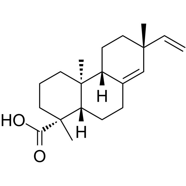
Continentalic acid is a diterpenoid organic acid. Continentalic acid exhibits multiple activities including anti-inflammatory, antioxidant, neuroprotective, antibacterial and antitumor effects. Continentalic acid alleviates oxidative stress, reduces pro-inflammatory cytokine production, inhibits MAPK phosphorylation and neutrophil infiltration, and induces growth inhibition and apoptosis of cancer cells. Continentalic acid can be used in research related to traumatic brain injury, cancer, inflammation and infections.
Para uso exclusivo en investigación. No vendemos a pacientes.
- Pureza : 99.72%
- No. CAS: 19889-23-7
- Fòrmula: C20H30O2
- Peso molecular:302.45
-
Almacenamiento:
4°C, protect from light
* In solvent : -80°C, 6 months; -20°C, 1 month (protect from light)

To place orders, for customer services and technical support, please contact: MedChemExpress USA
Tel: 609-228-6898 E-mail: [email protected] [email protected]


-
 Actividad biológica
-
Chemical Information
-
Solvente y solubilidad
- Protocolo
- Pureza y Documentación
- Referencias
-
 Help & FAQs



Ver todos los productos específicos de isoformas Caspase
More

Actividad biológica
Descripciòn
In Vitro
Continentalic acid (10-100 μM; 24-72 h) inhibits the growth of human hepatocellular carcinoma HepG2 cells in a time- and dose-dependent manner, with an IC50 of 50 μM[1].
Continentalic acid (25-100 μM; 72 h) induces apoptosis in human hepatocellular carcinoma HepG2 cells, including morphological changes and DNA fragmentation[1].
Continentalic acid (10-100 μM; 48 h) regulates the expression of apoptosis-related proteins in human hepatoma HepG2 cells, upregulates the levels of caspase-3, Bak and Bax, induces PARP cleavage, and downregulates the level of Bcl-2[1].
Continentalic acid (1-20 μM) dose-dependently inhibits the production of IL-6, IL-8, MMP-13, COX-2 and PGE2 stimulated by IL-1β in human osteoarthritic chondrocytes[3].
Continentalic acid (1-20 μM; 24 h) dose-dependently inhibits the phosphorylation of p38, ERK1/2 and JNK mitogen-activated protein kinases (MAP kinases) in IL-1β-stimulated human osteoarthritic chondrocytes[3].
Continentalic acid (10 μM) inhibits IL-1β-induced nuclear translocation of NF-κB/p65 in human osteoarthritic chondrocytes after 1 h of incubation[3].
Continentalic acid (8-16 µg/mL; 24 h) potently inhibits the growth of methicillin-sensitive Staphylococcus aureus ATCC 25923, methicillin-resistant Staphylococcus aureus ATCC 33591, and 12 clinical methicillin-resistant Staphylococcus aureus (MRSA) isolates, with an MIC value of 8-16 µg/mL[4].
MedChemExpress (MCE) has not independently confirmed the accuracy of these methods. They are for reference only.
-
Cell Line:human hepatocarcinoma HepG2 cells
-
Concentration:10, 25, 50, 100 μM
-
Incubation Time:72 h
-
Result:Induced distinct apoptotic morphological changes in HepG2 cells by 72 h, including cell shrinkage, nuclear and cytoplasmic condensation, chromatin condensation, and polylobation or fragmentation of nuclei.
Caused visible apoptotic changes in cells treated with 25, 50, and 100 μM continentalic acid.\nInduced dose-dependent DNA ladder formation in HepG2 cells after 72 h of incubation.
Caused clear laddering observed at concentrations of 25, 50, and 100 μM.
-
Cell Line:IL-1β-stimulated human osteoarthritis (OA) chondrocytes
-
Concentration:1, 3, 5, 10, 20 μM
-
Incubation Time:24 h
-
Result:Significantly inhibited IL-1β-stimulated phosphorylation of p38, ERK1/2, and JNK protein kinases in a dose-dependent manner.
In Vivo
Continentalic acid (0.97 mg/kg; i.p.; once daily; for 30 consecutive days) exerts significant anti-arthritic efficacy in MIA-induced osteoarthritis rats, restores weight-bearing balance, and prevents cartilage damage and proteoglycan loss[3].
Continentalic acid (1-50 mg/kg; i.p.; once daily for 14 consecutive days) exerts potent neuroprotective activity in a mouse traumatic brain injury (TBI) model[5].
MedChemExpress (MCE) has not independently confirmed the accuracy of these methods. They are for reference only.
-
Animal Model:BALB/c mice (6-week-old male, 27-33 g, traumatic brain injury weight-drop model)[5]
-
Dosage:1 mg/kg; 10 mg/kg; 50 mg/kg
-
Administration:i.p.; daily; 14 days
-
Result:Reduced neurological severity score.
Restored paw withdrawal threshold.
Reduced cortical histopathological damage, increased neuronal count, and reduced cerebral microbleeds compared to the TBI group.
Downregulated mRNA and protein expression of GFAP, Iba1, TLR4, NF-κB, and cleaved caspase-3; upregulated mRNA and protein expression of IκB-α and Bcl-2 compared to the TBI group.
Reduced TBI-induced DNA damage compared to the TBI group.
Reduced levels of NO, LPO, IL-1β, and TNF-α; increased levels of GST, GSH, catalase, and SOD compared to the TBI group.
-
Animal Model:BALB/c (male, 3-4 weeks old, 22-26 g, LPS-induced acute lung injury)[2]
-
Dosage:10 mg/kg; 50 mg/kg; 100 mg/kg
-
Administration:i.p.; single dose 30 minutes pre-LPS
-
Result:Achieved an 80% 24-hour survival rate at 100 mg/kg (vs. 20% in LPS-only group); showed no protective effect on mortality at 10 mg/kg.
Significantly reduced LPS-induced pyrexia at 50 mg/kg and 100 mg/kg).
Reduced lung wet/dry ratio dose-dependently at all three doses, with marked inhibition at 100 mg/kg.
Significantly elevated lung tissue levels of GSH, GST, catalase, and SOD at all three doses, with largest increases at 100 mg/kg.
Significantly reduced lung tissue MDA levels and both plasma and lung tissue NO levels at all three doses, with effects increasing with dose.
Significantly inhibited LPS-induced increases in lung tissue IL-1β, IL-6, and TNF-α at all three doses.
Normalized total neutrophil counts (NEU) to near control levels at 50 mg/kg and 100 mg/kg; normalized WBC counts to control levels at 100 mg/kg.
Reduced lung injury scores dose-dependently; reduced neutrophilic infiltration and improved tracheal architecture at these doses.
Reduced lung tissue MPO activity at all three doses, with significant inhibition at 100 mg/kg.
Significantly reduced lung tissue iNOS expression and significantly increased Nrf2 protein expression at 100 mg/kg.
- Kinase activity and phosphorylation assays
- Phagocytosis Functional Assay
- Research Protocol for Infectious Diseases
- Western Blot
- Detection of Apoptosis
- TUNEL staining for apoptotic DNA fragmentation
- Annexin V plus membrane-impermeant dye apoptosis staining
- ROS/oxidative-stress fluorescent staining
- Apoptosis Solutions
- Research Protocol for Inflammation-related Diseases
- Protocol for Kinase activity and phosphorylation assays


Chemical Information
-
No. CAS 19889-23-7
-
Appearance Solid
-
Peso molecular 302.45
-
Fòrmula C20H30O2
-
Color White to off-white
-
SMILES
C[C@]12[C@@](CCC3=C[C@](C=C)(C)CC[C@@]23[H])([H])[C@@](C)(CCC1)C(O)=O
-
Structure Classification
-
Initial Source
-
Envío
Room temperature in continental US; may vary elsewhere.
-
Almacenamiento
4°C, protect from light
* In solvent : -80°C, 6 months; -20°C, 1 month (protect from light)


Solvente y solubilidad
In Vitro:
DMSO : 100 mg/mL (330.63 mM; Need ultrasonic; Hygroscopic DMSO has a significant impact on the solubility of product, please use newly opened DMSO)
Please refer to the solubility information to select the appropriate solvent. Once prepared, please aliquot and store the solution to prevent product inactivation from repeated freeze-thaw cycles.
Storage method and period of stock solution: -80°C, 6 months; -20°C, 1 month (protect from light). When stored at -80°C, please use it within 6 months. When stored at -20°C, please use it within 1 month.
Please refer to the solubility information to select the appropriate solvent. Once prepared, please aliquot and store the solution to prevent product inactivation from repeated freeze-thaw cycles.
Storage method and period of stock solution: -80°C, 6 months; -20°C, 1 month (protect from light). When stored at -80°C, please use it within 6 months. When stored at -20°C, please use it within 1 month.
Concentration (start) × Volume (start) = Concentration (final) × Volume (final)
In Vivo:
Select the appropriate dissolution method based on your experimental animal and administration route.
- For the following dissolution methods, please ensure to first prepare a clear stock solution using an In Vitro approach and then sequentially add co-solvents:
- To ensure reliable experimental results, the clarified stock solution can be appropriately stored based on storage conditions. As for the working solution for In Vivo experiments, it is recommended to prepare freshly and use it on the same day.
- The percentages shown for the solvents indicate their volumetric ratio in the final prepared solution. If precipitation or phase separation occurs during preparation, heat and/or sonication can be used to aid dissolution.
Add each solvent one by one: 10% DMSO 40% PEG300 5% Tween-80 45% Saline
Solubility: 2.5 mg/mL (8.27 mM); Suspended solution; Need ultrasonic
This protocol yields a suspended solution of 2.5 mg/mL. Suspended solution can be used for oral and intraperitoneal injection.
Taking 1 mL working solution as an example, add 100 μL DMSO stock solution (25.0 mg/mL) to 400 μL PEG300, and mix evenly; then add 50 μL Tween-80 and mix evenly; then add 450 μL Saline to adjust the volume to 1 mL.
Preparation of Saline: Dissolve 0.9 g sodium chloride in ddH₂O and dilute to 100 mL to obtain a clear Saline solution.
Add each solvent one by one: 10% DMSO 90% (20% SBE-β-CD in Saline)
Solubility: ≥ 2.5 mg/mL (8.27 mM); Clear solution
This protocol yields a clear solution of ≥ 2.5 mg/mL (saturation unknown).
Taking 1 mL working solution as an example, add 100 μL DMSO stock solution (25.0 mg/mL) to 900 μL 20% SBE-β-CD in Saline, and mix evenly.
Preparation of 20% SBE-β-CD in Saline (4°C, storage for one week): 2 g SBE-β-CD powder is dissolved in 10 mL Saline, completely dissolve until clear.
In Vivo Dissolution Calculator
Please enter the basic information of animal experiments:
-
-
-
-
Recommended: Prepare an additional quantity of animals to account for potential losses during experiments.
Please enter your animal formula composition:
-
%DMSO +
Recommended: Keep the proportion of DMSO in working solution below 2% if your animal is weak.
-
%+
-
+%Tween-80 + +
-
%Saline +
The co-solvents required include: DMSO, . All of co-solvents are available by MedChemExpress (MCE). , Tween 80. All of co-solvents are available by MedChemExpress (MCE).
Working solution concentration: 0.22 mg/mL
Method for preparing stock solution: mg drug dissolved in μL DMSO. Stock solution concentration: mg/mL. * In solvent : -80°C, 6 months; -20°C, 1 month (protect from light)
1. Take μL DMSO stock solution;
2. Add μL .
μL , mix evenly;
3. Then add μL Tween 80, mix evenly;
4. Then add μL
Please ensure that the stock solution in the first step is dissolved to a clear state, and add co-solvents in sequence. You can use ultrasonic heating (ultrasonic cleaner, recommended frequency 20-40 kHz), vortexing, etc. to assist dissolution.


Pureza y Documentación
-
Ficha de datos (285 KB)
-
SDS (398 KB)
- English - EN (398 KB)
- Français - FR (398 KB)
- Deutsch - DE (398 KB)
- Norwegian - NO (398 KB)
- Español - ES (398 KB)
- Swedish - SV (398 KB)
- Italian - IT (398 KB)
- Korean - KR (398 KB)
- Portuguese - PT (398 KB)
-
Instrucciones de manejo (2659 KB)
Referencias
[1]. Kwon TO, et al. Continentalic acid from Aralia continentalis induces growth inhibition and apoptosis in HepG2 cells. Arch Pharm Res. 2008;31(9):1172-1178. [Content Brief]
[2]. Ali H, et al. Attenuation of LPS-induced acute lung injury by continentalic acid in rodents through inhibition of inflammatory mediators correlates with increased Nrf2 protein expression. BMC Pharmacol Toxicol. 2020;21(1):81. Published 2020 Nov 25. [Content Brief]
[3]. Hong R, et al. Continentalic Acid Rather Than Kaurenoic Acid Is Responsible for the Anti-Arthritic Activity of Manchurian Spikenard In Vitro and In Vivo. Int J Mol Sci. 2019;20(21):5488. Published 2019 Nov 4. [Content Brief]
[4]. Jeong SI, et al. Continentalic acid from Aralia continentalis shows activity against methicillin-resistant Staphylococcus aureus. Phytother Res. 2006;20(6):511-514. [Content Brief]
[5]. Zafar S, et al. Neuroprotective effect of Continentalic acid by targeting NF-κB/Bcl-2 and oxidative stress signaling in traumatic brain injury in male BALB/c mice. Mol Biol Rep. 2025;53(1):135. Published 2025 Nov 28. [Content Brief]


Complete Stock Solution Preparation Table
Please refer to the solubility information to select the appropriate solvent. Once prepared, please aliquot and store the solution to prevent product inactivation from repeated freeze-thaw cycles.
Storage method and period of stock solution: -80°C, 6 months; -20°C, 1 month (protect from light). When stored at -80°C, please use it within 6 months. When stored at -20°C, please use it within 1 month.
| Optional Solvent | Concentration Solvent Mass | 1 mg | 5 mg | 10 mg | 25 mg |
|---|---|---|---|---|---|
| DMSO | 1 mM | 3.3063 mL | 16.5317 mL | 33.0633 mL | 82.6583 mL |
| 5 mM | 0.6613 mL | 3.3063 mL | 6.6127 mL | 16.5317 mL | |
| 10 mM | 0.3306 mL | 1.6532 mL | 3.3063 mL | 8.2658 mL | |
| 15 mM | 0.2204 mL | 1.1021 mL | 2.2042 mL | 5.5106 mL | |
| 20 mM | 0.1653 mL | 0.8266 mL | 1.6532 mL | 4.1329 mL | |
| 25 mM | 0.1323 mL | 0.6613 mL | 1.3225 mL | 3.3063 mL | |
| 30 mM | 0.1102 mL | 0.5511 mL | 1.1021 mL | 2.7553 mL | |
| 40 mM | 0.0827 mL | 0.4133 mL | 0.8266 mL | 2.0665 mL | |
| 50 mM | 0.0661 mL | 0.3306 mL | 0.6613 mL | 1.6532 mL | |
| 60 mM | 0.0551 mL | 0.2755 mL | 0.5511 mL | 1.3776 mL | |
| 80 mM | 0.0413 mL | 0.2066 mL | 0.4133 mL | 1.0332 mL | |
| 100 mM | 0.0331 mL | 0.1653 mL | 0.3306 mL | 0.8266 mL |


Continentalic acid Related Classifications
HY-N6908 Related Classifications
 Powered by Bioz