



Crebanine
Based on 5 publication(s) in Google Scholar
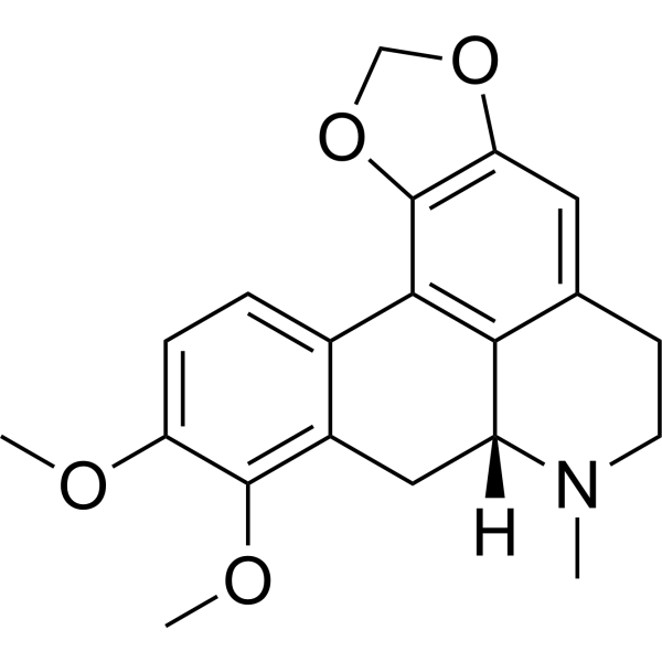
Crebanine is an isoquinoline-like alkaloid that can be derived from Stephania. Crebanine is an antagonist of the α7-nAChR with an IC50 of 19.1 μM. Crebanine suppresses the proliferation, migration, and invasion of cancer cells, triggers reactive oxygen species (ROS) burst, and promotes apoptosis. Crebanine inhibits the AKT/FoxO3a, NF-κB and MAPK signaling pathways. Crebanine attenuates NOX2 hyperactivation, exhibits antioxidant properties by reducing reactive oxygen species and peroxidation in microglia cells. Crebanine inhibits voltage-dependent Na+ current in guinea-pig ventricular myocytes. Crebanine has high inhibitory activity against gram-positive animal pathogenic bacteria. Crebanine ameliorates ischemia-reperfusion brain damage in middle cerebral artery occlusion and reperfusion (MCAO/R) rats. Crebanine significantly improves Scopolamine (HY-N0296)-induced cognitive deficits in ICR mice. Crebanine can be used for the study of hepatocellular carcinoma (HCC), cerebral ischemia and Alzheimer's disease.
For research use only. We do not sell to patients.
- Purity : 99.83%
- CAS No.: 25127-29-1
- Formula: C20H21NO4
- Molecular Weight:339.39
-
Storage:
4°C, protect from light
* In solvent : -80°C, 6 months; -20°C, 1 month (protect from light)

To place orders, for customer services and technical support, please contact: MedChemExpress USA
Tel: 609-228-6898 E-mail: [email protected] [email protected]


-
 Biological Activity
-
Chemical Information
-
Solvent & Solubility
- Protocols
- Purity & Documentation
- References
-
 Help & FAQs


-
HY-15431
CapivasertibAkt Inhibitor -
HY-10358
MK-2206 dihydrochlorideAllosteric Akt Inhibitor -
HY-50909
PerifosineAkt Inhibitor -
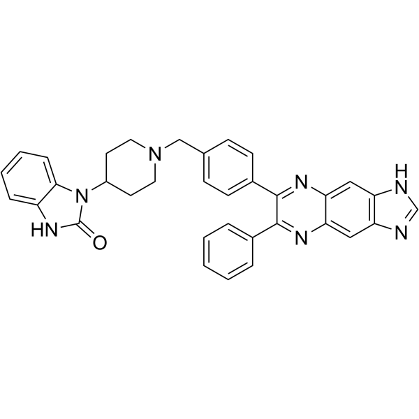
HY-10355
AKT inhibitor VIIIAKT Inhibitor -
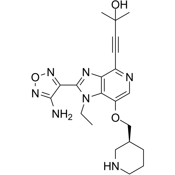
HY-10249
GSK-690693Akt Inhibitor -
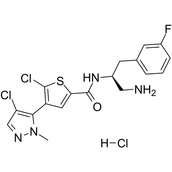
HY-15727A
Afuresertib hydrochlorideAkt Inhibitor -
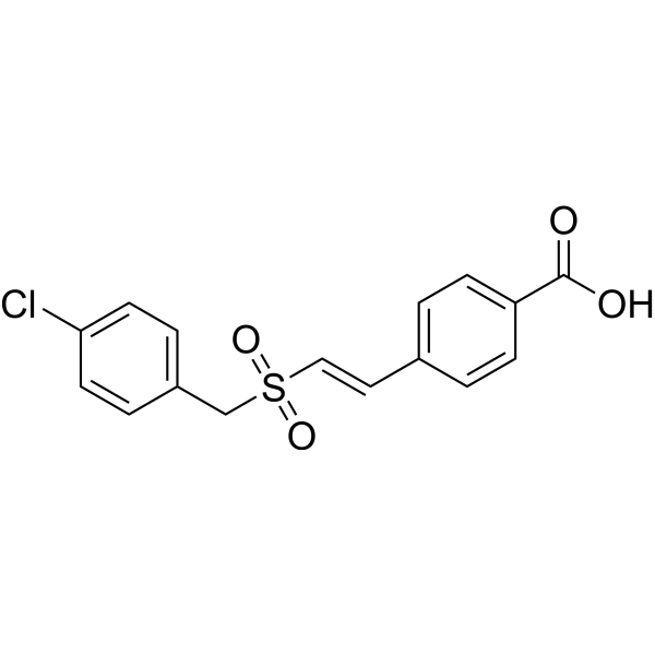
HY-101625
RecilisibRadioprotectant -
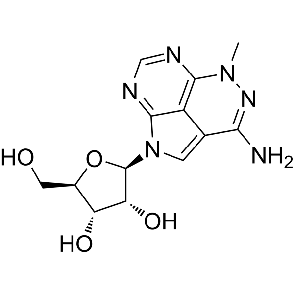
HY-15457
TriciribineHIV Inhibitor -
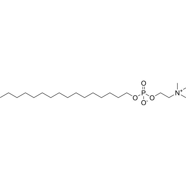
HY-13685
MiltefosinePI3K/Akt Inhibitor -
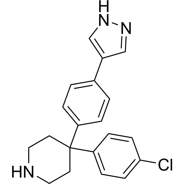
HY-12059
AT7867 -
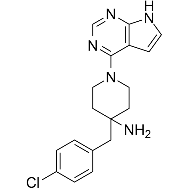
HY-13260
CCT128930AKT Inhibitor -
HY-18749
SC79Akt Activator
Publications Citing Use of MedChemExpress (MCE) Crebanine
More

Biological Activity
Description
Cellular Effect
|
Cell Line

|
Type | Value | Description | References |
|---|---|---|---|---|
| BV-2 | IC50 |
5.26 μM
Compound: 22
|
Inhibition of NO production in LPS-induced mouse BV-2 cells incubated for 24 hrs by Griess reagent based microplate reader analysis
Inhibition of NO production in LPS-induced mouse BV-2 cells incubated for 24 hrs by Griess reagent based microplate reader analysis
|
[PMID: 37172474] |
| SH-SY5Y | IC50 |
5.26 μM
Compound: 22
|
Neuroprotective activity against human SH-SY5Y cells assessed as increase in BDNF level measured after 48 hrs
Neuroprotective activity against human SH-SY5Y cells assessed as increase in BDNF level measured after 48 hrs
|
[PMID: 37172474] |
In Vitro
Crebanine (0-280 μM, 24-72 h) suppresses cell growth, reduces colony formation ability and inhibits migration and invasion of HepG2 cells[1].
Crebanine (35-175 μM, 24 h) reduces mitochondrial membrane potential (MMP) and induces apoptosis rate in HepG2 cells[1].
Crebanine (35-175 μM, 24 h) increases intracellular ROS levels, enhances MDA production, and decreases SOD and GSH-PX activities in HepG2 cells, an effect that can be reversed by Acetylcysteine (NAC) (HY-B0215) pretreatment[1].
Crebanine (35-175 μM, 24 h) down-regulates p-AKT and p-FoxO3a (Ser253) in HepG2 cells in a dose-dependent manner without affecting total AKT and FoxO3a levels[1].
Crebanine (1-20 μM, pretreated for 2 h, then co-treated with LPS for 24 h) inhibits NOX2 activation in BV-2 cells by reducing NADP⁺/NADPH levels, downregulating gp91phox and p47phox expression, and decreasing p47phox membrane translocation[2].
Crebanine (1-20 μM, pretreated for 2 h, then co-treated with LPS (HY-D1056) for 24 h) reduces intracellular superoxide anion, ROS, and MDA levels, suppresses NO, iNOS, IL-1β, IL-6, and TNF-α expression, and inhibits NF-κB and MAPK signaling pathways[2].
Crebanine (0-30 µg/mL, 48 h) inhibits proliferation of HL-60, U937, K562, HT1080, KB-3-1 and KB-V1 cells in a concentration-dependent manner with IC50 values of 9, 12, 13, 20, 24 and 16 µg/mL respectively, and shows slight toxicity to normal fibroblast cells (72% survival at 30 µg/mL)[3].
Crebanine (0-30 µg/mL, 24 h) induces G0/G1 phase arrest and induces apoptosis in HL-60 and U937 cells[3].
Crebanine (20 µg/mL, 0-24 h) down-regulates the expression of cyclins A, D1, PCNA, Bcl-2 and Bcl-xl and enhances Bax expression in HL-60 cells, while cyclin E expression remains unchanged[3].
Crebanine (0.0368-0.7366 mM) reversibly inhibits voltage-dependent Na⁺ current in guinea-pig ventricular myocytes in a concentration-dependent manner, with an IC50 of 0.283 mM; the effect partially recovers after 5 min washout[4].
Crebanine (10 μM) significantly inhibits the binding of (±)-[³H]-epibatidine to Ls-AChBP, Ac-AChBP and AcY55W-AChBP, with Ki values of 179 nM, 538 nM and >1000 nM respectively[5].
Crebanine (10.3 μM) exhibits antagonistic effects on α7-nAChR, α4β2-nAChR, (α1)2β1δγ-nAChR and 5-HT3A receptor in mammalian cells, with inhibition percentages of 100.7%, 29.0%, 81.7% and 55.3% respectively[5].
Crebanine exhibits high inhibitory activity against gram-positive animal pathogenic bacteria, with an MIC of 0.312 g/L against Micrococcus lysodeikticus, Bacillus megaterium, Bacillus subtilis and Staphylococcus aureus, and an MIC of 0.213 g/L against Bacillus cereus[6].
Crebanine inhibits hyphal growth of these 5 plant pathogenic fungi (EC50: 0.111 g/L for Cercospora kaki; 6.47×10⁻² g/L for Gymnosporangium haraeanum; 1.89×10⁻² g/L for Pyricularia oryzae; 4.25×10⁻² g/L for Rhizoctonia solani; 4.05×10⁻² g/L for Colletotrichum graminicola)[6].
Crebanine (0.4 g/L) inhibits spore germination of Thielaviopsis paradoxa (94.96%), Fusarium oxysporum f. sp. niveum (100%), Sphaceloma fawcettii (100%) and Gymnosporangium haraeanum (100%)[6].
MedChemExpress (MCE) has not independently confirmed the accuracy of these methods. They are for reference only. Further protocols information, click here.
-
Cell Line:HepG2 cells
-
Concentration:0, 35, 70, 105, 140, 175, 280 µM
-
Incubation Time:24, 48, 72 h
-
Result:Inhibited viability of HepG2 cells in a dose- and time-dependent manner, with IC50 values of 111.77 μM (24 h), 65.07 μM (48 h), and 23.68 μM (72 h).
-
Cell Line:HepG2 cells
-
Concentration:35, 105, 175 µM
-
Incubation Time:24 h
-
Result:Inhibited invasion of HepG2 cells in a dose-dependent manner.
-
Cell Line:HepG2 cells
-
Concentration:35, 105, 175 µM
-
Incubation Time:24 h
-
Result:Inhibited migration of HepG2 cells in a dose-dependent manner.
-
Cell Line:HepG2 cells
-
Concentration:35, 105, 175 µM
-
Incubation Time:24 h
-
Result:Increased apoptosis rate (up to 43.5% at highest dose) detected by flow cytometry with Annexin V-FITC/PI staining.
-
Cell Line:HepG2 cells
-
Concentration:35, 105, 175 µM
-
Incubation Time:24 h
-
Result:Up-regulated Bax, cleaved-PARP, cleaved-caspase-3, and cleaved-caspase-9.
Down-regulated Bcl-2, p-AKT and p-FoxO3a (Ser253).
-
Cell Line:BV2 cells
-
Concentration:1, 10, 20 μM
-
Incubation Time:Pretreated for 2 h, then co-treated with LPS for 24 h
-
Result:Suppressed IL-1β, IL-6, and TNF-α expression.
In Vivo
Crebanine (8.5 mg/kg (25 μmol/kg), i.p., single dose) significantly improves Scopolamine (HY-N0296)-induced cognitive deficits in ICR mice[5].
MedChemExpress (MCE) has not independently confirmed the accuracy of these methods. They are for reference only.
-
Animal Model:Adult Sprague-Dawley (SD) rats were used to establish the middle cerebral artery occlusion and reperfusion (MCAO/R) model by a modified monofilament nylon suture embolism method[1]
-
Dosage:250-500 μg/kg
-
Administration:After 2 h MCAO occlusion followed by 24 h reperfusion
-
Result:Reduced brain water content and infarct volume in MCAO/R rats, with 500 μg/kg exhibiting obvious efficacy.
Up-regulated NeuN⁺ fluorescence density and down-regulated FJB⁺ cell count in the ischemic cortex of MCAO/R rats.
Mitigated synaptic damage induced by MCAO/R operation.
Attenuated NOX2 hyperactivation by reducing NADP⁺ and NADPH levels, suppressing gp91phox and p47phox expressions, and decreasing p47phox membrane translocation in Iba-1⁺ cells of MCAO/R rats.
Reduced the quantity of Iba-1⁺ cells and protein expression in MCAO/R rats.
Exhibited antioxidant properties by down-regulating superoxide anion and intracellular ROS, and reducing lipid and DNA peroxidation in MCAO/R rats.
Exerted anti-inflammatory effects by reducing NO, IL-1β, TNF-α, IL-6, and iNOS expressions in MCAO/R rats.
Inhibited NF-κB and MAPK signaling pathways in MCAO/R rats, as evidenced by reduced NF-κB p65 promoter activity and nucleus translocation, suppressed IκBα phosphorylation and degradation, and inhibited phosphorylation of ERK, JNK, and p38 MAPKs.
-
Animal Model:Male ICR mice, weighing 25-35 g and 8 weeks of age, were used to establish scopolamine-induced amnesic models[5]
-
Dosage:8.5 mg/kg (25 μmol/kg)
-
Administration:i.p. 1 hour before Scopolamine (0.5 mg/kg, i.p.)
-
Result:Improved scopolamine-induced cognitive deficits in male ICR mice.
Increased the time spent in the target quadrant (Q1) in the Morris water maze test.


Chemical Information
-
CAS No. 25127-29-1
-
Appearance Solid
-
Molecular Weight 339.39
-
Formula C20H21NO4
-
Color Off-white to light yellow
-
SMILES
CN1CCC(C2=C3C4=CC=C(OC)C(OC)=C4C[C@@]12[H])=CC5=C3OCO5
-
Structure Classification
-
Initial Source
-
Shipping
Room temperature in continental US; may vary elsewhere.
-
Storage
4°C, protect from light
* In solvent : -80°C, 6 months; -20°C, 1 month (protect from light)


Publications (5)
-
Journal Impact Factor
-
Most Recent
-
-
Cell Biol Toxicol
EZH2/DUSP1/Akt signaling axis mediates the inhibitory effect of crebanine on hepatocellular carcinoma progression. [Abstract]2026 Jun 2. PMID: 42228265 -
Int J Mol Sci
Crebanine Induces Cell Death and Alters the Mitotic Process in Renal Cell Carcinoma In Vitro. [Abstract]2025 Jul 18;26(14):6896. PMID: 40725144 -
Biomol Ther (Seoul)
Crebanine Protects HUVECs from LPS-Induced Inflammation and Oxidative Stress by Suppressing NF-κB Pathway. [Abstract]2026 Mar 1;34(2):423-433. PMID: 41755781 -
J Cell Mol Med
Crebanine mitigates glucocorticoid-induced osteonecrosis of the femoral head by restoring bone remodelling homeostasis via attenuating oxidative stress. [Abstract]2024 Aug;28(16):e70044. PMID: 39205463


Solvent & Solubility
In Vitro:
DMSO : 100 mg/mL (294.65 mM; Need ultrasonic; Hygroscopic DMSO has a significant impact on the solubility of product, please use newly opened DMSO)
Please refer to the solubility information to select the appropriate solvent. Once prepared, please aliquot and store the solution to prevent product inactivation from repeated freeze-thaw cycles.
Storage method and period of stock solution: -80°C, 6 months; -20°C, 1 month (protect from light). When stored at -80°C, please use it within 6 months. When stored at -20°C, please use it within 1 month.
Please refer to the solubility information to select the appropriate solvent. Once prepared, please aliquot and store the solution to prevent product inactivation from repeated freeze-thaw cycles.
Storage method and period of stock solution: -80°C, 6 months; -20°C, 1 month (protect from light). When stored at -80°C, please use it within 6 months. When stored at -20°C, please use it within 1 month.
Concentration (start) × Volume (start) = Concentration (final) × Volume (final)
In Vivo:
Select the appropriate dissolution method based on your experimental animal and administration route.
- For the following dissolution methods, please ensure to first prepare a clear stock solution using an In Vitro approach and then sequentially add co-solvents:
- To ensure reliable experimental results, the clarified stock solution can be appropriately stored based on storage conditions. As for the working solution for In Vivo experiments, it is recommended to prepare freshly and use it on the same day.
- The percentages shown for the solvents indicate their volumetric ratio in the final prepared solution. If precipitation or phase separation occurs during preparation, heat and/or sonication can be used to aid dissolution.
Add each solvent one by one: 10% DMSO 40% PEG300 5% Tween-80 45% Saline
Solubility: ≥ 2.5 mg/mL (7.37 mM); Clear solution
This protocol yields a clear solution of ≥ 2.5 mg/mL (saturation unknown).
Taking 1 mL working solution as an example, add 100 μL DMSO stock solution (25.0 mg/mL) to 400 μL PEG300, and mix evenly; then add 50 μL Tween-80 and mix evenly; then add 450 μL Saline to adjust the volume to 1 mL.
Preparation of Saline: Dissolve 0.9 g sodium chloride in ddH₂O and dilute to 100 mL to obtain a clear Saline solution.
Add each solvent one by one: 10% DMSO 90% (20% SBE-β-CD in Saline)
Solubility: ≥ 2.5 mg/mL (7.37 mM); Clear solution
This protocol yields a clear solution of ≥ 2.5 mg/mL (saturation unknown).
Taking 1 mL working solution as an example, add 100 μL DMSO stock solution (25.0 mg/mL) to 900 μL 20% SBE-β-CD in Saline, and mix evenly.
Preparation of 20% SBE-β-CD in Saline (4°C, storage for one week): 2 g SBE-β-CD powder is dissolved in 10 mL Saline, completely dissolve until clear.
In Vivo Dissolution Calculator
Please enter the basic information of animal experiments:
-
-
-
-
Recommended: Prepare an additional quantity of animals to account for potential losses during experiments.
Please enter your animal formula composition:
-
%DMSO +
Recommended: Keep the proportion of DMSO in working solution below 2% if your animal is weak.
-
%+
-
+%Tween-80 + +
-
%Saline +
The co-solvents required include: DMSO, . All of co-solvents are available by MedChemExpress (MCE). , Tween 80. All of co-solvents are available by MedChemExpress (MCE).
Working solution concentration: 0.22 mg/mL
Method for preparing stock solution: mg drug dissolved in μL DMSO. Stock solution concentration: mg/mL. * In solvent : -80°C, 6 months; -20°C, 1 month (protect from light)
1. Take μL DMSO stock solution;
2. Add μL .
μL , mix evenly;
3. Then add μL Tween 80, mix evenly;
4. Then add μL
Please ensure that the stock solution in the first step is dissolved to a clear state, and add co-solvents in sequence. You can use ultrasonic heating (ultrasonic cleaner, recommended frequency 20-40 kHz), vortexing, etc. to assist dissolution.

Protocols
-
Annexin V plus membrane-impermeant dye apoptosis staining
Annexin V-based apoptosis assays rely on the detection of phosphatidylserine (PS) externalization from the inner leaflet of the plasma membrane to the outer leaflet, an early biochemical hallmark of apoptosis. Fluorescently labeled Annexin V binds PS in a calcium-dependent manner, enabling identification of early apoptotic cells by flow cytometry or fluorescence microscopy. When combined with a membrane-impermeant DNA-binding dye (e. g. , propidium iodide), this approach allows discrimination between viable (Annexin V−/dye−), early apoptotic (Annexin V+/dye−), and late apoptotic or necrotic (Annexin V+/dye+) cell populations by assessing membrane integrity and PS exposure.
-
Cell migration
Cell migration is a method that plays an important role in wound healing, cell differentiation, embryonic development, etc.
-
ROS/oxidative-stress fluorescent staining
ROS/oxidative-stress fluorescent staining uses cell-permeant fluorogenic probes that become fluorescent after oxidation inside cells or tissues; commonly used examples include DCFH-DA/DCFDA for broad cellular oxidant detection, DHE for superoxide-related signal detection, MitoSOX for mitochondrial superoxide-related signal detection, and CellROX probes for oxidative-stress-associated fluorescence readouts. The assay detects probe oxidation rather than a single ROS species unless the probe and analysis method have been chemically validated for that species. DCFH-DA enters cells, is deacetylated by intracellular esterases to DCFH, and produces fluorescent DCF after oxidation, so the readout is used as an operational measure of total cellular oxidative stress rather than a species-specific ROS measurement. DHE and MitoSOX can report superoxide-related oxidation, but red fluorescence alone can include non-specific ethidium-like oxidation products; HPLC or optimized spectral approaches are
-
Research Protocol for Inflammation-related Diseases
The NLRP3 inflammasome is a cytosolic innate immune signaling platform that integrates priming signals and danger-signal activation to promote caspase-1 activation, maturation of IL-1β and IL-18, and gasdermin D-mediated pyroptotic cell death. The core experimental logic is to determine whether inflammatory disease phenotypes are driven by increased NLRP3 expression, ASC-containing inflammasome assembly, caspase-1 cleavage, GSDMD cleavage, and extracellular release of IL-1β/IL-18 rather than by nonspecific cell injury alone. The pathway is strongly linked to inflammation-related disease phenotypes because monosodium urate crystals activate NALP3/NLRP3 inflammasome signaling in gout-like crystal inflammation, cholesterol crystals activate NLRP3 inflammasomes in atherogenesis models, and DSS-induced intestinal inflammation has been reported to involve NLRP3 inflammasome activity. However, experimental colitis studies also show context-dependent protective effects of NLRP3 inflammasome co
-
Gram Staining of Tissue Sections
Gram staining of tissue sections is a histochemical technique used to differentiate Gram-positive and Gram-negative bacteria within histological specimens based on differences in bacterial cell wall structure and dye retention, adapted from classical bacteriological Gram staining into tissue-compatible “histological Gram stain” variants. In tissue applications, modifications of the Brown-Hopps and Brown-Brenn methods are commonly used to improve differentiation of microorganisms embedded within host connective tissue and to reduce overstaining or loss of Gram-negative signal, which are known limitations of earlier approaches. The principle relies on crystal violet-iodine complex retention in Gram-positive organisms and subsequent decolorization and counterstaining steps that allow contrast visualization of Gram-negative organisms against tissue background.
-
Liver Cancer Modeling
Liver cancer can be classified into primary liver cancer and secondary liver cancer. Secondary liver cancer is the metastatic liver cancer. Primary liver cancer includes hepatocellular carcinoma (HCC), intrahepatic cholangiocarcinoma (ICC) and fibrolamellar HCC, of which HCC is the most common form, accounting for approximately 90% of primary liver cancers[1]. HCC mouse models include chemical agent-induced models, transplanted tumor models, and genetic engineered models.
-
Cell Viability Determination by MTT Colorimetric Assay
The following protocol uses the MTT colorimetric assay as a classic literature-established method for assessing cell viability/metabolic activity in cultured mammalian cells. MTT[3-(4,5-dimethylthiazol-2-yl)-2,5-diphenyltetrazolium bromide] is reduced by metabolically active cells to a colored formazan product; the amount of formazan is quantified spectrophotometrically and provides an indirect measure of metabolically active viable cells. Importantly, MTT reduction reflects cellular oxidoreductase/metabolic activity rather than an absolute direct count of living cells, so changes in cellular metabolism can alter the signal independently of cell number.
-
Alzheimer’s Disease Modeling
Alzheimer’s Disease (AD) is a neurodegenerative disorder characterized by a progressive decline in cognitive functions and loss of specific types of neurons and synapses. Alzheimer's symptoms can be simulated in mice by injecting drugs (such as Aβ) or genetically modified.
-
Apoptosis
Apoptosis, also called programmed cell death, is generally characterized by distinct morphological characteristics.
-
TUNEL staining for apoptotic DNA fragmentation
TUNEL staining detects DNA strand breaks by using terminal deoxynucleotidyl transferase to add labeled nucleotides to exposed 3′-OH DNA termini, generating either microscopic staining in fixed cells or tissue sections, or fluorescence/cytometric signal in cell suspensions. TUNEL positivity reflects DNA fragmentation but should not be interpreted alone as definitive apoptosis, because TUNEL can also label necrotic, autolytic, mechanically damaged, or DNA-repair-associated DNA breaks.
-
Cell Cytotoxicity Assay
Cytotoxicity assays are usually based on the assessment of cell membrane damage, which can also be indirectly detected by measuring cell viability. Detection methods include MTT assay, CKK-8 assay, LDH assay and ATP assay, etc.
-
Apoptosis Solutions
Apoptosis is a regulated, generally non-lytic cell-death pathway that removes unwanted, damaged, infected, or abnormal cells through coordinated morphological changes, caspase activation, DNA fragmentation, and membrane remodeling. The intrinsic apoptosis pathway is controlled mainly by mitochondrial outer membrane permeabilization, BCL-2 family proteins, cytochrome c release, apoptosome formation, caspase-9 activation, and downstream executioner caspase-3/7 activation. The extrinsic apoptosis pathway is initiated by death receptors such as Fas, TNFR, and TRAIL receptors, which recruit adaptor proteins and activate caspase-8 before engaging executioner caspases or mitochondrial amplification through BID cleavage. Apoptosis is linked to many phenotypes, including cancer cell killing, tissue homeostasis, immune regulation, neurodegeneration, infection response, and treatment-induced cytotoxicity; unresolved questions include how apoptosis interacts with necroptosis, pyroptosis, ferroptos
-
Cell invasion
Cell invasion is the ability of cells to migrate from one area to another via the extracellular matrix. Cell invasion is the response of normal and cancer cells to chemical and mechanical stimuli. Before migrating to a new region, the extracellular matrix is degraded by proteases within the cell. Cell invasion often occurs during wound repair, vascularization and inflammation, abnormal tissue invasion, and tumor cell metastasis.


Purity & Documentation
-
Data Sheet (309 KB)
-
SDS (394 KB)
- English - EN (394 KB)
- Français - FR (394 KB)
- Deutsch - DE (394 KB)
- Norwegian - NO (394 KB)
- Español - ES (394 KB)
- Swedish - SV (394 KB)
- Italian - IT (394 KB)
- Korean - KR (394 KB)
- Portuguese - PT (394 KB)
-
Handling Instructions (2659 KB)
References
[1]. Tan J, et al. Crebanine induces ROS-dependent apoptosis in human hepatocellular carcinoma cells via the AKT/FoxO3a signaling pathway. Front Pharmacol. 2023 Feb 16;14:1069093. [Content Brief]
[2]. Yang Y, et al. Crebanine ameliorates ischemia-reperfusion brain damage by inhibiting oxidative stress and neuroinflammation mediated by NADPH oxidase 2 in microglia. Phytomedicine. 2023 Nov;120:155044. [Content Brief]
[3]. Wongsirisin P, et al. Induction of G1 arrest and apoptosis in human cancer cells by crebanine, an alkaloid from Stephania venosa. Chem Pharm Bull (Tokyo). 2012;60(10):1283-9. [Content Brief]
[4]. Xiao-Shan H, et al. Crebanine inhibits voltage-dependent Na+ current in guinea-pig ventricular myocytes. Chin J Nat Med. 2014 Jan;12(1):20-3. [Content Brief]
[5]. Rojsanga P, et al. The effect of crebanine on memory and cognition impairment via the alpha-7 nicotinic acetylcholine receptor. Life Sci. 2012 Aug 21;91(3-4):107-14. [Content Brief]


Complete Stock Solution Preparation Table
Please refer to the solubility information to select the appropriate solvent. Once prepared, please aliquot and store the solution to prevent product inactivation from repeated freeze-thaw cycles.
Storage method and period of stock solution: -80°C, 6 months; -20°C, 1 month (protect from light). When stored at -80°C, please use it within 6 months. When stored at -20°C, please use it within 1 month.
| Optional Solvent | Concentration Solvent Mass | 1 mg | 5 mg | 10 mg | 25 mg |
|---|---|---|---|---|---|
| DMSO | 1 mM | 2.9465 mL | 14.7323 mL | 29.4646 mL | 73.6616 mL |
| 5 mM | 0.5893 mL | 2.9465 mL | 5.8929 mL | 14.7323 mL | |
| 10 mM | 0.2946 mL | 1.4732 mL | 2.9465 mL | 7.3662 mL | |
| 15 mM | 0.1964 mL | 0.9822 mL | 1.9643 mL | 4.9108 mL | |
| 20 mM | 0.1473 mL | 0.7366 mL | 1.4732 mL | 3.6831 mL | |
| 25 mM | 0.1179 mL | 0.5893 mL | 1.1786 mL | 2.9465 mL | |
| 30 mM | 0.0982 mL | 0.4911 mL | 0.9822 mL | 2.4554 mL | |
| 40 mM | 0.0737 mL | 0.3683 mL | 0.7366 mL | 1.8415 mL | |
| 50 mM | 0.0589 mL | 0.2946 mL | 0.5893 mL | 1.4732 mL | |
| 60 mM | 0.0491 mL | 0.2455 mL | 0.4911 mL | 1.2277 mL | |
| 80 mM | 0.0368 mL | 0.1842 mL | 0.3683 mL | 0.9208 mL | |
| 100 mM | 0.0295 mL | 0.1473 mL | 0.2946 mL | 0.7366 mL |


Crebanine Related Classifications
HY-N2255 Related Classifications
 Powered by Bioz