



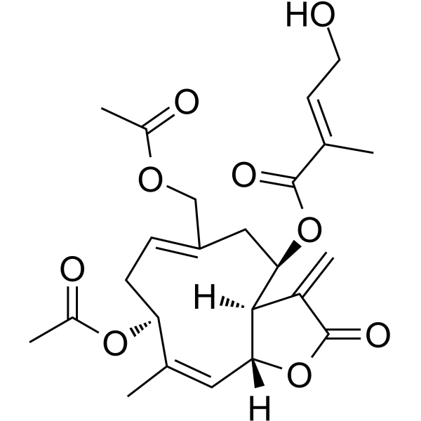
Eupalinolide A

Based on 1 Customer Validation
Eupalinolide A is a Yes-associated protein (YAP) degrader and HSP70 inducer. Eupalinolide A inhibits osteogenic differentiation of tendon-derived stem cells (TDSCs). Eupalinolide A induces autophagy in hepatocellular carcinoma cells via activating the ROS/ERK signaling pathway. Eupalinolide A protects PAM212 cells from UVB-, Menadione (HY-B0332)-, or heat shock-induced apoptosis. Eupalinolide A alleviates trauma-induced heterotopic ossification (HO) of Achilles tendon and inhibits growth of MHCC97-L and HCCLM3 hepatocellular carcinoma xenograft tumors in mice. Eupalinolide A can be used for the study of traumatic heterotopic ossification of tendons and hepatocellular carcinoma.
For research use only. We do not sell to patients.
- Purity : 99.92%
- CAS No.: 877822-40-7
- Formula: C24H30O9
- Molecular Weight:462.49
-
Storage:
4°C, protect from light
* In solvent : -80°C, 6 months; -20°C, 1 month (protect from light)

To place orders, for customer services and technical support, please contact: MedChemExpress USA
Tel: 609-228-6898 E-mail: [email protected] [email protected]


-
 Biological Activity
-
Chemical Information
-
Solvent & Solubility
- Protocols
- Purity & Documentation
- References
-
 Help & FAQs


-
HY-10941
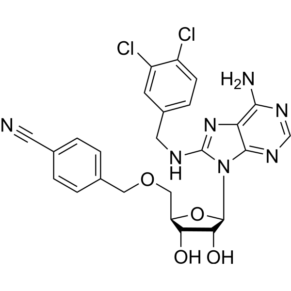
VER-155008Hsp70 Inhibitor -
HY-N0753
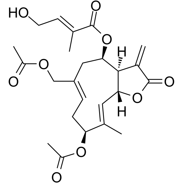
Eupalinolide BGerm Sesquiterpene -
HY-15785
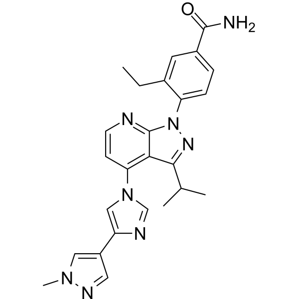
PimitespibHSP90α/HSP90β Inhibitor -
HY-10211
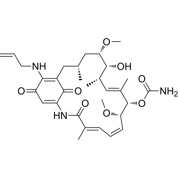
TanespimycinHSP90 Inhibitor -
HY-N8187
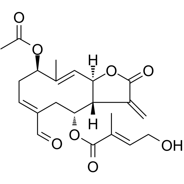
Eupalinolide OSesquiterpene Lactone -
HY-101712
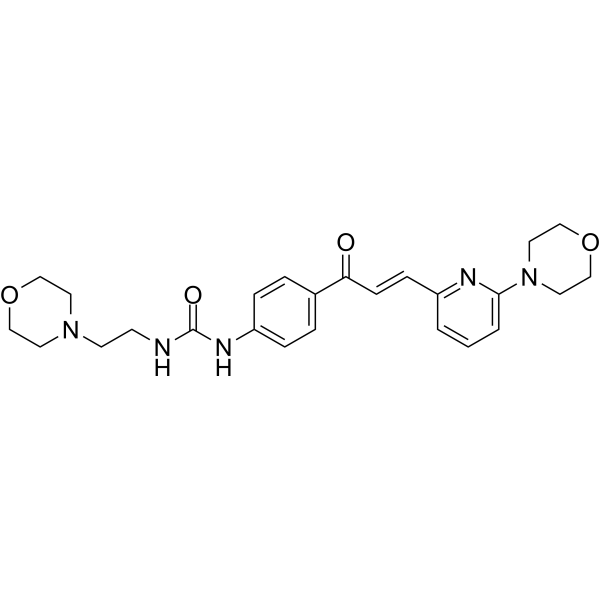
TRC051384HSP70 Inducer -
HY-100110
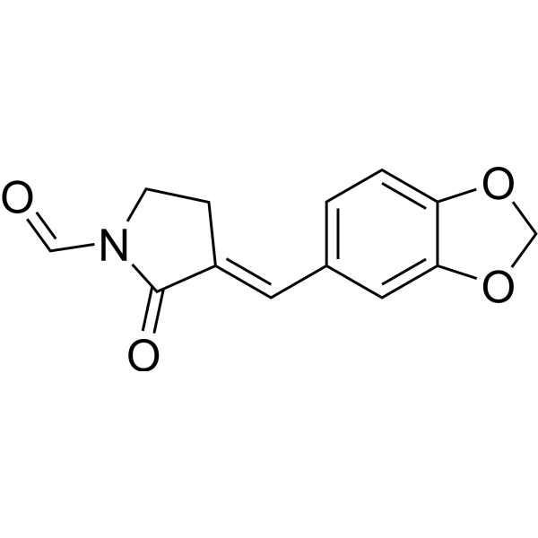
KNK437HSP Inhibitor -
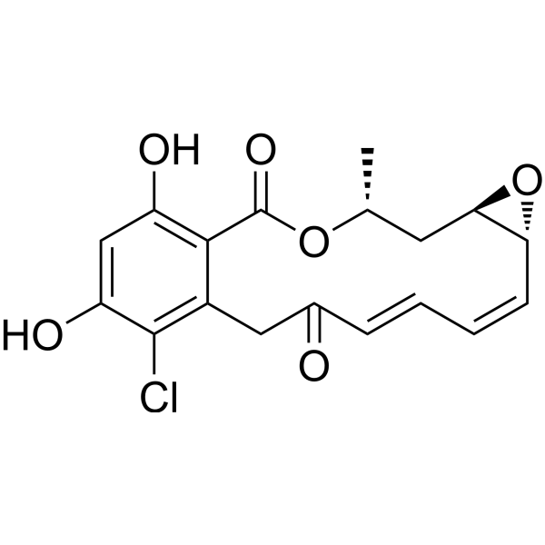
HY-N6769
RadicicolHsp90 Inhibitor -
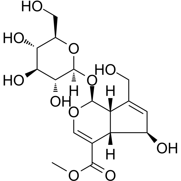
HY-N6249
FeretosideHSP Activator -

HY-10211R
Tanespimycin (Standard)HSP90 Inhibitor -
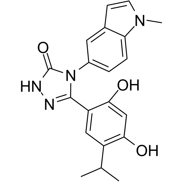
HY-15205
GanetespibHSP90 Inhibitor -
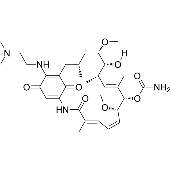
HY-10389
AlvespimycinHsp90 Inhibitor


Biological Activity
Description
In Vitro
Eupalinolide A (1-10 μM, 7-14 d) dose-dependently inhibits osteogenic differentiation of tendon-derived stem cells (TDSCs), as evidenced by decreased alkaline phosphatase (ALP) activity, reduced calcium deposition, downregulated mRNA expression of osteogenic genes (Alp, Col1a1, Osx, Ocn, Opn), and decreased protein levels of OSX and OCN[1].
Eupalinolide A (10 μM) induces YAP degradation via TOLLIP-mediated selective autophagy and Promotes K27-linked polyubiquitination of YAP in HEK293T cells[1].
Eupalinolide A (28 μM, 48 h) reduces BrdU-positive cells and inhibits DNA synthesis in MHCC97-L and HCCLM3 cells via BrdU staining[2].
Eupalinolide A (28 μM, 21 d) decreases colony number and size of MHCC97-L and HCCLM3 cells[2].
Eupalinolide A (7-28 μM, 24-48 h) suppresses migration and reverses EMT of MHCC97-L and HCCLM3 cells[2].
Eupalinolide A (7-28 μM, 48 h) induces G1 phase arrest and autophagy, and increases ROS production in MHCC97-L and HCCLM3 cells[2].
Eupalinolide A (5-10 μg/mL, 24 h) induces HSP70 expression in B16 mouse melanoma cells and PAM212 mouse squamous cell carcinoma cells[3].
Eupalinolide A (2.5-10 μg/mL, 24 h) induces the expression of HSP25, HSP40, and HSP90 (HSP90α) at both protein and mRNA levels in PAM212 cells[3].
Eupalinolide A (5-10 μg/mL, 3 h) promotes phosphorylation and nuclear translocation of HSF1 in PAM212 cells and inhibits the interaction between HSF1 and HSP90[3].
Eupalinolide A (5-10 μg/mL, 24 h) suppresses IBMX (HY-12318)-induced melanin production, tyrosinase activity and downregulates tyrosinase, Tyrp1, and Dct mRNA/protein levels in B16 cells[3].
MedChemExpress (MCE) has not independently confirmed the accuracy of these methods. They are for reference only.
-
Cell Line:MHCC97-L and HCCLM3 cells
-
Concentration:7, 14, 28 μM
-
Incubation Time:48 h
-
Result:Upregulated E-cadherin, N-cadherin, fibronectin, p-ERK and ZEB1.
Downregulated Vimentin in MHCC97-L and HCCLM3 cells.
Downregulated CDK2, CDK4, Cyclin E1, and Cyclin D1.
Upregulated LC3 II/I/Atg5 and downregulated p62.
-
Cell Line:MHCC97-L and HCCLM3 cells
-
Concentration:14, 28 μM
-
Incubation Time:48 h
-
Result:Induced G1 phase arrest in MHCC97-L and HCCLM3 cells.
In Vivo
Eupalinolide A (30-60 mg/kg, i.p., once daily, 3 weeks) inhibits growth of MHCC97-L and HCCLM3 hepatocellular carcinoma xenograft tumors in female nude mice[2].
MedChemExpress (MCE) has not independently confirmed the accuracy of these methods. They are for reference only.
-
Animal Model:8-week-old male C57BL/6 mice[1]
-
Dosage:1, 5, 10 μM
-
Administration:local injection, 20 μL/mouse, once weekly for 8 weeks
-
Result:Rduced ectopic bone volume in Achilles tendon.
Dcreased calcified areas.
Dwnregulated mRNA expression of osteogenic genes (Runx2, Alp, Osx, Ocn, Opn, Col1a1) and protein levels of OSX and OCN.
Ihibited YAP protein expression in Achilles tendon.
-
Animal Model:MHCC97-L cells and HCCLM3 cells (1 × 10⁶) were subcutaneously implanted into the flanks of 4-week-old female BALB/c nude mice[2]
-
Dosage:30, 60 mg/kg
-
Administration:i.p., once daily, 3 weeks
-
Result:Achieved tumor growth inhibition (TGI).
- Annexin V plus membrane-impermeant dye apoptosis staining
- ROS/oxidative-stress fluorescent staining
- Liver Cancer Modeling
- Detection of Apoptosis
- TUNEL staining for apoptotic DNA fragmentation
- Mesenchymal stromal/stem cell osteogenic differentiation
- Autophagy
- Patient-Derived Xenograft (PDX)
- Apoptosis Solutions
- Splenic/Portal-Vein Liver Metastasis Xenograft
- Lysosome and acidic-vesicle live-cell staining
- Subcutaneous Cell-Line-Derived Xenograft
- Macroautophagy Solutions
- Orthotopic Cell-Line Xenograft
- Intraperitoneal/Peritoneal Dissemination Xenograft


Chemical Information
-
CAS No. 877822-40-7
-
Appearance Solid
-
Molecular Weight 462.49
-
Formula C24H30O9
-
Color White to off-white
-
SMILES
O=C(O[C@@H]1C/C(COC(C)=O)=C\C[C@H](OC(C)=O)/C(C)=C\[C@@]([C@]1([H])C2=C)([H])OC2=O)/C(C)=C/CO
-
Structure Classification
-
Shipping
Room temperature in continental US; may vary elsewhere.
-
Storage
4°C, protect from light
* In solvent : -80°C, 6 months; -20°C, 1 month (protect from light)


Solvent & Solubility
In Vitro:
DMSO : 100 mg/mL (216.22 mM; Need ultrasonic; Hygroscopic DMSO has a significant impact on the solubility of product, please use newly opened DMSO)
Please refer to the solubility information to select the appropriate solvent. Once prepared, please aliquot and store the solution to prevent product inactivation from repeated freeze-thaw cycles.
Storage method and period of stock solution: -80°C, 6 months; -20°C, 1 month (protect from light). When stored at -80°C, please use it within 6 months. When stored at -20°C, please use it within 1 month.
Please refer to the solubility information to select the appropriate solvent. Once prepared, please aliquot and store the solution to prevent product inactivation from repeated freeze-thaw cycles.
Storage method and period of stock solution: -80°C, 6 months; -20°C, 1 month (protect from light). When stored at -80°C, please use it within 6 months. When stored at -20°C, please use it within 1 month.
Concentration (start) × Volume (start) = Concentration (final) × Volume (final)
In Vivo:
Select the appropriate dissolution method based on your experimental animal and administration route.
- For the following dissolution methods, please ensure to first prepare a clear stock solution using an In Vitro approach and then sequentially add co-solvents:
- To ensure reliable experimental results, the clarified stock solution can be appropriately stored based on storage conditions. As for the working solution for In Vivo experiments, it is recommended to prepare freshly and use it on the same day.
- The percentages shown for the solvents indicate their volumetric ratio in the final prepared solution. If precipitation or phase separation occurs during preparation, heat and/or sonication can be used to aid dissolution.
Add each solvent one by one: 10% DMSO 40% PEG300 5% Tween-80 45% Saline
Solubility: ≥ 2.5 mg/mL (5.41 mM); Clear solution
This protocol yields a clear solution of ≥ 2.5 mg/mL (saturation unknown).
Taking 1 mL working solution as an example, add 100 μL DMSO stock solution (25.0 mg/mL) to 400 μL PEG300, and mix evenly; then add 50 μL Tween-80 and mix evenly; then add 450 μL Saline to adjust the volume to 1 mL.
Preparation of Saline: Dissolve 0.9 g sodium chloride in ddH₂O and dilute to 100 mL to obtain a clear Saline solution.
Add each solvent one by one: 10% DMSO 90% (20% SBE-β-CD in Saline)
Solubility: ≥ 2.5 mg/mL (5.41 mM); Clear solution
This protocol yields a clear solution of ≥ 2.5 mg/mL (saturation unknown).
Taking 1 mL working solution as an example, add 100 μL DMSO stock solution (25.0 mg/mL) to 900 μL 20% SBE-β-CD in Saline, and mix evenly.
Preparation of 20% SBE-β-CD in Saline (4°C, storage for one week): 2 g SBE-β-CD powder is dissolved in 10 mL Saline, completely dissolve until clear.
In Vivo Dissolution Calculator
Please enter the basic information of animal experiments:
-
-
-
-
Recommended: Prepare an additional quantity of animals to account for potential losses during experiments.
Please enter your animal formula composition:
-
%DMSO +
Recommended: Keep the proportion of DMSO in working solution below 2% if your animal is weak.
-
%+
-
+%Tween-80 + +
-
%Saline +
The co-solvents required include: DMSO, . All of co-solvents are available by MedChemExpress (MCE). , Tween 80. All of co-solvents are available by MedChemExpress (MCE).
Working solution concentration: 0.22 mg/mL
Method for preparing stock solution: mg drug dissolved in μL DMSO. Stock solution concentration: mg/mL. * In solvent : -80°C, 6 months; -20°C, 1 month (protect from light)
1. Take μL DMSO stock solution;
2. Add μL .
μL , mix evenly;
3. Then add μL Tween 80, mix evenly;
4. Then add μL
Please ensure that the stock solution in the first step is dissolved to a clear state, and add co-solvents in sequence. You can use ultrasonic heating (ultrasonic cleaner, recommended frequency 20-40 kHz), vortexing, etc. to assist dissolution.


Purity & Documentation
-
Data Sheet (290 KB)
-
SDS (393 KB)
- English - EN (393 KB)
- Français - FR (393 KB)
- Deutsch - DE (393 KB)
- Norwegian - NO (393 KB)
- Español - ES (393 KB)
- Swedish - SV (393 KB)
- Italian - IT (393 KB)
- Korean - KR (393 KB)
- Portuguese - PT (393 KB)
-
Handling Instructions (2659 KB)
References
[1]. Jiang H, et al. Eupalinolide A attenuates trauma-induced heterotopic ossification of tendon in mice by promoting YAP degradation through TOLLIP-mediated selective autophagy. Phytomedicine. 2024 Dec;135:156163. [Content Brief]
[2]. Zhang Y, et al. Eupalinolide A induces autophagy via the ROS/ERK signaling pathway in hepatocellular carcinoma cells in vitro and in vivo. Int J Oncol. 2022 Nov;61(5):131. [Content Brief]
[3]. Yamashita Y, et al. Purification and characterization of HSP-inducers from Eupatorium lindleyanum. Biochem Pharmacol. 2012;83(7):909-922. [Content Brief]


Complete Stock Solution Preparation Table
Please refer to the solubility information to select the appropriate solvent. Once prepared, please aliquot and store the solution to prevent product inactivation from repeated freeze-thaw cycles.
Storage method and period of stock solution: -80°C, 6 months; -20°C, 1 month (protect from light). When stored at -80°C, please use it within 6 months. When stored at -20°C, please use it within 1 month.
| Optional Solvent | Concentration Solvent Mass | 1 mg | 5 mg | 10 mg | 25 mg |
|---|---|---|---|---|---|
| DMSO | 1 mM | 2.1622 mL | 10.8110 mL | 21.6221 mL | 54.0552 mL |
| 5 mM | 0.4324 mL | 2.1622 mL | 4.3244 mL | 10.8110 mL | |
| 10 mM | 0.2162 mL | 1.0811 mL | 2.1622 mL | 5.4055 mL | |
| 15 mM | 0.1441 mL | 0.7207 mL | 1.4415 mL | 3.6037 mL | |
| 20 mM | 0.1081 mL | 0.5406 mL | 1.0811 mL | 2.7028 mL | |
| 25 mM | 0.0865 mL | 0.4324 mL | 0.8649 mL | 2.1622 mL | |
| 30 mM | 0.0721 mL | 0.3604 mL | 0.7207 mL | 1.8018 mL | |
| 40 mM | 0.0541 mL | 0.2703 mL | 0.5406 mL | 1.3514 mL | |
| 50 mM | 0.0432 mL | 0.2162 mL | 0.4324 mL | 1.0811 mL | |
| 60 mM | 0.0360 mL | 0.1802 mL | 0.3604 mL | 0.9009 mL | |
| 80 mM | 0.0270 mL | 0.1351 mL | 0.2703 mL | 0.6757 mL | |
| 100 mM | 0.0216 mL | 0.1081 mL | 0.2162 mL | 0.5406 mL |


Eupalinolide A Related Classifications
HY-N0754 Related Classifications
 Powered by Bioz