



Isofraxidin
Based on 4 publication(s) in Google Scholar
Isofraxidin, a coumarin component from Acanthopanax senticosus, inhibits MMP-7 expression and cell invasion of human hepatoma cells. Isofraxidin inhibits the phosphorylation of ERK1/2 in hepatoma cells. Isofraxidin attenuates the expression of iNOS and COX-2, Isofraxidinalso inhibits TLR4/myeloid differentiation protein-2 (MD-2) complex formation.
For research use only. We do not sell to patients.
- Purity : 99.94%
- CAS No.: 486-21-5
- Formula: C11H10O5
- Molecular Weight:222.19
-
Storage:Powder -20°C, 3 years , 4°C, 2 years ; In solvent -80°C, 6 months , -20°C, 1 month
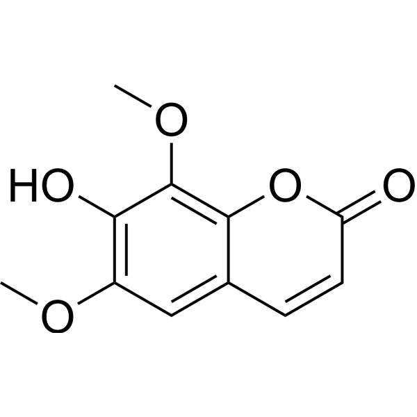

To place orders, for customer services and technical support, please contact: MedChemExpress USA
Tel: 609-228-6898 E-mail: [email protected] [email protected]


-
 Biological Activity
-
Chemical Information
-
Solvent & Solubility
- Protocols
- Purity & Documentation
- References
-
 Help & FAQs


-
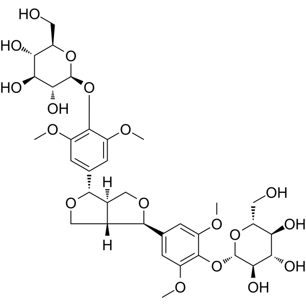
HY-N0272
Eleutheroside EAnti-Inflammatory Agent -
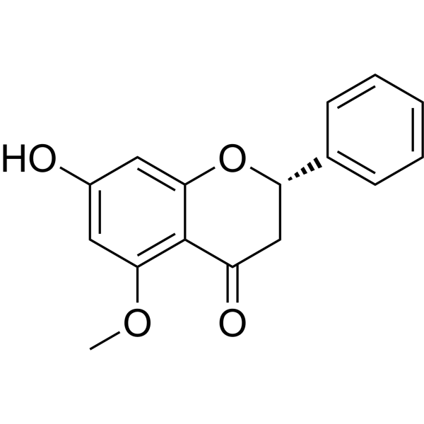
HY-N0625A
AlpinetinPPAR Activator -

HY-10071
Y-27632ROCK Inhibitor -
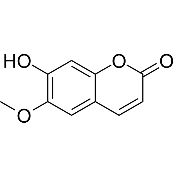
HY-N0342
ScopoletinCholinesterase (ChE) Inhibitor -
HY-N0351
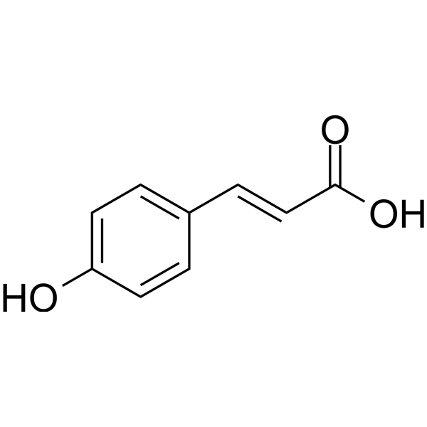
p-Coumaric acidAntibacterial agent -
HY-B0099
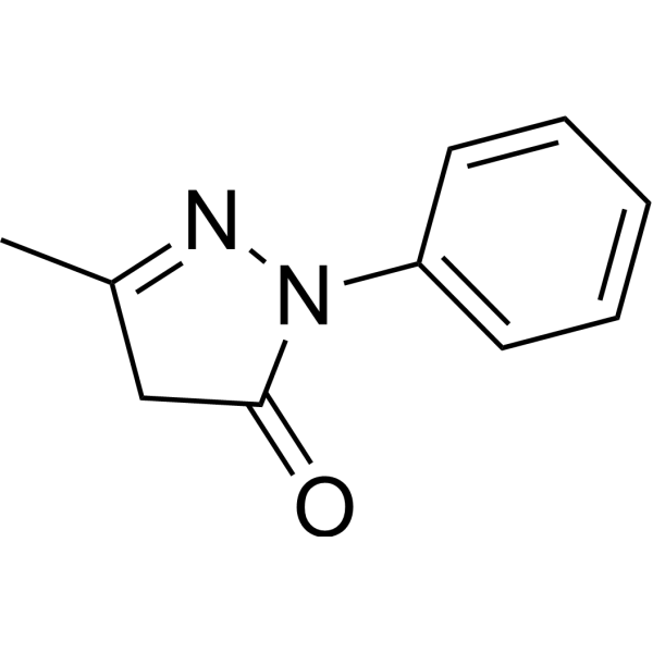
EdaravoneApoptosis Inducer -
HY-N0565B
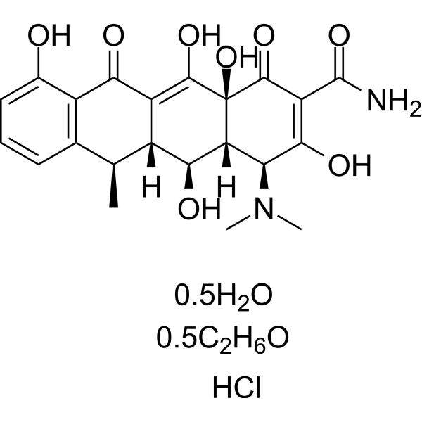
Doxycycline hyclateMMP Inhibitor -

HY-N0774R
Isofraxidin (Standard)COX-2/MMP-7/TLR4 Inhibitor -
HY-N0232
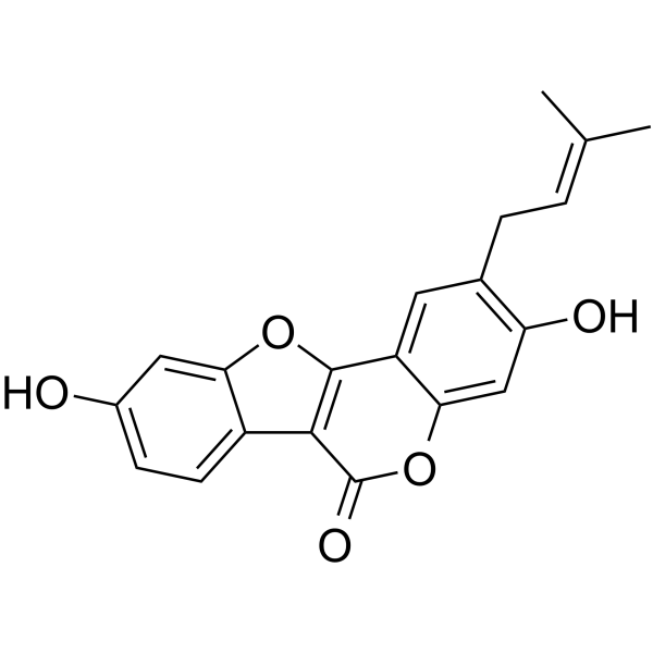
PsoralidinCOX-2/5-LOX Inhibitor -

HY-N0565BR
Doxycycline hyclate (Standard)Reference Standard -

HY-B0099R
Edaravone (Standard)Apoptosis Inducer -
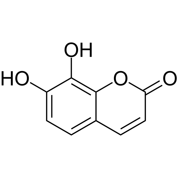
HY-N0281
DaphnetinProtein Kinase Inhibitor
Publications Citing Use of MedChemExpress (MCE) Isofraxidin
More Customer Validation & Images

-
Histological Imaging/Staining
-
In Vivo Efficacy Study
-
ELISA
-
IF
-
WB


Biological Activity
Description
|
COX-2 |
TLR4 |
MMP-7 |
Cellular Effect
|
Cell Line

|
Type | Value | Description | References |
|---|---|---|---|---|
| A549 | IC50 |
0.4 μg/mL
Compound: Isofraxidin
|
Cytotoxicity against human A549 cells by SRB assay
Cytotoxicity against human A549 cells by SRB assay
|
[PMID: 31784199] |
| BV-2 | IC50 |
>100 μM
Compound: 12
|
Antineuroinflammatory activity in mouse BV2 cells assessed as inhibition of LPS-induced NO production after 24 hrs in presence of LPS by Griess reaction based assay
Antineuroinflammatory activity in mouse BV2 cells assessed as inhibition of LPS-induced NO production after 24 hrs in presence of LPS by Griess reaction based assay
|
[PMID: 27919656] |
| HCT-15 | IC50 |
0.4 μg/mL
Compound: Isofraxidin
|
Cytotoxicity against human HCT15 cells by SRB assay
Cytotoxicity against human HCT15 cells by SRB assay
|
[PMID: 31784199] |
| KB | ED50 |
>100 μg/mL
Compound: 1, isofraxidin, NSC-324637
|
Cytotoxicity against human KB cells
Cytotoxicity against human KB cells
|
[PMID: 7463096] |
| P388 | ED50 |
1.7 μg/mL
Compound: 1, isofraxidin, NSC-324637
|
Cytotoxicity against mouse P388 cells
Cytotoxicity against mouse P388 cells
|
[PMID: 7463096] |
| RAW264.7 | IC50 |
>100 μM
Compound: 14
|
Antiinflammatory activity in mouse RAW264.7 cells assessed as decrease in LPS-induced NO production after 24 hrs by Griess assay
Antiinflammatory activity in mouse RAW264.7 cells assessed as decrease in LPS-induced NO production after 24 hrs by Griess assay
|
[PMID: 25666824] |
| SK-MEL-2 | IC50 |
0.4 μg/mL
Compound: Isofraxidin
|
Cytotoxicity against human SK-MEL-2 cells by SRB assay
Cytotoxicity against human SK-MEL-2 cells by SRB assay
|
[PMID: 31784199] |
| SK-OV-3 | IC50 |
0.4 μg/mL
Compound: Isofraxidin
|
Cytotoxicity against human SKOV3 cells by SRB assay
Cytotoxicity against human SKOV3 cells by SRB assay
|
[PMID: 31784199] |
| XF498 | IC50 |
0.4 μg/mL
Compound: Isofraxidin
|
Cytotoxicity against human XF498 cells by SRB assay
Cytotoxicity against human XF498 cells by SRB assay
|
[PMID: 31784199] |
In Vitro
Isofraxidin inhibits expression of MMP-7 and in vitro cell invasion at a non-toxic level through inhibiting ERK1/2 phosphorylation in hepatoma cell lines[1].
Isofraxidin competitively inhibits TLR4/MD-2 complex formation, and thus TLR4/NF-κB signalling cascades.Isofraxidin has potential in the treatment of Osteoarthritis (OA)[2].
MedChemExpress (MCE) has not independently confirmed the accuracy of these methods. They are for reference only.
- Spheroid/Tumor Organoid Invasion Assay
- RNA extraction experimental
- Ex Vivo Tissue Slice/Explant Invasion Assay
- Western Blot
- HL-60 granulocytic/neutrophil-like differentiation
- Mitochondrial membrane-potential fluorescent assay
- Patient-Derived Organoid Invasion Assay
- Liver Cancer Modeling
- 3D Tumor Spheroid Invasion Assay
- Matrigel Transwell/Boyden Chamber Invasion Assay
- Invadopodia/Fluorescent Gelatin Degradation Assay
- Kinase activity and phosphorylation assays
- Mitochondrial membrane-potential and mitochondrial mass staining
- Organotypic 3D Invasion Assay
- Cell invasion
- Matrigel/ECM Transwell Invasion Assay
- Protocol for Kinase activity and phosphorylation assays
- 3D Collagen/Hydrogel Matrix Invasion Assay


Chemical Information
-
CAS No. 486-21-5
-
Appearance Solid
-
Molecular Weight 222.19
-
Formula C11H10O5
-
Color Off-white to yellow
-
SMILES
O=C1C=CC2=CC(OC)=C(O)C(OC)=C2O1
-
Structure Classification
-
Shipping
Room temperature in continental US; may vary elsewhere.
-
Storage
Powder -20°C 3 years 4°C 2 years In solvent -80°C 6 months -20°C 1 month


Publications (4)
-
Journal Impact Factor
-
Most Recent
-
-
J Ethnopharmacol
2025 Jun 26:350:120002. PMID: 40412780 -
Int Immunopharmacol
The coumarin component isofraxidin targets the G-protein-coupled receptor S1PR1 to modulate IL-17 signaling and alleviate ulcerative colitis. [Abstract]2024 Apr 20:131:111814. PMID: 38479159
Isofraxidin purchased from MedChemExpress. Usage Cited in: Int Immunopharmacol. 2024 Apr 20:131:111814. [Abstract]
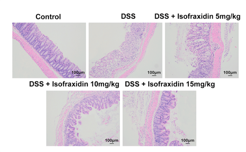
H&E staining revealed the pathology of the colon tissues treated with Isofraxidin (5, 10, 15 mg/kg, p.o.).
Isofraxidin purchased from MedChemExpress. Usage Cited in: Int Immunopharmacol. 2024 Apr 20:131:111814. [Abstract]
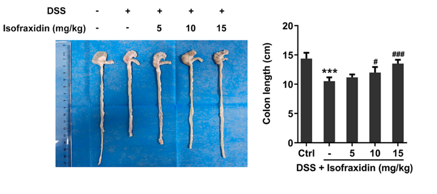
Colon lengths in mice treated with Isofraxidin (5, 10, 15 mg/kg, p.o.).
Isofraxidin purchased from MedChemExpress. Usage Cited in: Int Immunopharmacol. 2024 Apr 20:131:111814. [Abstract]
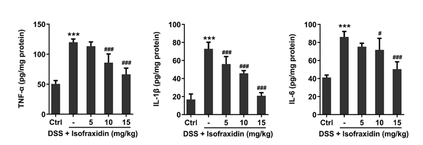
The levels of the inflammatory factors TNFα, IL-1β and IL-6 in colon tissue treated with Isofraxidin (5, 10, 15 mg/kg, p.o.).
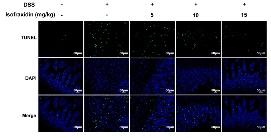
Isofraxidin purchased from MedChemExpress. Usage Cited in: Int Immunopharmacol. 2024 Apr 20:131:111814. [Abstract]
TUNEL staining showing apoptosis in tissues treated with Isofraxidin (5, 10, 15 mg/kg, p.o.).
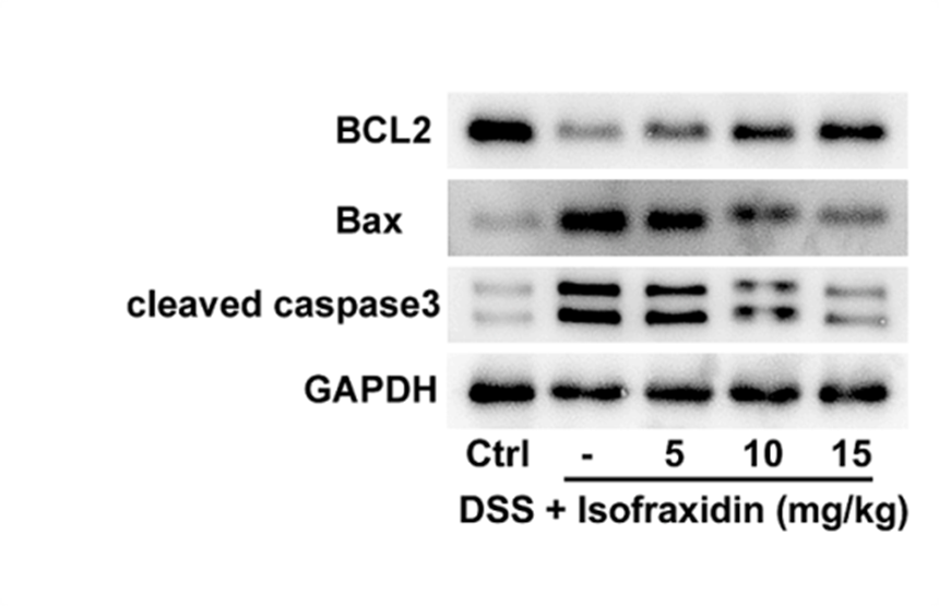
Isofraxidin purchased from MedChemExpress. Usage Cited in: Int Immunopharmacol. 2024 Apr 20:131:111814. [Abstract]
Western blotting was used to determine the levels of apoptosis-related proteins treated with Isofraxidin (5, 10, 15 mg/kg, p.o.).
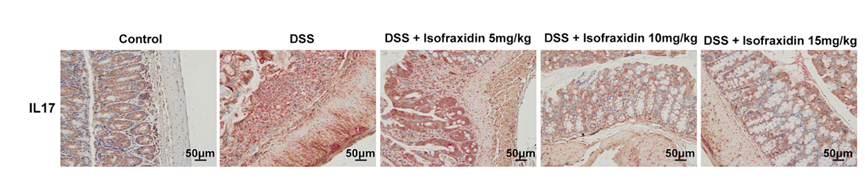
Isofraxidin purchased from MedChemExpress. Usage Cited in: Int Immunopharmacol. 2024 Apr 20:131:111814. [Abstract]
IHC revealed the positive rate of IL-17 in colon tissue, ZO-1 as epithelial cell localization treated with Isofraxidin (5, 10, 15 mg/kg, p.o.).
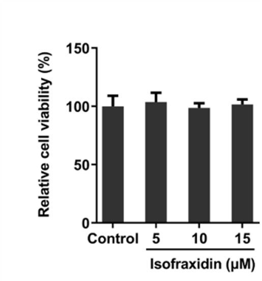
Isofraxidin purchased from MedChemExpress. Usage Cited in: Int Immunopharmacol. 2024 Apr 20:131:111814. [Abstract]
The effect of isofraxidin on the viability of NCM460 cells treated with Isofraxidin (5, 10, 15 μM).
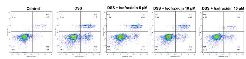
Isofraxidin purchased from MedChemExpress. Usage Cited in: Int Immunopharmacol. 2024 Apr 20:131:111814. [Abstract]
Flow cytometry revealed the proportion of apoptotic cells treated with Isofraxidin (5, 10, 15 μM).
-
J Cent Nerv Syst Dis
Neuroprotective potential of isofraxidin: Alleviating parkinsonian symptoms, inflammation and microglial activation. [Abstract]2025 Jan 9:17:11795735241312661. PMID: 39790641


Solvent & Solubility
In Vitro:
DMSO : 250 mg/mL (1125.16 mM; Need ultrasonic; Hygroscopic DMSO has a significant impact on the solubility of product, please use newly opened DMSO)
Please refer to the solubility information to select the appropriate solvent. Once prepared, please aliquot and store the solution to prevent product inactivation from repeated freeze-thaw cycles.
Storage method and period of stock solution: -80°C, 6 months; -20°C, 1 month. When stored at -80°C, please use it within 6 months. When stored at -20°C, please use it within 1 month.
Please refer to the solubility information to select the appropriate solvent. Once prepared, please aliquot and store the solution to prevent product inactivation from repeated freeze-thaw cycles.
Storage method and period of stock solution: -80°C, 6 months; -20°C, 1 month. When stored at -80°C, please use it within 6 months. When stored at -20°C, please use it within 1 month.
Concentration (start) × Volume (start) = Concentration (final) × Volume (final)
In Vivo:
Select the appropriate dissolution method based on your experimental animal and administration route.
- For the following dissolution methods, please ensure to first prepare a clear stock solution using an In Vitro approach and then sequentially add co-solvents:
- To ensure reliable experimental results, the clarified stock solution can be appropriately stored based on storage conditions. As for the working solution for In Vivo experiments, it is recommended to prepare freshly and use it on the same day.
- The percentages shown for the solvents indicate their volumetric ratio in the final prepared solution. If precipitation or phase separation occurs during preparation, heat and/or sonication can be used to aid dissolution.
Add each solvent one by one: 10% DMSO 40% PEG300 5% Tween-80 45% Saline
Solubility: ≥ 2.08 mg/mL (9.36 mM); Clear solution
This protocol yields a clear solution of ≥ 2.08 mg/mL (saturation unknown).
Taking 1 mL working solution as an example, add 100 μL DMSO stock solution (20.8 mg/mL) to 400 μL PEG300, and mix evenly; then add 50 μL Tween-80 and mix evenly; then add 450 μL Saline to adjust the volume to 1 mL.
Preparation of Saline: Dissolve 0.9 g sodium chloride in ddH₂O and dilute to 100 mL to obtain a clear Saline solution.
Add each solvent one by one: 10% DMSO 90% (20% SBE-β-CD in Saline)
Solubility: ≥ 2.08 mg/mL (9.36 mM); Clear solution
This protocol yields a clear solution of ≥ 2.08 mg/mL (saturation unknown).
Taking 1 mL working solution as an example, add 100 μL DMSO stock solution (20.8 mg/mL) to 900 μL 20% SBE-β-CD in Saline, and mix evenly.
Preparation of 20% SBE-β-CD in Saline (4°C, storage for one week): 2 g SBE-β-CD powder is dissolved in 10 mL Saline, completely dissolve until clear.
In Vivo Dissolution Calculator
Please enter the basic information of animal experiments:
-
-
-
-
Recommended: Prepare an additional quantity of animals to account for potential losses during experiments.
Please enter your animal formula composition:
-
%DMSO +
Recommended: Keep the proportion of DMSO in working solution below 2% if your animal is weak.
-
%+
-
+%Tween-80 + +
-
%Saline +
The co-solvents required include: DMSO, . All of co-solvents are available by MedChemExpress (MCE). , Tween 80. All of co-solvents are available by MedChemExpress (MCE).
Working solution concentration: 0.22 mg/mL
Method for preparing stock solution: mg drug dissolved in μL DMSO. Stock solution concentration: mg/mL.
1. Take μL DMSO stock solution;
2. Add μL .
μL , mix evenly;
3. Then add μL Tween 80, mix evenly;
4. Then add μL
Please ensure that the stock solution in the first step is dissolved to a clear state, and add co-solvents in sequence. You can use ultrasonic heating (ultrasonic cleaner, recommended frequency 20-40 kHz), vortexing, etc. to assist dissolution.


Purity & Documentation
-
Data Sheet (277 KB)
-
SDS (396 KB)
- English - EN (396 KB)
- Français - FR (396 KB)
- Deutsch - DE (396 KB)
- Norwegian - NO (396 KB)
- Español - ES (396 KB)
- Swedish - SV (396 KB)
- Italian - IT (396 KB)
- Korean - KR (396 KB)
- Portuguese - PT (396 KB)
-
Handling Instructions (2659 KB)
References
[1]. Yamazaki T, et al. Isofraxidin, a coumarin component from Acanthopanax senticosus, inhibits matrix metalloproteinase-7 expression and cell invasion of human hepatoma cells.Biol Pharm Bull. 2010;33(10):1716-22. [Content Brief]
[2]. Jin J , et al. Isofraxidin targets the TLR4/MD-2 axis to prevent osteoarthritis development.Food Funct. 2018 Nov 14;9(11):5641-5652. [Content Brief]


Complete Stock Solution Preparation Table
Please refer to the solubility information to select the appropriate solvent. Once prepared, please aliquot and store the solution to prevent product inactivation from repeated freeze-thaw cycles.
Storage method and period of stock solution: -80°C, 6 months; -20°C, 1 month. When stored at -80°C, please use it within 6 months. When stored at -20°C, please use it within 1 month.
| Optional Solvent | Concentration Solvent Mass | 1 mg | 5 mg | 10 mg | 25 mg |
|---|---|---|---|---|---|
| DMSO | 1 mM | 4.5007 mL | 22.5033 mL | 45.0065 mL | 112.5163 mL |
| 5 mM | 0.9001 mL | 4.5007 mL | 9.0013 mL | 22.5033 mL | |
| 10 mM | 0.4501 mL | 2.2503 mL | 4.5007 mL | 11.2516 mL | |
| 15 mM | 0.3000 mL | 1.5002 mL | 3.0004 mL | 7.5011 mL | |
| 20 mM | 0.2250 mL | 1.1252 mL | 2.2503 mL | 5.6258 mL | |
| 25 mM | 0.1800 mL | 0.9001 mL | 1.8003 mL | 4.5007 mL | |
| 30 mM | 0.1500 mL | 0.7501 mL | 1.5002 mL | 3.7505 mL | |
| 40 mM | 0.1125 mL | 0.5626 mL | 1.1252 mL | 2.8129 mL | |
| 50 mM | 0.0900 mL | 0.4501 mL | 0.9001 mL | 2.2503 mL | |
| 60 mM | 0.0750 mL | 0.3751 mL | 0.7501 mL | 1.8753 mL | |
| 80 mM | 0.0563 mL | 0.2813 mL | 0.5626 mL | 1.4065 mL | |
| 100 mM | 0.0450 mL | 0.2250 mL | 0.4501 mL | 1.1252 mL |


Isofraxidin Related Classifications
HY-N0774 Related Classifications
 Powered by Bioz