



MCB-36

Based on 1 Customer Validation
MCB-36 is a pan-KRAS PROTAC degrader that recruits VHL, with a Kd value of approximately 1 pM against KRAS, and it does not affect KRAS transcription. MCB-36 continuously degrades various KRAS mutants, inhibits oncogenic KRAS signaling pathways, reduces p-ERK levels, and induces apoptosis, thereby suppressing the growth of KRAS-dependent cancer cells. In vivo, MCB-36 inhibits tumor growth, overcomes resistance to KRAS G12C inhibitors, remodels the tumor immune microenvironment and enhances immune cell infiltration. MCB-36 can be used in research related to colorectal cancer and lung cancer.
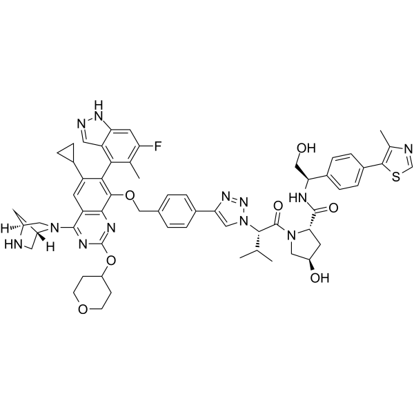
(Pink: Target protein ligand; Blue: VHL ligand (HY-112078); Black: linker (HY-W091879)).
For research use only. We do not sell to patients.
- Purity : 98.43%
- CAS No.: 3104382-00-2
- Formula: C60H71F2N9O7S
- Molecular Weight:1100.32
-
Storage:Powder -20°C, 3 years , 4°C, 2 years ; In solvent -80°C, 6 months , -20°C, 1 month


To place orders, for customer services and technical support, please contact: MedChemExpress USA
Tel: 609-228-6898 E-mail: [email protected] [email protected]


-
 Biological Activity
-
Chemical Information
-
Solvent & Solubility
- Protocols
- Purity & Documentation
- References
-
 Help & FAQs





Biological Activity
Description
IC50 & Target
|
KRas G12C |
KRas G12D |
KRas G12V |
KRAS G13D |
K-Ras WT |
Caspase 3 |
Cellular Effect
|
Cell Line

|
Type | Value | Description | References |
|---|---|---|---|---|
| MIA PaCa-2 | IC50 |
131.10 nM
|
Inhibition of cell viability against parental MIA PaCa-2 human cancer cells incubated for 5 days via 2D cell viability assay.
Inhibition of cell viability against parental MIA PaCa-2 human cancer cells incubated for 5 days via 2D cell viability assay.
|
40780213 |
In Vitro
MCB-36 (0.3125-5 nM; 120 s association, 300 s dissociation) binds to GDP-bound KRASG12D, KRASG12C, KRASG12V, and wild-type proteins with high affinity (Kd ≈ 1 pM) in cell-free SPR assays[1].
MCB-36 (100 nM; 12 h) significantly reduces the abundance of KRAS protein in AsPC-1 and H358 human cancer cells[1].
MCB-36 (0-10 μM; 3-48 h) induces concentration- and time-dependent degradation of KRAS and shortens the half-life of KRAS protein in human cancer cell lines AsPC-1, H358, Capan-2 and Caco-2[1].
MCB-36 (12 h) potently degrades wild-type and multiple mutant KRAS variants (KRASG12D, KRASG12C, KRASG12V, KRASG12S, KRASG13D, KRASQ61H) in transfected 293T cells[1].
MCB-36 forms a cooperative ternary complex with purified VCB, KRASG12D or KRASG12C proteins in cell-free fluorescence polarization (FP) assays[1].
MCB-36 forms a ternary complex with purified VCB and KRAS proteins in cell-free HTRF assays[1].
MCB-36 induces the formation of an intracellular ternary complex between KRAS and VHL in transfected 293T cells[1].
MCB-36 (administered for 5 consecutive days) inhibits the growth of KRAS-dependent human cancer cells with a mean IC50 of approximately 1 μM, whereas it exerts no significant inhibitory effect on KRAS-independent cancer cells and normal human cells (IC50 >10 μM)[1].
MCB-36 (administered for 5 consecutive days) inhibits the growth of MIA PaCa-2/SR and MIA PaCa-2/AR cells that are resistant to KRASG12C inhibitors, with IC50 values of 319.10 nM and 438.60 nM respectively after 5 days of treatment[1].
MCB-36 (administered for 5 consecutive days) inhibits the growth of MIA PaCa-2 cells harboring KRASG12C/Y96C or KRASG12C/H95D second-site mutations, with IC50 values of 186.50 nM and 185.70 nM respectively after 5 days of treatment, and retains activity against these KRASG12C inhibitor-resistant mutants[1].
MCB-36 (0-10 μM; 3-48 h) induces concentration- and time-dependent inhibition of KRAS signaling (decreased p-ERK levels) and apoptosis (increased levels of activated caspase-3 and activated PARP) in KRAS-dependent human cancer cells[1].
MCB-36 (0.3-0.6 μM; 6 days) inhibits the growth of patient-derived organoids from human colorectal cancer with KRAS mutations, while suppressing the MAPK signaling pathway and inducing apoptosis[1].
MedChemExpress (MCE) has not independently confirmed the accuracy of these methods. They are for reference only. Further protocols information, click here.
-
Cell Line:KRAS-mutant colorectal cancer organoids
-
Concentration:0, 1.25 and 2.5 μM
-
Incubation Time:6 days
-
Result:Decreased p-ERK, cleaved caspase-3, and cleaved PARP.
In Vivo
MCB-36 (60 mg/kg; i.p.; twice daily; for 16 consecutive days) exhibits potent antitumor activity in KRASG12V-mutant lung cancer PDX models, reduces tumor volume and weight, and downregulates the expression levels of KRAS and Ki67[1].
MCB-36 improves survival outcomes in a genetically engineered mouse model of pancreatic cancer driven by KRASG12D, extending the median survival to 58 days[1].
MCB-36 can remodel the tumor immune microenvironment in syngeneic CT26 tumors, increasing effector CD8+ T cells and reducing exhausted CD8+ T cells[1].
MCB-36 (60 mg/kg; i.p.; twice daily; 12 or 21 days) acts as a monotherapy to inhibit the growth of syngeneic CT26 tumors, and enhances the efficacy of combination therapy with anti-PD-1 antibody (HY-P990169) without inducing obvious toxicity[1].
MedChemExpress (MCE) has not independently confirmed the accuracy of these methods. They are for reference only.
-
Animal Model:BALB/c nude mice (6-8-week-old male)[1]
-
Dosage:60 mg/kg
-
Administration:i.p.; twice daily; 16 days
-
Result:Significantly reduced tumor volume and tumor weight compared to vehicle control.
Decreased KRAS and Ki67 expression in treated tumors.
Maintained sustained tumor growth inhibition following 16 days of treatment and a 12-day treatment-free period.
-
Animal Model:BALB/c nude mice (6-8-week-old male)[1]
-
Dosage:60 mg/kg
-
Administration:i.p.; twice daily; 16 days
-
Result:Significantly reduced tumor volume and tumor weight compared to vehicle control.
Decreased KRAS and Ki67 expression in treated tumors.
-
Animal Model:BALB/c mice (4-5-week-old male)[1]
-
Dosage:60 mg/kg (monotherapy); 60 mg/kg + 25 μg/dose (combination with anti-PD-1 antibody (HY-P990169))
-
Administration:i.p.; twice daily; 12 or 21 days (monotherapy); i.p.; twice weekly (anti-PD-1 antibody component)
-
Result:Significantly suppressed tumor growth compared to vehicle control as monotherapy.
Enhanced tumor growth suppression compared to either single agent alone when combined with anti-PD-1 antibody.
Caused no significant body weight loss in treated mice.


Chemical Information
-
CAS No. 3104382-00-2
-
Appearance Solid
-
Molecular Weight 1100.32
-
Formula C60H71F2N9O7S
-
Color Off-white to yellow
-
SMILES
C#CC1=C(C=CC2=CC(O)=CC(C3=NC=C4C(N5CCC[C@@](O)(C5)C)=NC(OC[C@@H]6CCCN6CCCCCCC(N[C@@H](C(C)(C)C)C(N7C[C@@H](C[C@H]7C(N[C@H](C8=CC=C(C=C8)C9=C(N=CS9)C)C)=O)O)=O)=O)=NC4=C3F)=C21)F
-
Shipping
Room temperature in continental US; may vary elsewhere.
-
Storage
Powder -20°C 3 years 4°C 2 years In solvent -80°C 6 months -20°C 1 month


Solvent & Solubility
In Vitro:
DMSO : 100 mg/mL (90.88 mM; Need ultrasonic; Hygroscopic DMSO has a significant impact on the solubility of product, please use newly opened DMSO)
Please refer to the solubility information to select the appropriate solvent. Once prepared, please aliquot and store the solution to prevent product inactivation from repeated freeze-thaw cycles.
Storage method and period of stock solution: -80°C, 6 months; -20°C, 1 month. When stored at -80°C, please use it within 6 months. When stored at -20°C, please use it within 1 month.
Please refer to the solubility information to select the appropriate solvent. Once prepared, please aliquot and store the solution to prevent product inactivation from repeated freeze-thaw cycles.
Storage method and period of stock solution: -80°C, 6 months; -20°C, 1 month. When stored at -80°C, please use it within 6 months. When stored at -20°C, please use it within 1 month.
Concentration (start) × Volume (start) = Concentration (final) × Volume (final)
In Vivo:
Select the appropriate dissolution method based on your experimental animal and administration route.
- For the following dissolution methods, please ensure to first prepare a clear stock solution using an In Vitro approach and then sequentially add co-solvents:
- To ensure reliable experimental results, the clarified stock solution can be appropriately stored based on storage conditions. As for the working solution for In Vivo experiments, it is recommended to prepare freshly and use it on the same day.
- The percentages shown for the solvents indicate their volumetric ratio in the final prepared solution. If precipitation or phase separation occurs during preparation, heat and/or sonication can be used to aid dissolution.
In Vivo Dissolution Calculator
Please enter the basic information of animal experiments:
-
-
-
-
Recommended: Prepare an additional quantity of animals to account for potential losses during experiments.
Please enter your animal formula composition:
-
%DMSO +
Recommended: Keep the proportion of DMSO in working solution below 2% if your animal is weak.
-
%+
-
+%Tween-80 + +
-
%Saline +
The co-solvents required include: DMSO, . All of co-solvents are available by MedChemExpress (MCE). , Tween 80. All of co-solvents are available by MedChemExpress (MCE).
Working solution concentration: 0.22 mg/mL
Method for preparing stock solution: mg drug dissolved in μL DMSO. Stock solution concentration: mg/mL.
1. Take μL DMSO stock solution;
2. Add μL .
μL , mix evenly;
3. Then add μL Tween 80, mix evenly;
4. Then add μL
Please ensure that the stock solution in the first step is dissolved to a clear state, and add co-solvents in sequence. You can use ultrasonic heating (ultrasonic cleaner, recommended frequency 20-40 kHz), vortexing, etc. to assist dissolution.

Protocols
-
Multiplex immunofluorescence IHC
Multiplex immunofluorescence IHC detects multiple protein biomarkers in one tissue section by sequential antibody staining, HRP-mediated tyramide fluorophore deposition, heat-mediated antibody stripping, nuclear counterstaining, multispectral imaging, spectral unmixing, and digital cell phenotyping; TSA deposits fluorophore near the antigen so the fluorescence signal remains after primary and secondary antibodies are removed, enabling repeated staining cycles, including with antibodies from the same host species. Classic FFPE tumor immune-profiling applications use panels such as CD3, CD8, CD68/CD163, FOXP3, PD-1, PD-L1, pancytokeratin, Ki67, and DAPI to identify tumor cells, immune-cell subsets, checkpoint-marker expression, co-expression phenotypes, cell density, and spatial relationships in the tumor microenvironment.
-
Apoptosis
Apoptosis, also called programmed cell death, is generally characterized by distinct morphological characteristics.
-
TUNEL staining for apoptotic DNA fragmentation
TUNEL staining detects DNA strand breaks by using terminal deoxynucleotidyl transferase to add labeled nucleotides to exposed 3′-OH DNA termini, generating either microscopic staining in fixed cells or tissue sections, or fluorescence/cytometric signal in cell suspensions. TUNEL positivity reflects DNA fragmentation but should not be interpreted alone as definitive apoptosis, because TUNEL can also label necrotic, autolytic, mechanically damaged, or DNA-repair-associated DNA breaks.
-
Annexin V plus membrane-impermeant dye apoptosis staining
Annexin V-based apoptosis assays rely on the detection of phosphatidylserine (PS) externalization from the inner leaflet of the plasma membrane to the outer leaflet, an early biochemical hallmark of apoptosis. Fluorescently labeled Annexin V binds PS in a calcium-dependent manner, enabling identification of early apoptotic cells by flow cytometry or fluorescence microscopy. When combined with a membrane-impermeant DNA-binding dye (e. g. , propidium iodide), this approach allows discrimination between viable (Annexin V−/dye−), early apoptotic (Annexin V+/dye−), and late apoptotic or necrotic (Annexin V+/dye+) cell populations by assessing membrane integrity and PS exposure.
-
Patient-Derived Xenograft (PDX)
Patient-derived xenograft (PDX) models are generated by engrafting primary human tumor tissue directly into immunodeficient mice, allowing in vivo propagation of patient tumor biology without initial in vitro adaptation. These models are used to preserve key histopathological and molecular characteristics of the original tumor and enable assessment of tumor growth dynamics and therapeutic response in a living organism. The biological readout is tumor engraftment and subsequent growth in the murine host, which reflects the ability of human tumor cells to survive, vascularize, and expand in an immunocompromised microenvironment.
-
Multiplex immunohistochemistry
Multiplex immunohistochemistry (mIHC), also known as tyramide dignal amplification (TSA), is an enzymatic detection method that uses horseradish peroxidase (HRP) to perform high-density in-situ labeling of target proteins or nucleic acids.
-
Apoptosis Solutions
Apoptosis is a regulated, generally non-lytic cell-death pathway that removes unwanted, damaged, infected, or abnormal cells through coordinated morphological changes, caspase activation, DNA fragmentation, and membrane remodeling. The intrinsic apoptosis pathway is controlled mainly by mitochondrial outer membrane permeabilization, BCL-2 family proteins, cytochrome c release, apoptosome formation, caspase-9 activation, and downstream executioner caspase-3/7 activation. The extrinsic apoptosis pathway is initiated by death receptors such as Fas, TNFR, and TRAIL receptors, which recruit adaptor proteins and activate caspase-8 before engaging executioner caspases or mitochondrial amplification through BID cleavage. Apoptosis is linked to many phenotypes, including cancer cell killing, tissue homeostasis, immune regulation, neurodegeneration, infection response, and treatment-induced cytotoxicity; unresolved questions include how apoptosis interacts with necroptosis, pyroptosis, ferroptos
-
Patient-Derived Orthotopic Xenograft (PDOX)
Patient-derived orthotopic xenograft (PDOX) modeling implants fresh patient tumor tissue or patient-derived tumor cells into the anatomically corresponding organ or tissue site of immunodeficient mice, usually by surgical orthotopic implantation, to preserve patient tumor histology, local microenvironmental context, invasion, metastatic behavior, and treatment-response features better than subcutaneous implantation. PDOX readouts include tumor engraftment, orthotopic tumor growth, local invasion, metastasis, recurrence after resection, histologic similarity to the donor tumor, biomarker retention, molecular concordance, survival, and response or resistance to therapy. PDOX models are used for preclinical drug testing and individualized therapy evaluation, but engraftment success varies by tumor type and specimen quality.
-
Subcutaneous Cell-Line-Derived Xenograft
Subcutaneous cell-line-derived xenograft (CDX) models are established by implanting cultured human cancer cell lines into immunodeficient mice, where the injected cells form localized tumors that can be monitored in vivo as a measure of tumorigenic potential, growth kinetics, and treatment response. These models are widely used in oncology research because they allow reproducible tumor formation and enable comparative assessment of tumor growth between different cell lines or genetic manipulations in a controlled in vivo microenvironment. Subcutaneous implantation of cancer cells in immunodeficient mice is a standard approach for evaluating tumor growth behavior and therapeutic response across multiple cancer types, including prostate, esophageal, pancreatic, and colon cancer models.
-
Orthotopic Cell-Line Xenograft
Orthotopic cell-line xenograft models involve implantation of human cancer cell lines into the anatomically corresponding organ of immunodeficient mice to reproduce tumor growth within a native microenvironment, enabling more clinically relevant tumor behavior compared with subcutaneous models. These models are widely used because orthotopic placement better recapitulates tumor progression, including invasion and metastatic spread, which are often underrepresented in heterotopic implantation systems. Compared with conventional xenografts, orthotopic implantation is described as more technically complex but provides improved simulation of tumor-microenvironment interactions and metastatic behavior, making it particularly valuable for translational oncology research. Surgical orthotopic implantation approaches have been emphasized as enabling faithful reproduction of clinical cancer features, including metastasis and disease progression patterns that align with the tumor’s organ of origi
-
Research Protocol for Cancer Immunology
Cancer immunology studies how the immune system recognizes, suppresses, edits, or fails to eliminate malignant cells through tumor antigen release, antigen presentation, T-cell priming, immune trafficking, tumor-cell killing, and feedback inhibition in the tumor microenvironment. The cancer-immunity cycle links tumor antigenicity, dendritic-cell priming, CD8+ T-cell infiltration, cytotoxic function, and immune-checkpoint regulation to tumor rejection or immune escape. Immune-checkpoint pathways such as PD-1/PD-L1 and CTLA-4 suppress antitumor T-cell activity and can be therapeutically blocked, but many tumors remain resistant because of poor antigen presentation, weak T-cell infiltration, suppressive myeloid cells, regulatory T cells, and tumor-intrinsic immune-exclusion programs. Unresolved questions include which immune-cell states predict response, how tumor-intrinsic pathways exclude immune cells, how myeloid suppression limits checkpoint blockade, and which combination strategies


Purity & Documentation
-
Data Sheet (295 KB)
-
SDS (254 KB)
- English - EN (254 KB)
- Français - FR (254 KB)
- Deutsch - DE (254 KB)
- Norwegian - NO (254 KB)
- Español - ES (254 KB)
- Swedish - SV (254 KB)
- Italian - IT (254 KB)
- Korean - KR (254 KB)
- Portuguese - PT (254 KB)
-
Handling Instructions (2659 KB)
References


Complete Stock Solution Preparation Table
Please refer to the solubility information to select the appropriate solvent. Once prepared, please aliquot and store the solution to prevent product inactivation from repeated freeze-thaw cycles.
Storage method and period of stock solution: -80°C, 6 months; -20°C, 1 month. When stored at -80°C, please use it within 6 months. When stored at -20°C, please use it within 1 month.
| Optional Solvent | Concentration Solvent Mass | 1 mg | 5 mg | 10 mg | 25 mg |
|---|---|---|---|---|---|
| DMSO | 1 mM | 0.9088 mL | 4.5441 mL | 9.0883 mL | 22.7207 mL |
| 5 mM | 0.1818 mL | 0.9088 mL | 1.8177 mL | 4.5441 mL | |
| 10 mM | 0.0909 mL | 0.4544 mL | 0.9088 mL | 2.2721 mL | |
| 15 mM | 0.0606 mL | 0.3029 mL | 0.6059 mL | 1.5147 mL | |
| 20 mM | 0.0454 mL | 0.2272 mL | 0.4544 mL | 1.1360 mL | |
| 25 mM | 0.0364 mL | 0.1818 mL | 0.3635 mL | 0.9088 mL | |
| 30 mM | 0.0303 mL | 0.1515 mL | 0.3029 mL | 0.7574 mL | |
| 40 mM | 0.0227 mL | 0.1136 mL | 0.2272 mL | 0.5680 mL | |
| 50 mM | 0.0182 mL | 0.0909 mL | 0.1818 mL | 0.4544 mL | |
| 60 mM | 0.0151 mL | 0.0757 mL | 0.1515 mL | 0.3787 mL | |
| 80 mM | 0.0114 mL | 0.0568 mL | 0.1136 mL | 0.2840 mL |


MCB-36 Related Classifications
HY-176786 Related Classifications
 Powered by Bioz