



Liriodendrin
Based on 1 publication(s) in Google Scholar
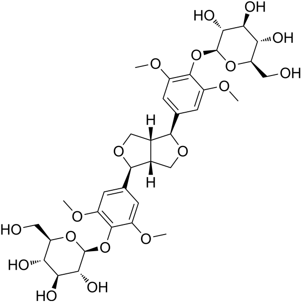
Liriodendrin is an orally effective lignan compound with anti-inflammatory, antioxidant, and anti-platelet aggregation activities. Liriodendrin is isolated and extracted from Sargentodoxae caulis and Linaria vulgaris. Liriodendrin regulates the expression of genes related to inflammation and cellular metabolism by inhibiting the NF-κB, PI3K/Akt, and TLR4 signaling pathways, or by activating the PI3K-DDX18 pathway. Liriodendrin increases the expression of HSF1 and HSP27 without cytotoxicity. Liriodendrin is used in studies on ulcerative colitis, myocardial ischemia-reperfusion injury, sepsis-induced acute lung injury, and chronic endometritis.
For research use only. We do not sell to patients.
- Purity: 99.02%
- CAS No.: 573-44-4
- Formula: C34H46O18
- Molecular Weight:742.72
-
Storage:
4°C, sealed storage, away from moisture and light
* In solvent : -80°C, 6 months; -20°C, 1 month (sealed storage, away from moisture and light)

To place orders, for customer services and technical support, please contact: MedChemExpress USA
Tel: 609-228-6898 E-mail: [email protected] [email protected]


-
 Biological Activity
-
Chemical Information
- Purity & Documentation
- References
-
 Help & FAQs


-
HY-N0293
Paeoniflorin -

HY-N0293R
Paeoniflorin (Standard) -
HY-N1986
Cucurbitacin D -
HY-N0830B
Palmitic acid sodium -
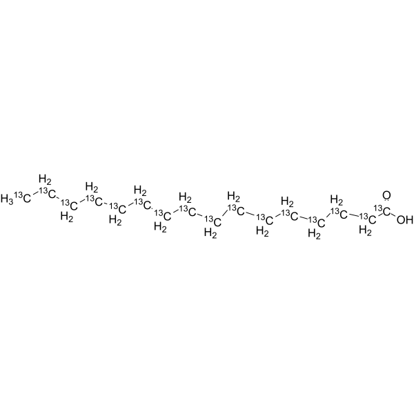
HY-N0830S6
Palmitic acid-13C16 -
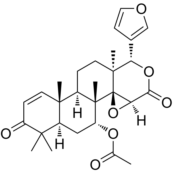
HY-107577
Gedunin -
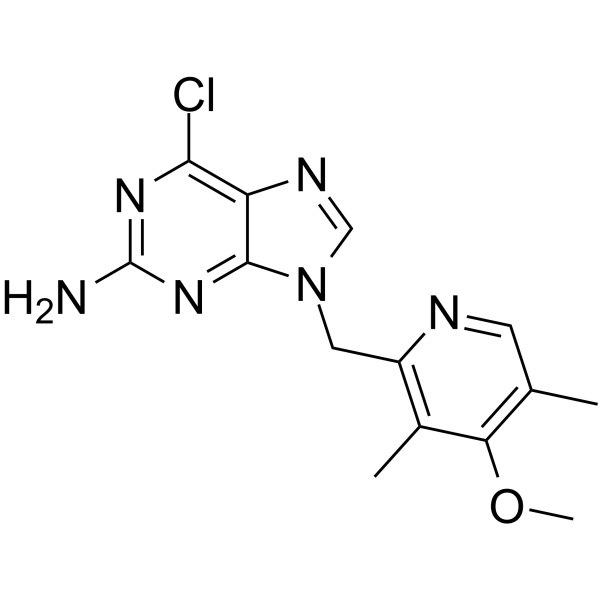
HY-10212
BIIB021 -

HY-N1986R
Cucurbitacin D (Standard) -
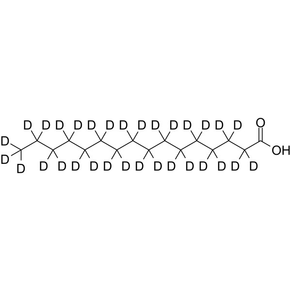
HY-N0830S2
Palmitic acid-d31 -
HY-12024
Alvespimycin hydrochloride -
HY-100872
KRIBB11 -
HY-15190
NVP-HSP990
Publications Citing Use of MedChemExpress (MCE) Liriodendrin
MoreAll Endogenous Metabolite Isoforms
More

Biological Activity
Description
|
HSF1 |
TLR4 |
Cellular Effect
|
Cell Line

|
Type | Value | Description | References |
|---|---|---|---|---|
| L132 | IC50 |
>5 μM
|
Cytotoxicity against human L132 cells assessed as reduction in cell viability by MTT assay.
Cytotoxicity against human L132 cells assessed as reduction in cell viability by MTT assay.
|
39134870 |
In Vitro
Liriodendrin (20-100 μM; pretreatment for 2 h) inhibits LPS (HY-D1056)-stimulated production of pro-inflammatory cytokines and suppresses NF-κB activation in RAW 264.7 macrophages, but does not affect Akt phosphorylation[5].
Liriodendrin (0.45-2.25 mM; 24 h) dose-dependently promotes cell proliferation, promotes cell cycle transition, and increases milk protein synthesis in bovine mammary epithelial cells (MECs)[6].
Liriodendrin (20-100 μM; 24 h) protects mitochondrial morphology, inhibits apoptosis, and suppresses NF-κB signaling pathway activation in rat cardiomyocytes H9C2 under hypoxia/reoxygenation conditions[3].
Liriodendrin (50 μM) inhibits pro-inflammatory cytokine secretion, reduces reactive oxygen species (ROS) production, and suppresses NF-κB (p65) phosphorylation in mouse macrophage RAW 264.7 cells[4].
Liriodendrin (3 μM; 12 h) induces HSF1 and HSP27 protein expression in L132 human lung fibroblasts[1].
Liriodendrin shows no cytotoxicity in L132 human lung fibroblasts at concentrations up to 5 μM[1].
MedChemExpress (MCE) has not independently confirmed the accuracy of these methods. They are for reference only.
-
Cell Line:RAW 264.7
-
Concentration:20 μM, 50 μM, 100 μM
-
Incubation Time:2 h pre-treatment prior to LPS exposure for 4 h
-
Result:Decreased the mRNA levels of TNF-α, IL-6, and IL-1β, and had no significant effect on ERβ mRNA expression.
-
Cell Line:RAW 264.7
-
Concentration:20 μM, 50 μM, 100 μM
-
Incubation Time:2 h pre-treatment prior to LPS exposure for 4 h
-
Result:Significantly reduced the phosphorylation of NF-κB and IκBα.
Had no effect on the phosphorylation of Akt.
-
Cell Line:Primary bovine mammary epithelial cells (MECs)
-
Concentration:0.45 mM, 0.9 mM, 1.35 mM, 1.8 mM, 2.25 mM
-
Incubation Time:24 h
-
Result:Reduced the proportion of cells in the G1 phase and increased the proportion of cells in the S and G2/M phases.
-
Cell Line:Primary bovine mammary epithelial cells (MECs)
-
Concentration:0.45 mM, 0.9 mM, 1.35 mM, 1.8 mM, 2.25 mM
-
Incubation Time:24 h
-
Result:Increased cell number and promoted cell proliferation in a dose-dependent manner, peaking at 1.35 mM.
-
Cell Line:H9C2 cardiomyocytes
-
Concentration:20, 50, 100 μM
-
Incubation Time:24 h
-
Result:Decreased protein expression levels of p-NF-κB and p-IκBα at all concentrations.
At 100 μM, decreased Bax protein expression levels and increased Bcl-2 protein expression.
-
Cell Line:RAW 264.7
-
Concentration:50 μM
-
Incubation Time:pre-treated for 6 h before LPS stimulation, co-treated with LPS for 18 h
-
Result:Reduced the secretion of TNF-α, IL-1β, MCP-1, and IL-6.
-
Cell Line:L132 human embryonic lung fibroblast cells
-
Concentration:3 μM
-
Incubation Time:12 h
-
Result:Induced an increase in the protein expression of HSF1 and HSP27
-
Cell Line:Rat H9C2 cardiomyocytes
-
Concentration:100 μM
-
Incubation Time:24 h
-
Result:Decreased the mRNA transcriptional activities of Bax and Caspase-3, and increased the Bcl-2/Bax ratio.
In Vivo
Liriodendrin (100 mg/kg; p.o.; once daily; from 5 days before surgery to 3 days after surgery) ameliorates ventricular remodeling, inhibits inflammatory cytokines, and attenuates cardiomyocyte apoptosis in a male Wistar rat model of myocardial ischemia-reperfusion injury induced by left anterior descending coronary artery ligation[3].
Liriodendrin (20 mg/kg; i.p.; administered at 0 and 6 h after CLP, days 1-5) improves survival, alleviates lung histopathological injury, and inhibits pro-inflammatory mediator production in a cecal ligation and puncture (CLP)-induced acute lung injury model of sepsis in male C57BL/6 mice[4].
Liriodendrin (50-200 mg/kg; p.o.; once daily; 21 days) alleviates uterine inflammation, modulates gut microbiota, and inhibits the L-arginine/NO metabolic pathway in a chronic endometritis model of female Sprague-Dawley rats[2].
Liriodendrin (1.45-13.05 mg/kg/d; p.o.; once daily; for 7 days) promotes β-casein and DDX18 expression in mammary tissue and increases the body weight of offspring mice in a maternal ICR mouse model on lactation day 10[6].
MedChemExpress (MCE) has not independently confirmed the accuracy of these methods. They are for reference only.
-
Animal Model:BALB/c (6-week-old, male, 20 ± 2 g)[5]
-
Dosage:100 mg/kg
-
Administration:p.o.; once daily; pretreated 3 days before DSS treatment and continued during the 7-day DSS treatment period
-
Result:Decreased the disease activity index (DAI) compared with the DSS group.
Significantly reversed the shortening in colon length induced by DSS.
Attenuated pathological changes induced by DSS treatment and markedly inhibited inflammation scores compared with the DSS group.
Significantly reduced the DSS-induced increase in MPO activity.
Significantly reduced the levels of pro-inflammatory cytokines IL-6, TNF-α, and IL-1β.
Significantly attenuated the decrease in the activities of antioxidant enzymes SOD and GPx induced by DSS treatment and significantly counteracted the DSS-induced elevated malondialdehyde (MDA).
Led to a significant reduction in the phosphorylation of NF-κB and Akt, and blocked the phosphorylation of IκBα in the colon.
Significantly up-regulated the mRNA expression of ERβ in the colon compared to the DSS group.
-
Animal Model:ICR (11-week-old lactating mothers)[6]
-
Dosage:1.45 mg/kg/d; 4.35 mg/kg/d; 13.05 mg/kg/d
-
Administration:p.o.; once daily; for 7 d
-
Result:Produced higher expression of β-casein and DDX18 in mouse mammary gland and greater weight gain of offspring mice at 4.35 mg/kg/d than the normal feeding group and the 1.45 mg/kg/d group.
Showed reduced effects at 13.05 mg/kg/d compared with the 4.35 mg/kg/d group.
-
Animal Model:Wistar rats (male, 8 weeks old, 200 g)[3]
-
Dosage:100 mg/kg
-
Administration:intragastric; daily; 5 days before surgery to 3 days after surgery
-
Result:Decreased I/R-induced elevation of CKMB and cTnT levels.
Reversed I/R-induced elevation of EDV and LVIDd.
Lowered EF, LVIDs, and ESV compared with the I/R group.
Reduced inflammatory cell infiltration and pathological alterations.
Decreased MCP-1 expression and ROS levels.
Increased SOD levels in peripheral blood.
Decreased expression of Bax and cleaved caspase-3.
Increased expression of Bcl-2.
Markedly decreased apoptosis index (AI), though not statistically significant.
Increased Bcl-2 protein expression and decreased Bax and cleaved caspase-3 protein expression.
Decreased expression of p-NF-κB and p-IκBα.
-
Animal Model:C57BL/6 mice (male, 6-8 weeks old)[4]
-
Dosage:20 mg/kg
-
Administration:i.p.; dose at 0 and 6 h after CLP (mortality study); 20 mg/kg for the 24-h lung injury study
-
Result:Improved survival rate to 30%.
Mitigated lung hemorrhage, edema, fibrin deposition, and neutrophil accumulation at 24 h.
Reduced CLP-induced increase in lung MPO activity.
Significantly reduced albumin content in BAL fluids.
Lowered levels of TNF-α, IL-1β, MCP-1, and IL-6 in BAL fluids at 24 h.
Suppressed expression of VEGF and phosphorylation of NF-kB (p65) in septic lungs.
-
Animal Model:Sprague-Dawley (Female, 200-220 g, 15% hydroxybenzene mucilage-induced chronic endometritis)[2]
-
Dosage:50 mg/kg ; 100 mg/kg; 200 mg/kg
-
Administration:p.o.; once daily; 21 days
-
Result:Suppressed CE-induced pathological changes in uterus tissues and decreased the number of inflammatory cells.
Did not adversely affect the tissue morphology or exert toxic effects on the liver and kidney.
Mitigated the CE-induced upregulation of TLR4 and P-p65 protein levels in uterine tissues.
Suppressed the CE-induced upregulation of Tlr4, Tnf-α, Il-1β, and Il-6 mRNA levels and downregulation of Il-10 mRNA levels in the uterus tissues.
Decreased the CE-induced upregulation of the Firmicutes/Bacteroidetes ratio.
Downregulated the abundances of Campilobacterota and Bacteroidetes and downregulated the abundances of Actinobacteria and Candidatus_Saccharibacteria in CE rats.
Mitigated the CE-induced upregulation of Lachnospiraceae_unclassified and Clostridiales_unclassified abundances, and suppressed the CE-induced downregulation of Ruminococcaceae_unclassified and Muribaculaceae_unclassified relative abundances.
Downregulated Arg-2 levels in a dose-dependent manner.
Mitigated the CE-induced downregulation of NO, eNOS, and L-arginine in the uterus tissues.


Chemical Information
-
CAS No. 573-44-4
-
Appearance Solid
-
Molecular Weight 742.72
-
Formula C34H46O18
-
Color White to off-white
-
SMILES
COC1=C(O[C@@H]2O[C@@H]([C@H]([C@@H]([C@H]2O)O)O)CO)C(OC)=CC([C@@H]3[C@]4([H])[C@@](CO3)([H])[C@@H](C5=CC(OC)=C(C(OC)=C5)O[C@@H]6O[C@@H]([C@H]([C@@H]([C@H]6O)O)O)CO)OC4)=C1
-
Structure Classification
-
Initial Source
-
Shipping
Room temperature in continental US; may vary elsewhere.
-
Storage
4°C, sealed storage, away from moisture and light
* In solvent : -80°C, 6 months; -20°C, 1 month (sealed storage, away from moisture and light)


Publications (1)
-
Journal Impact Factor
-
Most Recent
-
Int J Mol Med
Liriodendrin alleviates myocardial ischemia‑reperfusion injury via partially attenuating apoptosis, inflammation and mitochondria damage in rats. [Abstract]2025 Apr;55(4):65. PMID: 39981888


Purity & Documentation
-
Data Sheet (316 KB)
-
SDS (252 KB)
- English - EN (252 KB)
- Français - FR (252 KB)
- Deutsch - DE (252 KB)
- Norwegian - NO (252 KB)
- Español - ES (252 KB)
- Swedish - SV (252 KB)
- Italian - IT (252 KB)
- Korean - KR (252 KB)
- Portuguese - PT (252 KB)
-
Handling Instructions (2659 KB)
References
[1]. Nam JW, et al. Heat shock factor 1 inducers from the bark of Eucommia ulmoides as cytoprotective agents. Chemistry & biodiversity. 2013 Jul;10(7):1322-7. [Content Brief]
[3]. Li B, et al. Liriodendrin alleviates myocardial ischemia‑reperfusion injury via partially attenuating apoptosis, inflammation and mitochondria damage in rats. International journal of molecular medicine. 2025 Apr;55(4):65. [Content Brief]

Calculators
Concentration (start) × Volume (start) = Concentration (final) × Volume (final)


Liriodendrin Related Classifications
HY-N3377 Related Classifications
 Powered by Bioz
Keywords
- Liriodendrin
- 573-44-4
- HSP
- NF-κB
- PI3K
- Akt
- Apoptosis
- Toll-like Receptor (TLR)
- Endogenous Metabolite
- mice
- sepsis-induced acute lung injury
- bovine MECs
- H9C2 cells
- myocardial ischemia-reperfusion injury
- L132 human lung fibroblast cells
- rats
- RAW 264.7 macrophages
- ulcerative colitis
- chronic endometritis
- Inhibitor
- inhibitor
- inhibit