



YPX-C-05

Based on 1 Customer Validation
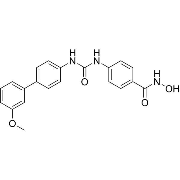
YPX-C-05 is an orally active HDAC inhibitor and vasodilator. YPX-C-05 inhibits HDAC enzymatic activity, increases histone H4 acetylation, activates the PI3K/Akt pathway, promotes Akt and eNOS phosphorylation, enhances eNOS activity, and boosts nitric oxide production. YPX-C-05 increases Tetrahydrobiopterin (HY-107383) levels, restores serum nitric oxide levels, reduces endothelin-1 levels, and improves endothelial function. YPX-C-05 reduces vascular remodeling and exerts antihypertensive effects in hypertensive Mus musculus mice. YPX-C-05 can be used for the research of hypertension.
For research use only. We do not sell to patients.
- Purity : 98.05%
- CAS No.: 2894823-79-9
- Formula: C21H19N3O4
- Molecular Weight:377.39
-
Storage:Powder -20°C, 3 years ; In solvent -80°C, 6 months , -20°C, 1 month

To place orders, for customer services and technical support, please contact: MedChemExpress USA
Tel: 609-228-6898 E-mail: [email protected] [email protected]


-
 Biological Activity
-
Chemical Information
-
Solvent & Solubility
- Protocols
- Purity & Documentation
- References
-
 Help & FAQs




Biological Activity
Description
IC50 & Target
[1]|
HDAC |
eNOS |
In Vitro
YPX-C-05 (5 μM; 18 h, or 50 μM phenylephrine pretreatment for 6 h followed by 5 μM for 18 h) activates the PI3K/Akt/eNOS pathway in HUVECs by increasing phosphorylation of Akt and eNOS, reversing Phenylephrine-induced inhibition of this pathway[1].
MedChemExpress (MCE) has not independently confirmed the accuracy of these methods. They are for reference only.
-
Cell Line:human umbilical vein endothelial cells (HUVECs)
-
Concentration:3, 10, 30, 100 μM
-
Incubation Time:24 h
-
Result:Caused a dose-dependent increase in histone H4 acetylation levels.
-
Cell Line:human umbilical vein endothelial cells (HUVECs)
-
Concentration:2.5, 5, 10, 20, 50, 100, 200 μM
-
Incubation Time:24 h
-
Result:Left cell viability unaffected at concentrations up to 10 μM.
Caused a small but statistically significant reduction in viability at 25 μM, though viability remained >70%.
-
Cell Line:human umbilical vein endothelial cells (HUVECs)
-
Concentration:5 μM (YPX-C-05); 50 μM phenylephrine (pretreatment) followed by 5 μM YPX-C-05
-
Incubation Time:18 h (YPX-C-05); 6 h (phenylephrine pretreatment) followed by 18 h (YPX-C-05)
-
Result:Reversed phenylephrine-induced reduction in P-eNOS/eNOS and P-Akt/Akt ratios.
Significantly increased both P-eNOS/eNOS and P-Akt/Akt ratios.
Increased total eNOS expression.
In Vivo
MedChemExpress (MCE) has not independently confirmed the accuracy of these methods. They are for reference only.
-
Animal Model:C57BL/6 (6-8-week-old male, 20-25 g, hypertension induced by intraperitoneal injections of 15 mg/kg N-omega-nitro-L-arginine twice daily for 21 days)[1]
-
Dosage:7.615 mg/kg
-
Administration:p.o.; daily; 4 weeks
-
Result:Reduced systolic blood pressure, diastolic blood pressure, mean arterial pressure, and heart rate compared to untreated hypertensive mice.
Restored serum nitric oxide (NO) levels from ~29.5 μM to ~35 μM.
Reduced serum endothelin-1 (ET-1) levels from ~146 μg/L to ~129 μg/L.
Improved endothelial cell layer integrity, increased the thoracic aorta inner/outer diameter ratio from ~0.79 to ~0.84, decreased aortic wall thickness from ~52 μm to ~49 μm, and reduced collagen deposition in the aortic wall from ~14.5% to ~12%.
Improved acetylcholine-induced vasorelaxation in isolated thoracic aortas, restoring the relaxation response from ~25% to ~55% of phenylephrine-induced tone.
Increased endothelial eNOS expression in thoracic aortas, with integrated optical density values increasing from ~8 to ~14.


Chemical Information
-
CAS No. 2894823-79-9
-
Appearance Solid
-
Molecular Weight 377.39
-
Formula C21H19N3O4
-
Color White to off-white
-
SMILES
COC1=CC(C2=CC=C(NC(NC3=CC=C(C(NO)=O)C=C3)=O)C=C2)=CC=C1
-
Shipping
Room temperature in continental US; may vary elsewhere.
-
Storage
Powder -20°C 3 years In solvent -80°C 6 months -20°C 1 month


Solvent & Solubility
In Vitro:
DMSO : 25 mg/mL (66.24 mM; Need ultrasonic; Hygroscopic DMSO has a significant impact on the solubility of product, please use newly opened DMSO)
Please refer to the solubility information to select the appropriate solvent. Once prepared, please aliquot and store the solution to prevent product inactivation from repeated freeze-thaw cycles.
Storage method and period of stock solution: -80°C, 6 months; -20°C, 1 month. When stored at -80°C, please use it within 6 months. When stored at -20°C, please use it within 1 month.
Please refer to the solubility information to select the appropriate solvent. Once prepared, please aliquot and store the solution to prevent product inactivation from repeated freeze-thaw cycles.
Storage method and period of stock solution: -80°C, 6 months; -20°C, 1 month. When stored at -80°C, please use it within 6 months. When stored at -20°C, please use it within 1 month.
Concentration (start) × Volume (start) = Concentration (final) × Volume (final)


Purity & Documentation
-
Data Sheet (276 KB)
-
SDS (252 KB)
- English - EN (252 KB)
- Français - FR (252 KB)
- Deutsch - DE (252 KB)
- Norwegian - NO (252 KB)
- Español - ES (252 KB)
- Swedish - SV (252 KB)
- Italian - IT (252 KB)
- Korean - KR (252 KB)
- Portuguese - PT (252 KB)
-
Handling Instructions (2659 KB)
References


Complete Stock Solution Preparation Table
Please refer to the solubility information to select the appropriate solvent. Once prepared, please aliquot and store the solution to prevent product inactivation from repeated freeze-thaw cycles.
Storage method and period of stock solution: -80°C, 6 months; -20°C, 1 month. When stored at -80°C, please use it within 6 months. When stored at -20°C, please use it within 1 month.
| Optional Solvent | Concentration Solvent Mass | 1 mg | 5 mg | 10 mg | 25 mg |
|---|---|---|---|---|---|
| DMSO | 1 mM | 2.6498 mL | 13.2489 mL | 26.4978 mL | 66.2445 mL |
| 5 mM | 0.5300 mL | 2.6498 mL | 5.2996 mL | 13.2489 mL | |
| 10 mM | 0.2650 mL | 1.3249 mL | 2.6498 mL | 6.6244 mL | |
| 15 mM | 0.1767 mL | 0.8833 mL | 1.7665 mL | 4.4163 mL | |
| 20 mM | 0.1325 mL | 0.6624 mL | 1.3249 mL | 3.3122 mL | |
| 25 mM | 0.1060 mL | 0.5300 mL | 1.0599 mL | 2.6498 mL | |
| 30 mM | 0.0883 mL | 0.4416 mL | 0.8833 mL | 2.2081 mL | |
| 40 mM | 0.0662 mL | 0.3312 mL | 0.6624 mL | 1.6561 mL | |
| 50 mM | 0.0530 mL | 0.2650 mL | 0.5300 mL | 1.3249 mL | |
| 60 mM | 0.0442 mL | 0.2208 mL | 0.4416 mL | 1.1041 mL |


YPX-C-05 Related Classifications
HY-162060 Related Classifications
 Powered by Bioz