



Z-FF-FMK

Based on 1 Customer Validation
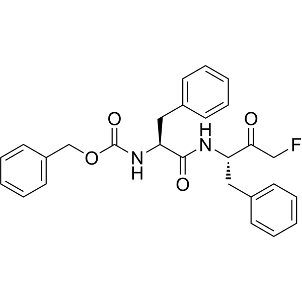
Z-FF-FMK (Z-Phe-Phe-FMK) is a cell-permeable, irreversible, and cysteine protease inhibitor targeting cathepsin-L. Z-FF-FMK inhibits angiotensin II-induced MEK activation in vascular walls, aortic medial remodeling, blood pressure elevation, and upregulation of cystatin C in aortic walls. Z-FF-FMK prevents β-amyloid-mediated caspase-3 activation, poly (ADP-ribose) polymerase cleavage, DNA fragmentation, and apoptosis of cortical neurons (apoptosis). Z-FF-FMK can be used in research related to hypertension and Alzheimer's disease.
For research use only. We do not sell to patients.
- Purity : 95.98%
- CAS No.: 105608-85-3
- Formula: C27H27FN2O4
- Molecular Weight:462.51
-
Storage:
Sealed storage, away from moisture.
Powder -80°C, 2 years , -20°C, 1 year* In solvent : -80°C, 6 months; -20°C, 1 month (sealed storage, away from moisture)

To place orders, for customer services and technical support, please contact: MedChemExpress USA
Tel: 609-228-6898 E-mail: [email protected] [email protected]


-
 Biological Activity
-
Chemical Information
-
Solvent & Solubility
- Protocols
- Purity & Documentation
- References
-
 Help & FAQs


-
HY-15282
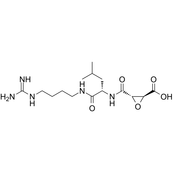
E-64Cysteine Peptidase Inhibitor -
HY-18234A
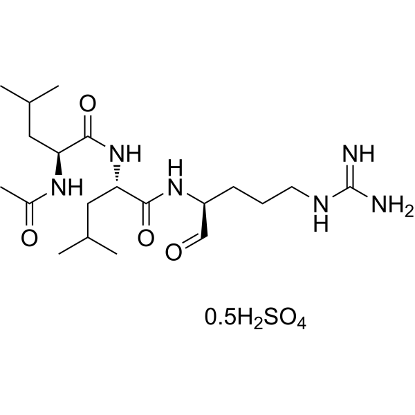
Leupeptin hemisulfateProtease Inhibitor -
HY-100350
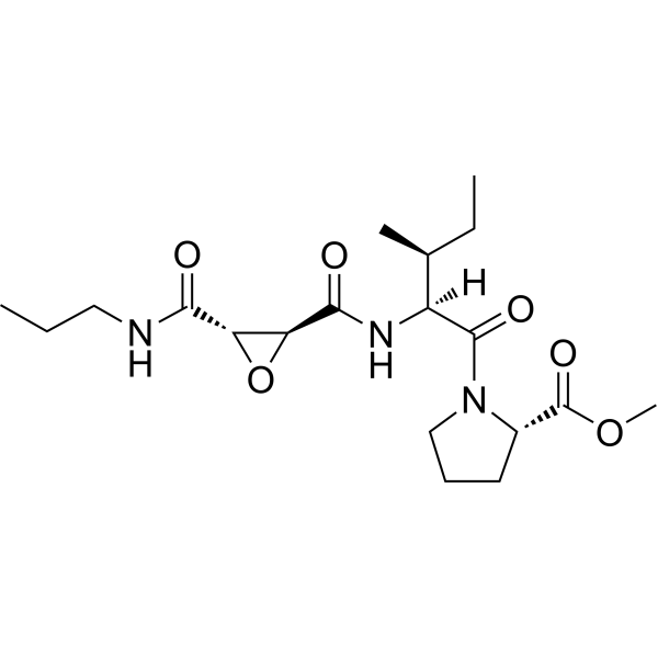
CA-074 methyl esterCathepsin B Inhibitor -
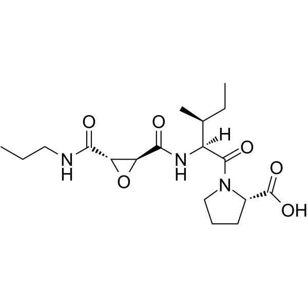
HY-103350
CA-074Cathepsin B Inhibitor -
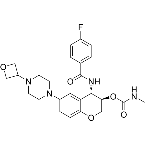
HY-15533
LY 3000328Cathepsin S Inhibitor -
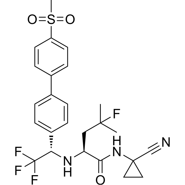
HY-10042
OdanacatibCathepsin K Inhibitor -
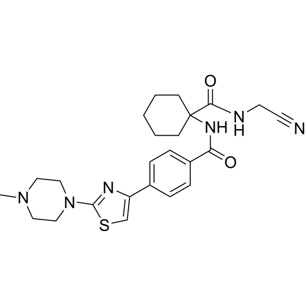
HY-103352
L-006235Cathepsin K Inhibitor -
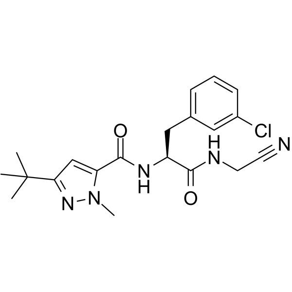
HY-100231
Cathepsin inhibitor 1Cathepsin Inhibitor -
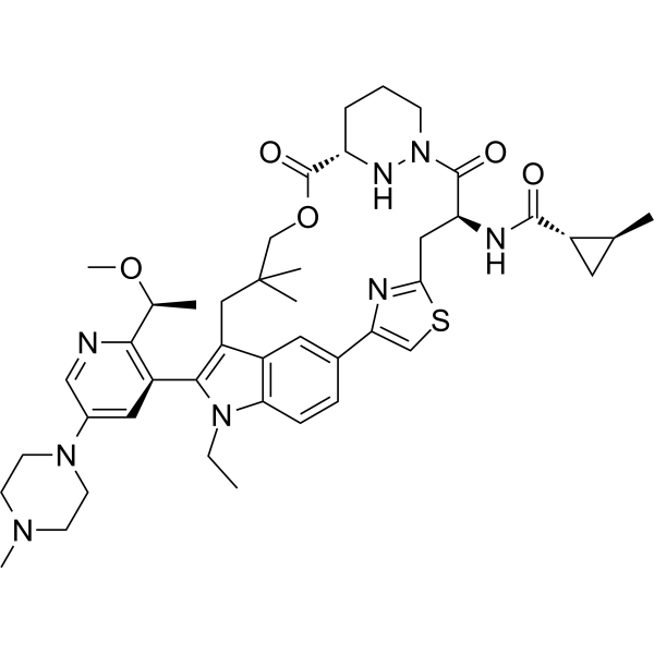
HY-148439
DaraxonrasibRAS(ON) Inhibitor -
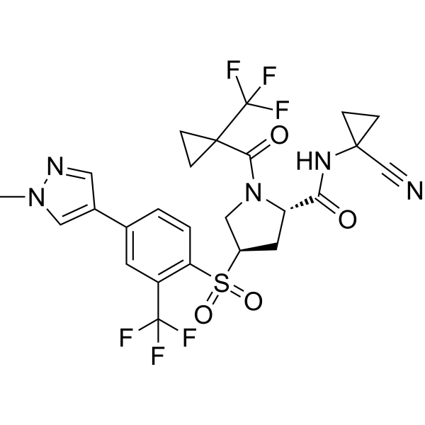
HY-109069
PetesicatibCathepsin S Inhibitor -
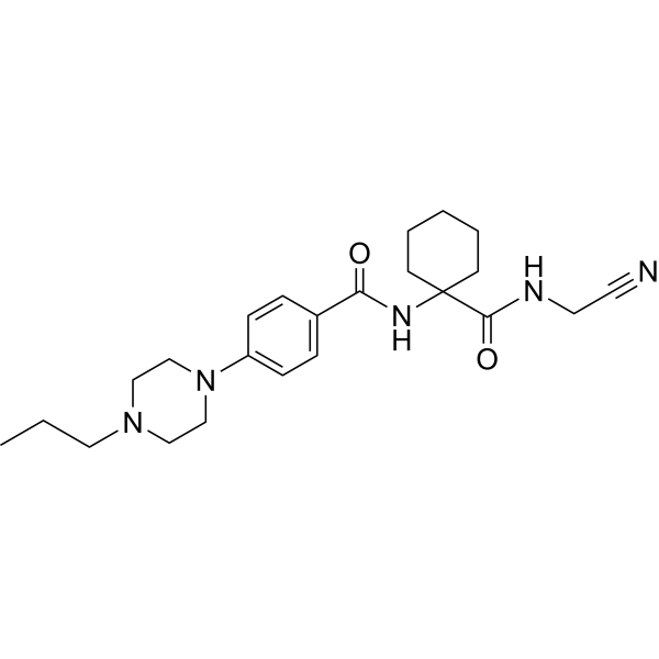
HY-15100
BalicatibCathepsin K Inhibitor -
HY-P0018
PepstatinAspartic Protease Inhibitor
All Cathepsin Isoforms
MoreAll MEK Isoforms
MoreAll Caspase Isoforms
MoreAll DNA/RNA Synthesis Isoforms
More

Biological Activity
Description
|
cathepsin L |
Caspase 3 |
In Vitro
Z-FF-FMK (10 μM; 6 h) potently inhibits basal and Aβ-stimulated cathepsin-L activity in primary cultured rat cortical neurones, reducing basal activity to near undetectable levels and eliminating the 6 h Aβ-induced activity increase[2].
Z-FF-FMK (10 μM; 24 h) completely prevents Aβ1-40-induced caspase-3 activation in primary cultured rat cortical neurones after 24 h of co-incubation[2].
Z-FF-FMK (10 μM; 24 h) fully blocks Aβ1-40-stimulated caspase-3 activity in primary cultured rat cortical neurones after 24 h of co-incubation[2].
Z-FF-FMK (10 μM) completely inhibits Aβ1-40-induced PARP cleavage in primary cultured rat cortical neurones[2].
Z-FF-FMK (10 μM; 72 h) fully prevents Aβ1-40-induced DNA fragmentation in primary cultured rat cortical neurones after 72 h of co-incubation[2].
MedChemExpress (MCE) has not independently confirmed the accuracy of these methods. They are for reference only. Further protocols information, click here.
In Vivo
MedChemExpress (MCE) has not independently confirmed the accuracy of these methods. They are for reference only.
-
Animal Model:C57BL/6 wild-type (8-12 weeks old; angiotensin II-induced hypertension model)[1]
-
Dosage:10 mg/kg
-
Administration:i.p.; every other day; 28 days
-
Result:Almost completely blocked angiotensin II-induced aortic artery media remodeling, reducing media thickness from a 2.1-fold increase (vs. control) to a 1.3-fold increase (vs. control).
Prevented angiotensin II-induced systolic blood pressure elevation, maintaining systolic blood pressure near baseline (~100 mmHg) compared to the ~160 mmHg peak in angiotensin II-only mice.
Suppressed angiotensin II-induced increases in aortic p-MEK expression, reducing from a 3.2-fold increase vs. control to a 1.2-fold increase vs. control.
Suppressed angiotensin II-induced increases in cystatin C expression, reducing from a 0.8-fold increase vs. control to a 0.4-fold increase vs. control.
Normalized angiotensin II-induced elevated arterial cathepsin L activity to control levels.


Chemical Information
-
CAS No. 105608-85-3
-
Appearance Solid
-
Molecular Weight 462.51
-
Formula C27H27FN2O4
-
Color White to off-white
-
Synonyms
Z-Phe-Phe-FMK
-
Sequence
Z-Phe-Phe-FMK
-
Sequence Shortening
Z-FF-FMK
-
Shipping
Room temperature in continental US; may vary elsewhere.
-
Storage
Sealed storage, away from moisture
Powder -80°C 2 years -20°C 1 year * In solvent : -80°C, 6 months; -20°C, 1 month (sealed storage, away from moisture)


Solvent & Solubility
In Vitro:
DMSO : 100 mg/mL (216.21 mM; Need ultrasonic; Hygroscopic DMSO has a significant impact on the solubility of product, please use newly opened DMSO)
Please refer to the solubility information to select the appropriate solvent. Once prepared, please aliquot and store the solution to prevent product inactivation from repeated freeze-thaw cycles.
Storage method and period of stock solution: -80°C, 6 months; -20°C, 1 month (sealed storage, away from moisture). When stored at -80°C, please use it within 6 months. When stored at -20°C, please use it within 1 month.
Please refer to the solubility information to select the appropriate solvent. Once prepared, please aliquot and store the solution to prevent product inactivation from repeated freeze-thaw cycles.
Storage method and period of stock solution: -80°C, 6 months; -20°C, 1 month (sealed storage, away from moisture). When stored at -80°C, please use it within 6 months. When stored at -20°C, please use it within 1 month.
Concentration (start) × Volume (start) = Concentration (final) × Volume (final)

Protocols
-
Apoptosis
Apoptosis, also called programmed cell death, is generally characterized by distinct morphological characteristics.
-
TUNEL staining for apoptotic DNA fragmentation
TUNEL staining detects DNA strand breaks by using terminal deoxynucleotidyl transferase to add labeled nucleotides to exposed 3′-OH DNA termini, generating either microscopic staining in fixed cells or tissue sections, or fluorescence/cytometric signal in cell suspensions. TUNEL positivity reflects DNA fragmentation but should not be interpreted alone as definitive apoptosis, because TUNEL can also label necrotic, autolytic, mechanically damaged, or DNA-repair-associated DNA breaks.
-
Annexin V plus membrane-impermeant dye apoptosis staining
Annexin V-based apoptosis assays rely on the detection of phosphatidylserine (PS) externalization from the inner leaflet of the plasma membrane to the outer leaflet, an early biochemical hallmark of apoptosis. Fluorescently labeled Annexin V binds PS in a calcium-dependent manner, enabling identification of early apoptotic cells by flow cytometry or fluorescence microscopy. When combined with a membrane-impermeant DNA-binding dye (e. g. , propidium iodide), this approach allows discrimination between viable (Annexin V−/dye−), early apoptotic (Annexin V+/dye−), and late apoptotic or necrotic (Annexin V+/dye+) cell populations by assessing membrane integrity and PS exposure.
-
Apoptosis Solutions
Apoptosis is a regulated, generally non-lytic cell-death pathway that removes unwanted, damaged, infected, or abnormal cells through coordinated morphological changes, caspase activation, DNA fragmentation, and membrane remodeling. The intrinsic apoptosis pathway is controlled mainly by mitochondrial outer membrane permeabilization, BCL-2 family proteins, cytochrome c release, apoptosome formation, caspase-9 activation, and downstream executioner caspase-3/7 activation. The extrinsic apoptosis pathway is initiated by death receptors such as Fas, TNFR, and TRAIL receptors, which recruit adaptor proteins and activate caspase-8 before engaging executioner caspases or mitochondrial amplification through BID cleavage. Apoptosis is linked to many phenotypes, including cancer cell killing, tissue homeostasis, immune regulation, neurodegeneration, infection response, and treatment-induced cytotoxicity; unresolved questions include how apoptosis interacts with necroptosis, pyroptosis, ferroptos
-
Primary Embryonic Cortical Neuron Culture
Primary embryonic cortical neuron culture isolates cortical tissue from prenatal rodents, dissociates it into single cells, and maintains neurons in vitro so that neurite extension, neuronal marker expression, synapse formation, survival, and treatment responses can be examined outside the intact brain.
-
Research Protocol for Cardiovascular Diseases
Cardiovascular disease can be modeled as maladaptive cardiac remodeling, where ischemic injury or pressure overload activates inflammatory signaling, fibroblast activation, extracellular-matrix deposition, cardiomyocyte hypertrophy, vascular remodeling, and progressive ventricular dysfunction. The TGF-β/SMAD axis is a central profibrotic pathway after myocardial injury and pressure overload, while innate immune and cytokine pathways regulate leukocyte recruitment, scar formation, and adverse remodeling. Key unresolved questions include which inflammatory signals are reparative versus harmful, when fibrosis is protective versus maladaptive, and whether pathway inhibition improves function without weakening necessary infarct healing or compensatory remodeling.
-
Amyloid: Congo Red Amyloid Staining
Congo red amyloid staining is a histochemical method used to detect extracellular amyloid deposits in tissue sections based on the affinity of Congo red dye for β-pleated sheet-rich protein aggregates. When bound to amyloid, Congo red produces characteristic apple-green birefringence under polarized light microscopy, which is widely regarded as a diagnostic feature of amyloid deposition in histopathology. The diagnostic principle relies on the combination of dye binding (congophilia) and optical anisotropy under polarized illumination, which distinguishes amyloid from most non-amyloid eosinophilic extracellular deposits in routine histological evaluation. Amyloid identification by Congo red staining remains a cornerstone in diagnostic pathology despite the availability of adjunct methods such as immunohistochemistry and mass spectrometry, particularly because of its ability to localize deposits directly within tissue architecture. The specificity of Congo red-positive deposits is incre
-
Alzheimer’s Disease Modeling
Alzheimer’s Disease (AD) is a neurodegenerative disorder characterized by a progressive decline in cognitive functions and loss of specific types of neurons and synapses. Alzheimer's symptoms can be simulated in mice by injecting drugs (such as Aβ) or genetically modified.


Purity & Documentation
-
Data Sheet (275 KB)
-
SDS (252 KB)
- English - EN (252 KB)
- Français - FR (252 KB)
- Deutsch - DE (252 KB)
- Norwegian - NO (252 KB)
- Español - ES (252 KB)
- Swedish - SV (252 KB)
- Italian - IT (252 KB)
- Korean - KR (252 KB)
- Portuguese - PT (252 KB)
-
Handling Instructions (2659 KB)
References


Complete Stock Solution Preparation Table
Please refer to the solubility information to select the appropriate solvent. Once prepared, please aliquot and store the solution to prevent product inactivation from repeated freeze-thaw cycles.
Storage method and period of stock solution: -80°C, 6 months; -20°C, 1 month (sealed storage, away from moisture). When stored at -80°C, please use it within 6 months. When stored at -20°C, please use it within 1 month.
| Optional Solvent | Concentration Solvent Mass | 1 mg | 5 mg | 10 mg | 25 mg |
|---|---|---|---|---|---|
| DMSO | 1 mM | 2.1621 mL | 10.8106 mL | 21.6212 mL | 54.0529 mL |
| 5 mM | 0.4324 mL | 2.1621 mL | 4.3242 mL | 10.8106 mL | |
| 10 mM | 0.2162 mL | 1.0811 mL | 2.1621 mL | 5.4053 mL | |
| 15 mM | 0.1441 mL | 0.7207 mL | 1.4414 mL | 3.6035 mL | |
| 20 mM | 0.1081 mL | 0.5405 mL | 1.0811 mL | 2.7026 mL | |
| 25 mM | 0.0865 mL | 0.4324 mL | 0.8648 mL | 2.1621 mL | |
| 30 mM | 0.0721 mL | 0.3604 mL | 0.7207 mL | 1.8018 mL | |
| 40 mM | 0.0541 mL | 0.2703 mL | 0.5405 mL | 1.3513 mL | |
| 50 mM | 0.0432 mL | 0.2162 mL | 0.4324 mL | 1.0811 mL | |
| 60 mM | 0.0360 mL | 0.1802 mL | 0.3604 mL | 0.9009 mL | |
| 80 mM | 0.0270 mL | 0.1351 mL | 0.2703 mL | 0.6757 mL | |
| 100 mM | 0.0216 mL | 0.1081 mL | 0.2162 mL | 0.5405 mL |


Z-FF-FMK Related Classifications
HY-141867 Related Classifications
 Powered by Bioz