



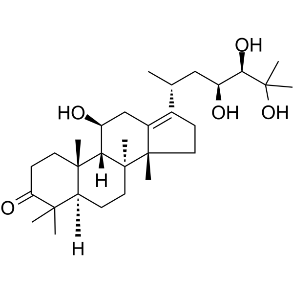
Alisol A
Based on 5 publication(s) in Google Scholar
Alisol A is an orally active tetracyclic triterpenoid compound of the prototerpane type. Alisol A can be extracted from the rhizome of Alisma orientale. Alisol A activates AMPK/ACC/SREBP-1c, SIRT1, PPARα, inhibits MMP-2/-9, decreases inflammatory cytokine expression (IL-1β, IL-6, IL-8). Alisol A has anti-tumor activity against breast cancer and colorectal cancer. Alisol A has anti-obesity and anti-atherosclerotic activities. Alisol A can be used in the research of hepatitis B, breast cancer, colorectal cancer, atherosclerosis, and obesity.
For research use only. We do not sell to patients.
- Purity : 99.59%
- CAS No.: 19885-10-0
- Formula: C30H50O5
- Molecular Weight:490.72
-
Storage:Powder -20°C, 3 years , 4°C, 2 years ; In solvent -80°C, 2 years , -20°C, 1 year

To place orders, for customer services and technical support, please contact: MedChemExpress USA
Tel: 609-228-6898 E-mail: [email protected] [email protected]


-
 Biological Activity
-
Chemical Information
-
Solvent & Solubility
- Protocols
- Purity & Documentation
- References
-
 Help & FAQs


-
HY-N1201
ApigeninCytochrome P450 Inhibitor -
HY-N0203
Atractylenolide IIIApoptosis Inducer -
HY-N0197
BaicalinAutophagy Inducer -
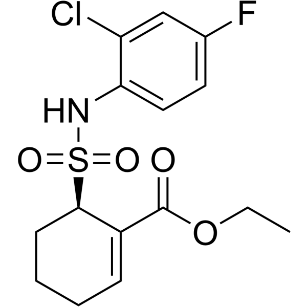
HY-11109
ResatorvidTLR4 Inhibitor -
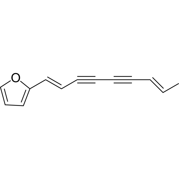
HY-N0238
AtractylodinInsecticide -
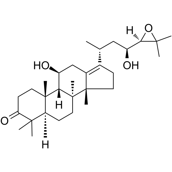
HY-N0805A
Alisol BTriterpene -
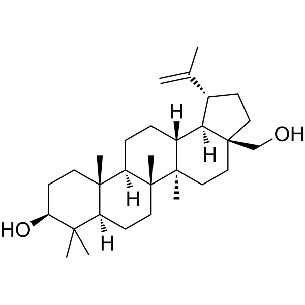
HY-N0083
BetulinEndogenous Metabolite -
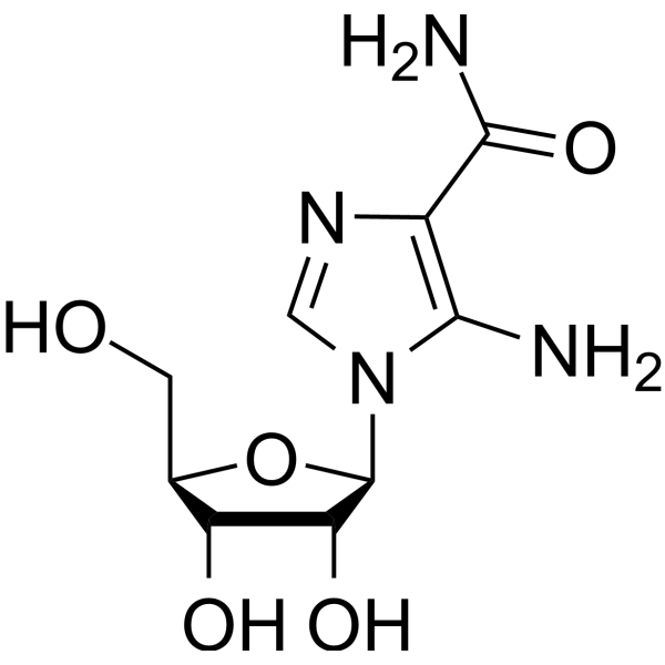
HY-13417
AICARAMPK Activator -
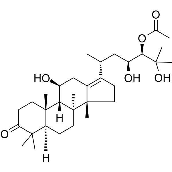
HY-N0853A
Alisol A 24-acetateNatural Product -

HY-13417R
AICAR (Standard)AMPK Activator -
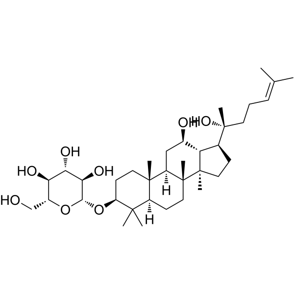
HY-N1401
20(R)-Ginsenoside Rh2Anti-tumor/Anti-viral/Anti-inflammatory Agent -

HY-N0853R
Alisol A (Standard)Standard Of Alisol A
Publications Citing Use of MedChemExpress (MCE) Alisol A
More Customer Validation & Images

-
Apoptosis Analysis

-
WB
-
IF

-
Cell Migration/Invasion Assay
-
Flow Cytometry

All AMPK Isoforms
More

Biological Activity
Description
|
MMP-9 |
MMP-2 |
PPARα |
IL-6 |
IL-1β |
IL-8 |
Cellular Effect
|
Cell Line

|
Type | Value | Description | References |
|---|---|---|---|---|
| Erythrocyte | IC50 |
13.8 μM
Compound: 57a
|
Antiplasmodial activity against Plasmodium falciparum K1 infected in human erythrocyte assessed as inhibition of parasite growth incubated for 72 hrs by [3H]-hypoxanthine incorporation based liquid scintillation counting method
Antiplasmodial activity against Plasmodium falciparum K1 infected in human erythrocyte assessed as inhibition of parasite growth incubated for 72 hrs by [3H]-hypoxanthine incorporation based liquid scintillation counting method
|
[PMID: 35985254] |
| HepG2 | EC50 |
10.16 μM
Compound: 23
|
Transactivation of FXR (unknown origin) transfected in HepG2 cells co-expressing pBSEP/pGL4.74 incubated for 24 hrs by luciferase reporter gene assay
Transactivation of FXR (unknown origin) transfected in HepG2 cells co-expressing pBSEP/pGL4.74 incubated for 24 hrs by luciferase reporter gene assay
|
[PMID: 31494470] |
| HepG2 2.2.15 | CC50 |
0.062 mM
Compound: 1
|
Cytotoxicity against human HepG2.2.15 cells
Cytotoxicity against human HepG2.2.15 cells
|
[PMID: 18644720] |
| HepG2 2.2.15 | IC50 |
>2.4 mM
Compound: 1
|
Antiviral activity against hepatitis B virus-infected human HepG2.2.15 cells assessed as inhibition of HBV e antigen secretion
Antiviral activity against hepatitis B virus-infected human HepG2.2.15 cells assessed as inhibition of HBV e antigen secretion
|
[PMID: 18644720] |
| HepG2 2.2.15 | IC50 |
0.039 mM
Compound: 1
|
Antiviral activity against hepatitis B virus-infected human HepG2.2.15 cells assessed as inhibition of HBV surface antigen secretion
Antiviral activity against hepatitis B virus-infected human HepG2.2.15 cells assessed as inhibition of HBV surface antigen secretion
|
[PMID: 18644720] |
In Vitro
Alisol A (2.5-40 μM; 24 h) suppresses proliferation, migration, and invasion in human breast cancer MDA-MB-231 cells[3].
Alisol A (1-5 μM) concentration-dependently restores the levels of p-AMPK and p-ACC in HepG2 cells treated with free fatty acids (FFA)[4].
Alisol A (40 μM; 24-72 h) inhibits the proliferation of human aortic endothelial cells (HAECs)[5].
Alisol A (5-160 µM; 24 h) inhibits the proliferation of HCT-116 and HT-29 cells in a dose-dependent manner[6].
MedChemExpress (MCE) has not independently confirmed the accuracy of these methods. They are for reference only. Further protocols information, click here.
-
Cell Line:MDA-MB-231
-
Concentration:0, 5 μM
-
Incubation Time:24 h
-
Result:Downregulated MMP-2/-9.
In Vivo
Alisol A (150 ppm; p.o.; in diet; 16 weeks) significantly reduces the aortic plaque area in ApoE-/- mice with atherosclerosis, without obvious effect on blood lipid levels[5].
Alisol A (25-100 mg/kg; i.g.; twice daily; 12 weeks) alleviates arterial plaque by activating AMPK/SIRT1 signaling pathway in apoE-deficient mice[7].
MedChemExpress (MCE) has not independently confirmed the accuracy of these methods. They are for reference only.
-
Animal Model:Male C57BL/6 mice (6 weeks old) with high-fat diet-induced obesity[4]
-
Dosage:100 mg/kg
-
Administration:Intraperitoneal injection, once daily, for 4 weeks
-
Result:Reduced body weight and abdominal fat mass, improved plasma lipid profiles (decreased TC, TG, LDL-C, NEFA, FABP4).
Enhanced glucose tolerance and insulin sensitivity, alleviated hepatic steatosis (reduced fat vacuoles and lipid deposition).
Decreased inflammatory cytokine expression (TNF-α, IL-1β, IL-6, IL-8) in adipose tissue, and activated the AMPK/ACC/SREBP-1c pathway (increased p-AMPK, p-ACC, decreased SREBP-1c) in liver, skeletal muscle, and adipose tissue.
-
Animal Model:Male C57BL/6 background ApoE-/- mice (6-week-old), high-fat diet-induced atherosclerosis model[7]
-
Dosage:25 mg/kg, 100 mg/kg
-
Administration:Intragastric administration, twice daily, 12 weeks
-
Result:Attenuated high-fat diet-induced body weight gain, reduced aortic plaque area, widened the inner diameter of the aortic arch.
Inhibited the expression of ICAM-1, IL-6, and MMP-9 in aortic plaques, and increased the expression of PPARα, PPARδ, p-AMPK, SIRT1, and IkBa in the liver.


Chemical Information
-
CAS No. 19885-10-0
-
Appearance Solid
-
Molecular Weight 490.72
-
Formula C30H50O5
-
Color White to light yellow
-
SMILES
C[C@]([C@@]1(C2=C([C@H](C)C[C@H](O)[C@@H](O)C(C)(O)C)CC1)C)(CC[C@@]3([H])C4(C)C)[C@]([C@H](C2)O)([H])[C@]3(CCC4=O)C
-
Structure Classification
-
Initial Source
-
Shipping
Room temperature in continental US; may vary elsewhere.
-
Storage
Powder -20°C 3 years 4°C 2 years In solvent -80°C 2 years -20°C 1 year


Publications (5)
-
Journal Impact Factor
-
Most Recent
-
Molecules
Alisol A Suppresses Proliferation, Migration, and Invasion in Human Breast Cancer MDA-MB-231 Cells. [Abstract]2019 Oct 10;24(20):3651. PMID: 31658635
Alisol A purchased from MedChemExpress. Usage Cited in: Molecules. 2019 Oct 10;24(20):3651. [Abstract]
Quantification of apoptotic cells was performed by flow cytometer. MDA-MB-231 cells were treated with different concentrations of Alisol A (0, 10, 20, 40 μM) for 24 h. Cells were stained with Annexin-V-FITC/7AAD according to the manufacturer’s instructions.
Alisol A purchased from MedChemExpress. Usage Cited in: Molecules. 2019 Oct 10;24(20):3651. [Abstract]
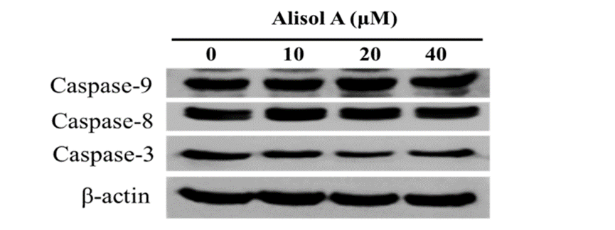
Effects of alisol A on the expression of caspases in the MDA-MB-231 cells. The cells were treated with Alisol A (0, 10, 20, 40 μM) for 24 h. The cell lysates were collected and subjected to Western blotting analysis.
Alisol A purchased from MedChemExpress. Usage Cited in: Molecules. 2019 Oct 10;24(20):3651. [Abstract]
Autophagy in Alisol A (20, 40 μM)-treated MDA-MB-231 cells were stained with AO and examined under a fluorescence microscope.
Alisol A purchased from MedChemExpress. Usage Cited in: Molecules. 2019 Oct 10;24(20):3651. [Abstract]
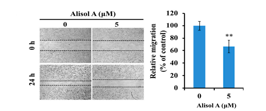
The effects of Alisol A (0, 5 μM) on cell migration was measured by wound healing assay.
Alisol A purchased from MedChemExpress. Usage Cited in: Molecules. 2019 Oct 10;24(20):3651. [Abstract]
Effects of Alisol A (0, 20, 40 μM) on cell cycle in MDA-MB-231 cells.
-
Vet Microbiol
Identification and evaluation of Nordihydroguaiaretic acid (NDGA) as an active traditional Chinese medicine compound inhibiting the 3C-like protease of feline infectious peritonitis virus. [Abstract]2025 Sep 15:310:110730. PMID: 40976146 -
Nephrology (Carlton)
Alisol A inhibits the circ_0001831/miR-346/LIN28B pathway to ameliorate high glucose-induced injury of human renal mesangial cells. [Abstract]2024 Mar;29(3):154-163. PMID: 38013222 -
Biomed Pharmacother
A novel Alisma orientale extract alleviates non-alcoholic steatohepatitis in mice via modulation of PPARα signaling pathway. [Abstract]2024 Jul:176:116908. PMID: 38850668 -


Solvent & Solubility
In Vitro:
DMSO : 100 mg/mL (203.78 mM; Need ultrasonic; Hygroscopic DMSO has a significant impact on the solubility of product, please use newly opened DMSO)
Please refer to the solubility information to select the appropriate solvent. Once prepared, please aliquot and store the solution to prevent product inactivation from repeated freeze-thaw cycles.
Storage method and period of stock solution: -80°C, 2 years; -20°C, 1 year. When stored at -80°C, please use it within 2 years. When stored at -20°C, please use it within 1 year.
Please refer to the solubility information to select the appropriate solvent. Once prepared, please aliquot and store the solution to prevent product inactivation from repeated freeze-thaw cycles.
Storage method and period of stock solution: -80°C, 2 years; -20°C, 1 year. When stored at -80°C, please use it within 2 years. When stored at -20°C, please use it within 1 year.
Concentration (start) × Volume (start) = Concentration (final) × Volume (final)
In Vivo:
Select the appropriate dissolution method based on your experimental animal and administration route.
- For the following dissolution methods, please ensure to first prepare a clear stock solution using an In Vitro approach and then sequentially add co-solvents:
- To ensure reliable experimental results, the clarified stock solution can be appropriately stored based on storage conditions. As for the working solution for In Vivo experiments, it is recommended to prepare freshly and use it on the same day.
- The percentages shown for the solvents indicate their volumetric ratio in the final prepared solution. If precipitation or phase separation occurs during preparation, heat and/or sonication can be used to aid dissolution.
Add each solvent one by one: 10% DMSO 40% PEG300 5% Tween-80 45% Saline
Solubility: ≥ 2.5 mg/mL (5.09 mM); Clear solution
This protocol yields a clear solution of ≥ 2.5 mg/mL (saturation unknown).
Taking 1 mL working solution as an example, add 100 μL DMSO stock solution (25.0 mg/mL) to 400 μL PEG300, and mix evenly; then add 50 μL Tween-80 and mix evenly; then add 450 μL Saline to adjust the volume to 1 mL.
Preparation of Saline: Dissolve 0.9 g sodium chloride in ddH₂O and dilute to 100 mL to obtain a clear Saline solution.
Add each solvent one by one: 10% DMSO 90% (20% SBE-β-CD in Saline)
Solubility: ≥ 2.5 mg/mL (5.09 mM); Clear solution
This protocol yields a clear solution of ≥ 2.5 mg/mL (saturation unknown).
Taking 1 mL working solution as an example, add 100 μL DMSO stock solution (25.0 mg/mL) to 900 μL 20% SBE-β-CD in Saline, and mix evenly.
Preparation of 20% SBE-β-CD in Saline (4°C, storage for one week): 2 g SBE-β-CD powder is dissolved in 10 mL Saline, completely dissolve until clear.
In Vivo Dissolution Calculator
Please enter the basic information of animal experiments:
-
-
-
-
Recommended: Prepare an additional quantity of animals to account for potential losses during experiments.
Please enter your animal formula composition:
-
%DMSO +
Recommended: Keep the proportion of DMSO in working solution below 2% if your animal is weak.
-
%+
-
+%Tween-80 + +
-
%Saline +
The co-solvents required include: DMSO, . All of co-solvents are available by MedChemExpress (MCE). , Tween 80. All of co-solvents are available by MedChemExpress (MCE).
Working solution concentration: 0.22 mg/mL
Method for preparing stock solution: mg drug dissolved in μL DMSO. Stock solution concentration: mg/mL.
1. Take μL DMSO stock solution;
2. Add μL .
μL , mix evenly;
3. Then add μL Tween 80, mix evenly;
4. Then add μL
Please ensure that the stock solution in the first step is dissolved to a clear state, and add co-solvents in sequence. You can use ultrasonic heating (ultrasonic cleaner, recommended frequency 20-40 kHz), vortexing, etc. to assist dissolution.

Protocols
-
RNA extraction experimental
By lysing cells, releasing RNA, and removing impurities such as proteins and DNA, high-purity RNA products are finally obtained. The commonly used traditional method is the guanidine isothiocyanate/phenol/chloroform method (Trizol), which is suitable for a variety of animal materials including animal tissues, microorganisms, cultured cells, etc., and most plant materials.
-
Mitochondrial membrane-potential fluorescent assay
Mitochondrial membrane potential fluorescent assays estimate ΔΨm in living cells using lipophilic cationic dyes such as TMRM, TMRE, rhodamine 123, and JC-1, which accumulate in mitochondria according to membrane polarization; loss of signal after FCCP or CCCP treatment is interpreted as mitochondrial depolarization. TMRM/TMRE and rhodamine 123 are commonly used for semi-quantitative live-cell microscopy or flow cytometry, while JC-1 can report a shift from red aggregate fluorescence to green monomer fluorescence during depolarization; interpretation requires controls because dye concentration, quenching mode, cell type, dye efflux, and mitochondrial mass can affect fluorescence independently of ΔΨm.
-
Mitochondrial membrane-potential and mitochondrial mass staining
Mitochondrial membrane potential staining measures the electrochemical polarization across the mitochondrial inner membrane in live cells using lipophilic cationic fluorescent probes; early rhodamine-based work showed that selective mitochondrial dye accumulation is lost when the mitochondrial transmembrane potential is dissipated. JC-1 reports mitochondrial polarization by shifting from green monomer fluorescence to red J-aggregate fluorescence as dye concentration increases within energized mitochondria; therefore, the red/green fluorescence ratio is used as a relative readout of mitochondrial membrane potential. TMRE or TMRM staining provides a single-channel relative readout because these cationic rhodamine esters accumulate in polarized mitochondria, and lower fluorescence indicates reduced mitochondrial polarization when acquisition and dye-loading conditions are controlled. Mitochondrial mass staining is commonly performed with MitoTracker Green FM or related MitoTracker dyes as
-
Research Protocol for Cardiovascular Diseases
Cardiovascular disease can be modeled as maladaptive cardiac remodeling, where ischemic injury or pressure overload activates inflammatory signaling, fibroblast activation, extracellular-matrix deposition, cardiomyocyte hypertrophy, vascular remodeling, and progressive ventricular dysfunction. The TGF-β/SMAD axis is a central profibrotic pathway after myocardial injury and pressure overload, while innate immune and cytokine pathways regulate leukocyte recruitment, scar formation, and adverse remodeling. Key unresolved questions include which inflammatory signals are reparative versus harmful, when fibrosis is protective versus maladaptive, and whether pathway inhibition improves function without weakening necessary infarct healing or compensatory remodeling.
-
Research Protocol for Inflammation-related Diseases
The NLRP3 inflammasome is a cytosolic innate immune signaling platform that integrates priming signals and danger-signal activation to promote caspase-1 activation, maturation of IL-1β and IL-18, and gasdermin D-mediated pyroptotic cell death. The core experimental logic is to determine whether inflammatory disease phenotypes are driven by increased NLRP3 expression, ASC-containing inflammasome assembly, caspase-1 cleavage, GSDMD cleavage, and extracellular release of IL-1β/IL-18 rather than by nonspecific cell injury alone. The pathway is strongly linked to inflammation-related disease phenotypes because monosodium urate crystals activate NALP3/NLRP3 inflammasome signaling in gout-like crystal inflammation, cholesterol crystals activate NLRP3 inflammasomes in atherogenesis models, and DSS-induced intestinal inflammation has been reported to involve NLRP3 inflammasome activity. However, experimental colitis studies also show context-dependent protective effects of NLRP3 inflammasome co
-
Breast Cancer Modeling
Breast cancer is a heterogeneous cancer, and it has been distinguished into four subtypes: luminal A, luminal B, HER2-positive and basal-like. Molecular mutations, epigenetic alterations, hormone exposure and immune microenvironment are related to the progression of breast cancer.
-
Research Protocol for Metabolic Diseases
AMP-activated protein kinase, AMPK, is a conserved cellular energy sensor that responds to reduced cellular energy status and coordinates metabolism by increasing ATP-generating catabolic pathways while suppressing ATP-consuming anabolic processes. In metabolic disease research, the AMPK pathway is experimentally relevant because it regulates hepatic lipid synthesis, fatty acid oxidation, glucose production, skeletal-muscle glucose disposal, mTORC1-linked biosynthesis, autophagy, mitochondrial homeostasis, and whole-body energy balance. The central pathway logic is that energy stress, metformin, exercise-like stimulation, or direct AMPK activators increase AMPKα Thr172 phosphorylation and downstream substrate phosphorylation, including ACC and RAPTOR. Phosphorylation of ACC suppresses lipogenesis and supports fatty acid oxidation, whereas phosphorylation of RAPTOR suppresses mTORC1 signaling and links cellular energy status to growth and protein synthesis control. The pathway is linked
-
Pyroptosis Solutions
Pyroptosis is a lytic inflammatory cell-death pathway executed by gasdermin pores, most classically through inflammasome-mediated activation of caspase-1, cleavage of gasdermin D, membrane pore formation, LDH release, and secretion of IL-1β and IL-18. The canonical pathway is commonly modeled by priming cells with an inflammatory signal such as LPS to induce pro-IL-1β and inflammasome components, followed by an activation signal such as ATP or nigericin to activate NLRP3, ASC speck formation, caspase-1 cleavage, GSDMD cleavage, cytokine release, and pyroptotic membrane rupture. The non-canonical pathway is triggered when cytosolic LPS activates mouse caspase-11 or human caspase-4/5, leading to GSDMD cleavage and pyroptosis, and this can secondarily activate NLRP3-dependent IL-1β release. Pyroptosis is linked to inflammatory injury, infection, cancer, liver disease, ocular disease, placental inflammation, and other disease phenotypes, but unresolved questions include which gasdermin fam


Purity & Documentation
-
Data Sheet (295 KB)
-
SDS (393 KB)
- English - EN (393 KB)
- Français - FR (393 KB)
- Deutsch - DE (393 KB)
- Norwegian - NO (393 KB)
- Español - ES (393 KB)
- Swedish - SV (393 KB)
- Italian - IT (393 KB)
- Korean - KR (393 KB)
- Portuguese - PT (393 KB)
-
Handling Instructions (2659 KB)
References
[1]. Matsuda H, et al. Studies on Alismatis Rhizoma. II. Anti-complementary activities of methanol extract and terpene components from Alismatis Rhizoma (dried rhizome of Alisma orientale). Biol Pharm Bull. 1998 Dec;21(12):1317-21. [Content Brief]
[2]. Zhang Q, et al. Anti-HBV agents. Part 1: Synthesis of alisol A derivatives: a new class of hepatitis B virus inhibitors. Bioorg Med Chem Lett. 2008 Aug 15;18(16):4647-50. [Content Brief]
[3]. Lou C, et al. Alisol A Suppresses Proliferation, Migration, and Invasion in Human Breast Cancer MDA-MB-231 Cells. Molecules. 2019 Oct 10;24(20):3651. [Content Brief]
[4]. Ho C, et al. Alisol A attenuates high-fat-diet-induced obesity and metabolic disorders via the AMPK/ACC/SREBP-1c pathway. J Cell Mol Med. 2019 Aug;23(8):5108-5118. [Content Brief]
[5]. Ma Y, et al. Alisol A inhibits and stabilizes atherosclerotic plaques by protecting vascular endothelial cells. Front Pharmacol. 2024 Oct 25;15:1493948. [Content Brief]
[6]. Han W, et al. Alisol A attenuates malignant phenotypes of colorectal cancer cells by inactivating PI3K/Akt signaling. Oncol Lett. 2022 Jun 7;24(2):249. [Content Brief]
[7]. Wang K, et al. Alisol A Alleviates Arterial Plaque by Activating AMPK/SIRT1 Signaling Pathway in apoE-Deficient Mice. Front Pharmacol. 2020 Nov 2;11:580073. [Content Brief]


Complete Stock Solution Preparation Table
Please refer to the solubility information to select the appropriate solvent. Once prepared, please aliquot and store the solution to prevent product inactivation from repeated freeze-thaw cycles.
Storage method and period of stock solution: -80°C, 2 years; -20°C, 1 year. When stored at -80°C, please use it within 2 years. When stored at -20°C, please use it within 1 year.
| Optional Solvent | Concentration Solvent Mass | 1 mg | 5 mg | 10 mg | 25 mg |
|---|---|---|---|---|---|
| DMSO | 1 mM | 2.0378 mL | 10.1891 mL | 20.3782 mL | 50.9455 mL |
| 5 mM | 0.4076 mL | 2.0378 mL | 4.0756 mL | 10.1891 mL | |
| 10 mM | 0.2038 mL | 1.0189 mL | 2.0378 mL | 5.0946 mL | |
| 15 mM | 0.1359 mL | 0.6793 mL | 1.3585 mL | 3.3964 mL | |
| 20 mM | 0.1019 mL | 0.5095 mL | 1.0189 mL | 2.5473 mL | |
| 25 mM | 0.0815 mL | 0.4076 mL | 0.8151 mL | 2.0378 mL | |
| 30 mM | 0.0679 mL | 0.3396 mL | 0.6793 mL | 1.6982 mL | |
| 40 mM | 0.0509 mL | 0.2547 mL | 0.5095 mL | 1.2736 mL | |
| 50 mM | 0.0408 mL | 0.2038 mL | 0.4076 mL | 1.0189 mL | |
| 60 mM | 0.0340 mL | 0.1698 mL | 0.3396 mL | 0.8491 mL | |
| 80 mM | 0.0255 mL | 0.1274 mL | 0.2547 mL | 0.6368 mL | |
| 100 mM | 0.0204 mL | 0.1019 mL | 0.2038 mL | 0.5095 mL |


Alisol A Related Classifications
HY-N0853 Related Classifications
 Powered by Bioz