



BV02
Based on 5 publication(s) in Google Scholar
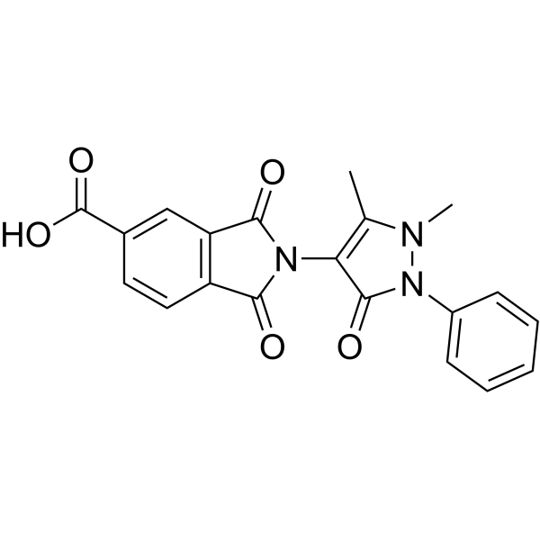
BV02 is a 14-3-3 inhibitor and an antibacterial agent. BV02 enhances the cleavage of PARP and caspase-3. BV02 induces Apoptosis, Autophagy, and enhances Akt activation. BV02 has anti-B. melitensis infection and epilepsy-promoting effects. BV02 can also be used in colitis research.
For research use only. We do not sell to patients.
- Purity : 99.75%
- CAS No.: 292870-53-2
- Formula: C20H15N3O5
- Molecular Weight:377.36
-
Storage:Powder -20°C, 3 years , 4°C, 2 years ; In solvent -80°C, 6 months , -20°C, 1 month

To place orders, for customer services and technical support, please contact: MedChemExpress USA
Tel: 609-228-6898 E-mail: [email protected] [email protected]


-
 Biological Activity
-
Chemical Information
-
Solvent & Solubility
- Protocols
- Purity & Documentation
- References
-
 Help & FAQs


-
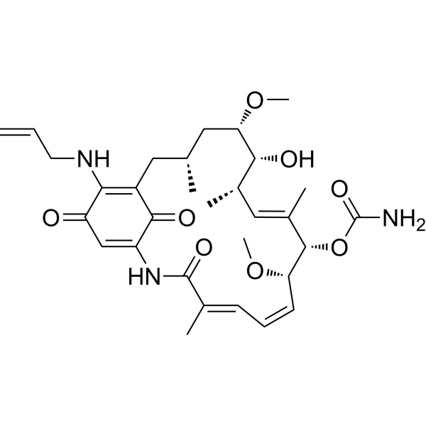
HY-10211
TanespimycinHSP90 Inhibitor -
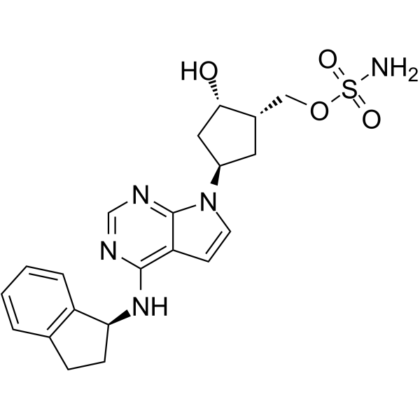
HY-70062
PevonedistatNAE Inhibitor -

HY-100579
Ferrostatin-1Ferroptosis Inhibitor -
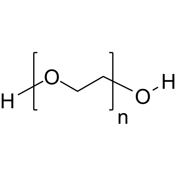
HY-Y0873A
PEG400 -

HY-10583
Y-27632 dihydrochlorideROCK Inhibitor -

HY-K1005
Ultra High Sensitivity ECL Kit -
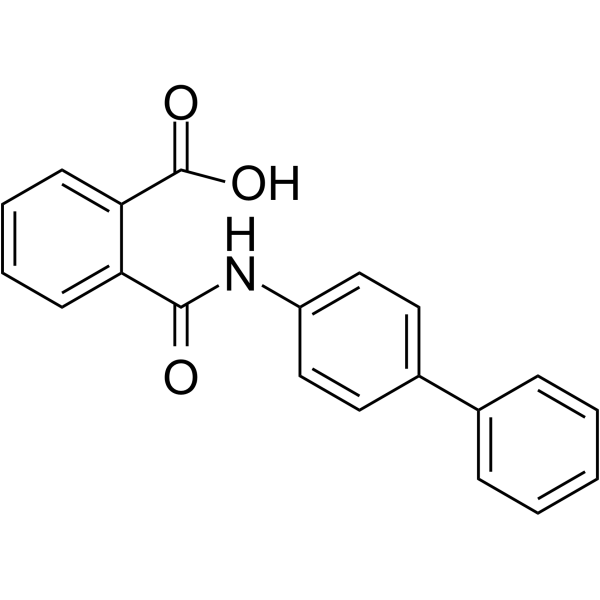
HY-16268
KartogeninChondroprotective Agent -
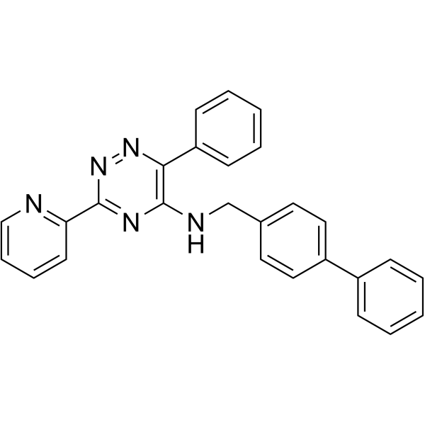
HY-12754
ML228HIF Activator -
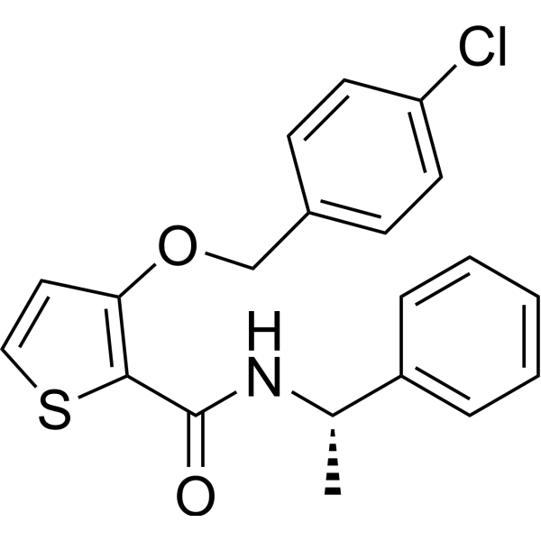
HY-18686
AS1949490SHIP2 Phosphatase Inhibitor -

HY-P1380A
Difopein TFA14-3-3 Protein Inhibitor -

HY-P1039A
R18 TFA14-3-3 Peptide Antagonist -

HY-101985R
BV02 (Standard)Reference Standard
Publications Citing Use of MedChemExpress (MCE) BV02
MoreAll Caspase Isoforms
More

Biological Activity
Description
IC50 & Target
[4]|
Caspase-3 |
In Vitro
BV02 (5 μM; 24 h) induces apoptotic death and recruits cells into the sub-G1 phase of the cell cycle in Bcr-Abl-expressing Ba/F3 cells, regardless of whether they express the wild-type Bcr-Abl or the T315I mutation[1].
BV02 (5-50 μM; 24 h) significantly reduces the surface expression of wild-type K2P5.1-GFP channels in HEK293T cells[2].
BV02 (5 μM; 6 h) induces autophagy in C33A cells, with increasing the LC3-II level and decreasing the SQSTM1 level[3].
BV02 (5 μM; 36 h) enhances Akt activation, as shown by increased phosphorylation of Akt at Thr308 and Ser473, and triggers apoptosis in SW480 cells[4].
BV02 (20 μM; 24 h) decreases the invasion of Brucella into macrophages[7].
MedChemExpress (MCE) has not independently confirmed the accuracy of these methods. They are for reference only. Further protocols information, click here.
-
Cell Line:SW480 cells
-
Concentration:5 μM
-
Incubation Time:36 h
-
Result:Increased the phosphorylation of Akt at Thr308 and Ser473.
Augmented the cleavage of PARP and caspase-3, indicating apoptosis.
Decreased the level of p-Hist3, a marker of cell proliferation.
In Vivo
BV02 (10 mg/kg; i.p.; 1 day prior to receiving PTZ) weakens the anti-epileptic effects of LV-14-3-3 and LV-HAP1 in Pentylenetetrazole (PTZ)-induced epileptic rats, increasing seizure severity, shortening latency and increasing generalized tonic clonic (GTC) values per rat[5].
BV02 (3 mg/kg; i.p.; once daily; for 3 days starting 10 days post - infection) reduces the splenic load of B. melitensis in infected mice[6].
MedChemExpress (MCE) has not independently confirmed the accuracy of these methods. They are for reference only.


Chemical Information
-
CAS No. 292870-53-2
-
Appearance Solid
-
Molecular Weight 377.36
-
Formula C20H15N3O5
-
Color White to off-white
-
SMILES
O=C(C1=CC2=C(C(N(C3=C(C)N(C)N(C4=CC=CC=C4)C3=O)C2=O)=O)C=C1)O
-
Shipping
Room temperature in continental US; may vary elsewhere.
-
Storage
Powder -20°C 3 years 4°C 2 years In solvent -80°C 6 months -20°C 1 month


Publications (5)
-
Journal Impact Factor
-
Most Recent
-
Cell Metab
Endometrial stromal cell-derived TMAO sustains decidualization to prevent recurrent spontaneous abortion. [Abstract]2026 Feb 3;38(2):316-330.e8. PMID: 41512878 -
Neuro Oncol
A disintegrin and metalloproteinase 22 activates integrin β1 through its disintegrin domain to promote the progression of pituitary adenoma. [Abstract]2024 Jan 5;26(1):137-152. PMID: 37555799 -
-
bioRxiv
Chronic TGFβ1 Signaling Drives Aberrant Alveolar-Basaloid Metaplasia through a KRT17-Stratifin migratory complex. [Abstract]2026 May 17:2026.05.17.724675. PMID: 42182417 -


Solvent & Solubility
In Vitro:
DMSO : 12.5 mg/mL (33.13 mM; ultrasonic and warming and heat to 60°C; Hygroscopic DMSO has a significant impact on the solubility of product, please use newly opened DMSO)
Please refer to the solubility information to select the appropriate solvent. Once prepared, please aliquot and store the solution to prevent product inactivation from repeated freeze-thaw cycles.
Storage method and period of stock solution: -80°C, 6 months; -20°C, 1 month. When stored at -80°C, please use it within 6 months. When stored at -20°C, please use it within 1 month.
Please refer to the solubility information to select the appropriate solvent. Once prepared, please aliquot and store the solution to prevent product inactivation from repeated freeze-thaw cycles.
Storage method and period of stock solution: -80°C, 6 months; -20°C, 1 month. When stored at -80°C, please use it within 6 months. When stored at -20°C, please use it within 1 month.
Concentration (start) × Volume (start) = Concentration (final) × Volume (final)
In Vivo:
Select the appropriate dissolution method based on your experimental animal and administration route.
- For the following dissolution methods, please ensure to first prepare a clear stock solution using an In Vitro approach and then sequentially add co-solvents:
- To ensure reliable experimental results, the clarified stock solution can be appropriately stored based on storage conditions. As for the working solution for In Vivo experiments, it is recommended to prepare freshly and use it on the same day.
- The percentages shown for the solvents indicate their volumetric ratio in the final prepared solution. If precipitation or phase separation occurs during preparation, heat and/or sonication can be used to aid dissolution.
Add each solvent one by one: 10% DMSO 40% PEG300 5% Tween-80 45% Saline
Solubility: ≥ 1.25 mg/mL (3.31 mM); Clear solution
This protocol yields a clear solution of ≥ 1.25 mg/mL (saturation unknown).
Taking 1 mL working solution as an example, add 100 μL DMSO stock solution (12.5 mg/mL) to 400 μL PEG300, and mix evenly; then add 50 μL Tween-80 and mix evenly; then add 450 μL Saline to adjust the volume to 1 mL.
Preparation of Saline: Dissolve 0.9 g sodium chloride in ddH₂O and dilute to 100 mL to obtain a clear Saline solution.
Add each solvent one by one: 10% DMSO 90% (20% SBE-β-CD in Saline)
Solubility: ≥ 1.25 mg/mL (3.31 mM); Clear solution
This protocol yields a clear solution of ≥ 1.25 mg/mL (saturation unknown).
Taking 1 mL working solution as an example, add 100 μL DMSO stock solution (12.5 mg/mL) to 900 μL 20% SBE-β-CD in Saline, and mix evenly.
Preparation of 20% SBE-β-CD in Saline (4°C, storage for one week): 2 g SBE-β-CD powder is dissolved in 10 mL Saline, completely dissolve until clear.
In Vivo Dissolution Calculator
Please enter the basic information of animal experiments:
-
-
-
-
Recommended: Prepare an additional quantity of animals to account for potential losses during experiments.
Please enter your animal formula composition:
-
%DMSO +
Recommended: Keep the proportion of DMSO in working solution below 2% if your animal is weak.
-
%+
-
+%Tween-80 + +
-
%Saline +
The co-solvents required include: DMSO, . All of co-solvents are available by MedChemExpress (MCE). , Tween 80. All of co-solvents are available by MedChemExpress (MCE).
Working solution concentration: 0.22 mg/mL
Method for preparing stock solution: mg drug dissolved in μL DMSO. Stock solution concentration: mg/mL.
1. Take μL DMSO stock solution;
2. Add μL .
μL , mix evenly;
3. Then add μL Tween 80, mix evenly;
4. Then add μL
Please ensure that the stock solution in the first step is dissolved to a clear state, and add co-solvents in sequence. You can use ultrasonic heating (ultrasonic cleaner, recommended frequency 20-40 kHz), vortexing, etc. to assist dissolution.

Protocols
-
Research Protocol for Infectious Diseases
Infectious-disease experiments test how pathogens interact with host barriers, innate immune receptors, inflammatory signaling, pathogen replication, and tissue injury; pattern-recognition receptors such as TLRs, RIG-I-like receptors, NOD-like receptors, and inflammasomes detect microbial molecules and activate NF-κB, interferon, and cytokine responses. The central hypothesis is that infection severity reflects the balance between pathogen burden and host response: protective inflammation restricts pathogen growth, whereas excessive or mislocalized inflammation contributes to tissue damage and disease phenotype. Unresolved questions include which host pathways are protective versus pathogenic, why some infection models fail to translate to human disease, and which combined readouts best predict clinically relevant infection outcomes.
-
Apoptosis
Apoptosis, also called programmed cell death, is generally characterized by distinct morphological characteristics.
-
TUNEL staining for apoptotic DNA fragmentation
TUNEL staining detects DNA strand breaks by using terminal deoxynucleotidyl transferase to add labeled nucleotides to exposed 3′-OH DNA termini, generating either microscopic staining in fixed cells or tissue sections, or fluorescence/cytometric signal in cell suspensions. TUNEL positivity reflects DNA fragmentation but should not be interpreted alone as definitive apoptosis, because TUNEL can also label necrotic, autolytic, mechanically damaged, or DNA-repair-associated DNA breaks.
-
Annexin V plus membrane-impermeant dye apoptosis staining
Annexin V-based apoptosis assays rely on the detection of phosphatidylserine (PS) externalization from the inner leaflet of the plasma membrane to the outer leaflet, an early biochemical hallmark of apoptosis. Fluorescently labeled Annexin V binds PS in a calcium-dependent manner, enabling identification of early apoptotic cells by flow cytometry or fluorescence microscopy. When combined with a membrane-impermeant DNA-binding dye (e. g. , propidium iodide), this approach allows discrimination between viable (Annexin V−/dye−), early apoptotic (Annexin V+/dye−), and late apoptotic or necrotic (Annexin V+/dye+) cell populations by assessing membrane integrity and PS exposure.
-
DSS-Induced Colitis
Dextran sulfate sodium (DSS)-induced colitis is generated by administering DSS in mouse drinking water, producing epithelial injury, barrier disruption, weight loss, diarrhea, fecal blood, colon shortening, histologic mucosal damage, and inflammatory mediator changes; the model is mainly used to study acute or chronic intestinal inflammation resembling selected features of ulcerative colitis. DSS injury is interpreted through clinical and tissue readouts rather than a single molecular endpoint: daily body weight, stool consistency, and bleeding are combined into a disease activity index, while colon length, histology, cytokines, myeloperoxidase activity, intestinal permeability, and tight-junction markers provide complementary measures of inflammation and barrier damage.
-
TNBS-Induced Colitis
TNBS-induced colitis is produced by intrarectal delivery of 2,4,6-trinitrobenzene sulfonic acid in ethanol, where ethanol disrupts the mucosal barrier and TNBS haptenates colonic proteins, generating immune-mediated colonic inflammation with weight loss, diarrhea, ulceration, transmural injury, inflammatory-cell infiltration, and cytokine responses. The model is used as an experimental intestinal inflammation model with Crohn’s disease–like features, especially when Th1-type responses, IL-12–dependent inflammation, chronic relapsing inflammation, or fibrosis-related endpoints are studied.
-
Autophagy
Autophagy is a process in which eukaryotic cells use lysosomes to degrade their own cytoplasmic proteins and damaged organelles under the regulation of autophagy related gene (Atg). Microtubule-associated proteins light chain 3 (LC3) is recognized as autophagy marker, which transfers from cytoplasmic LC3 (LC3-I) to membrane type (LC3-II). LC3-II/I ratio could be detected by Western Blot and fluorescence microscopy.
-
Apoptosis Solutions
Apoptosis is a regulated, generally non-lytic cell-death pathway that removes unwanted, damaged, infected, or abnormal cells through coordinated morphological changes, caspase activation, DNA fragmentation, and membrane remodeling. The intrinsic apoptosis pathway is controlled mainly by mitochondrial outer membrane permeabilization, BCL-2 family proteins, cytochrome c release, apoptosome formation, caspase-9 activation, and downstream executioner caspase-3/7 activation. The extrinsic apoptosis pathway is initiated by death receptors such as Fas, TNFR, and TRAIL receptors, which recruit adaptor proteins and activate caspase-8 before engaging executioner caspases or mitochondrial amplification through BID cleavage. Apoptosis is linked to many phenotypes, including cancer cell killing, tissue homeostasis, immune regulation, neurodegeneration, infection response, and treatment-induced cytotoxicity; unresolved questions include how apoptosis interacts with necroptosis, pyroptosis, ferroptos
-
Lysosome and acidic-vesicle live-cell staining
Lysosome and acidic-vesicle live-cell staining detects acidic intracellular compartments by using membrane-permeant acidotropic probes that accumulate in low-pH vesicles, including lysosomes, late endosomes, autolysosomes, and acidic phagosomes. LysoTracker staining is commonly used as an intensity-based readout of acidic lysosomal compartment abundance or enlargement, while acridine orange produces green fluorescence in less concentrated compartments and red fluorescence after concentration-dependent accumulation in acidic vesicular organelles. Loss or reduction of acridine-orange red signal can be used as a readout of lysosomal membrane permeabilization or reduced acidic-vesicle integrity. This protocol is designed for live cultured cells and can be adapted for fluorescence microscopy, high-content imaging, plate-reader readout, or flow cytometry when the selected literature supports the readout. Because these dyes report acidotropic accumulation rather than lysosome identity alone,
-
Macroautophagy Solutions
Macroautophagy is a conserved lysosome-dependent degradation pathway in which cytoplasmic material is sequestered into double-membrane autophagosomes and delivered to lysosomes for degradation and recycling. The pathway supports cellular homeostasis during nutrient limitation, organelle stress, protein-aggregate accumulation, infection, differentiation, and tissue remodeling by coupling cargo sequestration, autophagosome maturation, lysosomal fusion, and degradation of cargo-derived macromolecules. The core molecular sequence includes initiation by nutrient- and stress-regulated autophagy machinery, autophagosome nucleation, LC3/ATG8-family conjugation to autophagosomal membranes, cargo selection through receptors such as SQSTM1/p62, autophagosome-lysosome fusion, and lysosomal degradation. LC3 was identified as a mammalian homolog of yeast Atg8 that localizes to autophagosomal membranes after processing, and p62/SQSTM1 was shown to connect ubiquitinated cargo with autophagic degradati


Purity & Documentation
-
Data Sheet (281 KB)
-
SDS (399 KB)
- English - EN (399 KB)
- Français - FR (399 KB)
- Deutsch - DE (399 KB)
- Norwegian - NO (399 KB)
- Español - ES (399 KB)
- Swedish - SV (399 KB)
- Italian - IT (399 KB)
- Korean - KR (399 KB)
- Portuguese - PT (399 KB)
-
Handling Instructions (2659 KB)
References
[1]. Mancini M, et al. A new nonpeptidic inhibitor of 14-3-3 induces apoptotic cell death in chronic myeloid leukemia sensitive or resistant to imatinib. J Pharmacol Exp Ther. 2011 Mar;336(3):596-604. [Content Brief]
[2]. Fernández-Orth J, et al. 14-3-3 Proteins regulate K2P 5.1 surface expression on T lymphocytes. Traffic. 2017 Jan;18(1):29-43. [Content Brief]
[3]. Kim HJ, et al. Subcellular localization of FOXO3a as a potential biomarker of response to combined treatment with inhibitors of PI3K and autophagy in PIK3CA-mutant cancer cells. Oncotarget. 2017 Jan 24;8(4):6608-6622. [Content Brief]
[4]. Gómez-Suárez M, et al. 14-3-3 Proteins regulate Akt Thr308 phosphorylation in intestinal epithelial cells. Cell Death Differ. 2016 Jun;23(6):1060-72. [Content Brief]
[5]. Wen Y, et al. HAP1 interacts with 14-3-3 to regulate epileptic seizure via GABAAR-mediated inhibitory synaptic transmission in pentylenetetrazole rat model. Neurosci Res. 2022 Sep;182:7-14. [Content Brief]
[6]. Joshi K, et al. Brucella targets the host ubiquitin-specific protease, Usp8, through the effector protein, TcpB, for facilitating infection of macrophages. Infect Immun. 2024 Feb 13;92(2):e0028923. [Content Brief]


Complete Stock Solution Preparation Table
Please refer to the solubility information to select the appropriate solvent. Once prepared, please aliquot and store the solution to prevent product inactivation from repeated freeze-thaw cycles.
Storage method and period of stock solution: -80°C, 6 months; -20°C, 1 month. When stored at -80°C, please use it within 6 months. When stored at -20°C, please use it within 1 month.
| Optional Solvent | Concentration Solvent Mass | 1 mg | 5 mg | 10 mg | 25 mg |
|---|---|---|---|---|---|
| DMSO | 1 mM | 2.6500 mL | 13.2501 mL | 26.5002 mL | 66.2504 mL |
| 5 mM | 0.5300 mL | 2.6500 mL | 5.3000 mL | 13.2501 mL | |
| 10 mM | 0.2650 mL | 1.3250 mL | 2.6500 mL | 6.6250 mL | |
| 15 mM | 0.1767 mL | 0.8833 mL | 1.7667 mL | 4.4167 mL | |
| 20 mM | 0.1325 mL | 0.6625 mL | 1.3250 mL | 3.3125 mL | |
| 25 mM | 0.1060 mL | 0.5300 mL | 1.0600 mL | 2.6500 mL | |
| 30 mM | 0.0883 mL | 0.4417 mL | 0.8833 mL | 2.2083 mL |


BV02 Related Classifications
HY-101985 Related Classifications
 Powered by Bioz