



DZ2002
Based on 1 publication(s) in Google Scholar
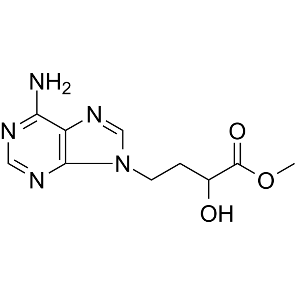
DZ2002 is an orally active, reversible and low-cytotoxic type III SAHH inhibitor (Ki=17.9 nM), with good immunosuppressive activity. DZ2002 prevents the development of experimental dermal fibrosis by reversing the profibrotic phenotype of various cell types. DZ2002 can be used in studies of autoimmune diseases such as lupus syndrome and systemic sclerosis.
For research use only. We do not sell to patients.
- Purity : 99.83%
- CAS No.: 33231-14-0
- Formula: C10H13N5O3
- Molecular Weight:251.25
-
Storage:Powder -20°C, 3 years , 4°C, 2 years ; In solvent -80°C, 2 years , -20°C, 1 year

To place orders, for customer services and technical support, please contact: MedChemExpress USA
Tel: 609-228-6898 E-mail: [email protected] [email protected]


-
 Biological Activity
-
Chemical Information
-
Solvent & Solubility
- Protocols
- Purity & Documentation
- References
-
 Help & FAQs


Publications Citing Use of MedChemExpress (MCE) DZ2002
MoreAll MEK Isoforms
MoreAll Antibiotic Isoforms
More

Biological Activity
Description
In Vitro
DZ2002 (0.1, 1, 10 μM; 96 h) inhibits the mixed lymphocyte reaction (MLR) response[1].
DZ2002 (0.1, 1, 10 μM; 24 h) inhibits IL-12 and TNF-α production from both mouse peritoneal exudate cells and humanTHP-1 Cells[1].
DZ2002 (0.1, 1, 10 μM; 64 h) inhibits expression of B7 (CD80/CD86) on differentiated THP-1 cells[1].
MedChemExpress (MCE) has not independently confirmed the accuracy of these methods. They are for reference only.
-
Cell Line:BALB/c and C57BL/6 splenocytes (Mitomycin C-pretreated; mixed lymphocyte)
-
Concentration:0.1, 1, 10 µM
-
Incubation Time:96 h
-
Result:Suppressed the MLR by 24.5, 42.3, and 46.0% at dosages of 0.1, 1, and 10 µM, respectively.
-
Cell Line:TG-stimulated mouse peritoneal macrophages and human THP-1 cells
-
Concentration:0.1, 1, 10 µM
-
Incubation Time:24 h
-
Result:Significantly blocked IL-12 p40 production from ~1800 pg/mL in untreated cells to ~850 pg/ml at 10 µM, and drastically reduced the active p70 form from ~1200 pg/mL in untreated cells to ~50 pg/mL.
Reduced TNF-α level by 45%.
-
Cell Line:THP-1 cells
-
Concentration:0.1, 1, 10 µM
-
Incubation Time:64 h
-
Result:Dramatically down-regulated CD80 and, in particular, CD86 expression in a dose-dependent manner.
In Vivo
DZ2002 (0.08, 2 mg/kg; i.p.; single daily for 7 days) significantly suppresses a delayed-type hypersensitivity reaction as well as antibody secretion[1].
DZ2002 (50, 100 mg/kg; p.o.; single daily for 4 weeks) exerts a potent anti-fibrotic effect on dermal fibrosis by reducing the production of collagen, facilitating its degradation and regulating expression of various soluble factors in SSc mice model[2].
MedChemExpress (MCE) has not independently confirmed the accuracy of these methods. They are for reference only.
-
Animal Model:Male and female BALB/c and C57BL/6 mice (6 to 8-week-old; DNFB-induced ear swelling model)[1].
-
Dosage:2, 10, 50 mg/kg
-
Administration:Intraperitoneal injection; twice (1 h before and 24 h after challenge)
-
Result:Suppressed ear swelling by 19.1, 28.7, and 33.1%, respectively and in a dose-dependent manner.
-
Animal Model:Male and female BALB/c and C57BL/6 mice (6 to 8-week-old; DNFB-induced ear swelling model)[1].
-
Dosage:0.08, 2 mg/kg
-
Administration:Intraperitoneal injection; single daily for 7 days.
-
Result:Inhibited hemolysis by 24.5 and 18.4% at doses of 0.08 and 2 mg/kg, respectively, thus decreasing anti-SRBC antibody production in vivo.
-
Animal Model:Wild-type C57BL/6 mice (8 to 12-week-old; BLM-induced mice model of SSc)[2].
-
Dosage:50, 100 mg/kg
-
Administration:Oral gavage; single daily for 4 weeks.
-
Result:Significantly decreased skin thickness and dermal thickness in BLM-induced mice.
Significantly reduced collagen accumulation and α-SMA expression in the dermis of mice and suppressed the mRNA expression of vascular endothelial growth factor (VEGF) in mice skin tissue.
Notably reduced collagen content and mRNA expression of the Col1a1 and Col1a2 while promoting that of the matrix metalloproteinase-13 (MMP-13) in the lesional skin of BLM-induced mice.


Chemical Information
-
CAS No. 33231-14-0
-
Appearance Solid
-
Molecular Weight 251.25
-
Formula C10H13N5O3
-
Color White to off-white
-
SMILES
OC(C(OC)=O)CCN1C(N=CN=C2N)=C2N=C1
-
Shipping
Room temperature in continental US; may vary elsewhere.
-
Storage
Powder -20°C 3 years 4°C 2 years In solvent -80°C 2 years -20°C 1 year


Publications (1)
-
Journal Impact Factor
-
Most Recent


Solvent & Solubility
In Vitro:
DMSO : ≥ 61 mg/mL (242.79 mM; Hygroscopic DMSO has a significant impact on the solubility of product, please use newly opened DMSO)
* "≥" means soluble, but saturation unknown.
Please refer to the solubility information to select the appropriate solvent. Once prepared, please aliquot and store the solution to prevent product inactivation from repeated freeze-thaw cycles.
Storage method and period of stock solution: -80°C, 2 years; -20°C, 1 year. When stored at -80°C, please use it within 2 years. When stored at -20°C, please use it within 1 year.
Please refer to the solubility information to select the appropriate solvent. Once prepared, please aliquot and store the solution to prevent product inactivation from repeated freeze-thaw cycles.
Storage method and period of stock solution: -80°C, 2 years; -20°C, 1 year. When stored at -80°C, please use it within 2 years. When stored at -20°C, please use it within 1 year.
Concentration (start) × Volume (start) = Concentration (final) × Volume (final)
In Vivo:
Select the appropriate dissolution method based on your experimental animal and administration route.
- For the following dissolution methods, please ensure to first prepare a clear stock solution using an In Vitro approach and then sequentially add co-solvents:
- To ensure reliable experimental results, the clarified stock solution can be appropriately stored based on storage conditions. As for the working solution for In Vivo experiments, it is recommended to prepare freshly and use it on the same day.
- The percentages shown for the solvents indicate their volumetric ratio in the final prepared solution. If precipitation or phase separation occurs during preparation, heat and/or sonication can be used to aid dissolution.
Add each solvent one by one: 10% DMSO 90% (20% SBE-β-CD in Saline)
Solubility: ≥ 2.5 mg/mL (9.95 mM); Clear solution
This protocol yields a clear solution of ≥ 2.5 mg/mL (saturation unknown).
Taking 1 mL working solution as an example, add 100 μL DMSO stock solution (25.0 mg/mL) to 900 μL 20% SBE-β-CD in Saline, and mix evenly.
Preparation of 20% SBE-β-CD in Saline (4°C, storage for one week): 2 g SBE-β-CD powder is dissolved in 10 mL Saline, completely dissolve until clear.
In Vivo Dissolution Calculator
Please enter the basic information of animal experiments:
-
-
-
-
Recommended: Prepare an additional quantity of animals to account for potential losses during experiments.
Please enter your animal formula composition:
-
%DMSO +
Recommended: Keep the proportion of DMSO in working solution below 2% if your animal is weak.
-
%+
-
+%Tween-80 + +
-
%Saline +
The co-solvents required include: DMSO, . All of co-solvents are available by MedChemExpress (MCE). , Tween 80. All of co-solvents are available by MedChemExpress (MCE).
Working solution concentration: 0.22 mg/mL
Method for preparing stock solution: mg drug dissolved in μL DMSO. Stock solution concentration: mg/mL.
1. Take μL DMSO stock solution;
2. Add μL .
μL , mix evenly;
3. Then add μL Tween 80, mix evenly;
4. Then add μL
Please ensure that the stock solution in the first step is dissolved to a clear state, and add co-solvents in sequence. You can use ultrasonic heating (ultrasonic cleaner, recommended frequency 20-40 kHz), vortexing, etc. to assist dissolution.


Purity & Documentation
-
Data Sheet (279 KB)
-
SDS (251 KB)
- English - EN (251 KB)
- Français - FR (251 KB)
- Deutsch - DE (251 KB)
- Norwegian - NO (251 KB)
- Español - ES (251 KB)
- Swedish - SV (251 KB)
- Italian - IT (251 KB)
- Korean - KR (251 KB)
- Portuguese - PT (251 KB)
-
Handling Instructions (2659 KB)
References
[1]. Wu QL, et al. Inhibition of S-adenosyl-L-homocysteine hydrolase induces immunosuppression. J Pharmacol Exp Ther. 2005 May;313(2):705-11. [Content Brief]
[2]. Zhang Z, et al. DZ2002 ameliorates fibrosis, inflammation, and vasculopathy in experimental systemic sclerosis models. Arthritis Res Ther. 2019 Dec 16;21(1):290. [Content Brief]


Complete Stock Solution Preparation Table
Please refer to the solubility information to select the appropriate solvent. Once prepared, please aliquot and store the solution to prevent product inactivation from repeated freeze-thaw cycles.
Storage method and period of stock solution: -80°C, 2 years; -20°C, 1 year. When stored at -80°C, please use it within 2 years. When stored at -20°C, please use it within 1 year.
| Optional Solvent | Concentration Solvent Mass | 1 mg | 5 mg | 10 mg | 25 mg |
|---|---|---|---|---|---|
| DMSO | 1 mM | 3.9802 mL | 19.9008 mL | 39.8016 mL | 99.5041 mL |
| 5 mM | 0.7960 mL | 3.9802 mL | 7.9603 mL | 19.9008 mL | |
| 10 mM | 0.3980 mL | 1.9901 mL | 3.9802 mL | 9.9504 mL | |
| 15 mM | 0.2653 mL | 1.3267 mL | 2.6534 mL | 6.6336 mL | |
| 20 mM | 0.1990 mL | 0.9950 mL | 1.9901 mL | 4.9752 mL | |
| 25 mM | 0.1592 mL | 0.7960 mL | 1.5921 mL | 3.9802 mL | |
| 30 mM | 0.1327 mL | 0.6634 mL | 1.3267 mL | 3.3168 mL | |
| 40 mM | 0.0995 mL | 0.4975 mL | 0.9950 mL | 2.4876 mL | |
| 50 mM | 0.0796 mL | 0.3980 mL | 0.7960 mL | 1.9901 mL | |
| 60 mM | 0.0663 mL | 0.3317 mL | 0.6634 mL | 1.6584 mL | |
| 80 mM | 0.0498 mL | 0.2488 mL | 0.4975 mL | 1.2438 mL | |
| 100 mM | 0.0398 mL | 0.1990 mL | 0.3980 mL | 0.9950 mL |


DZ2002 Related Classifications
HY-18620 Related Classifications
 Powered by Bioz