



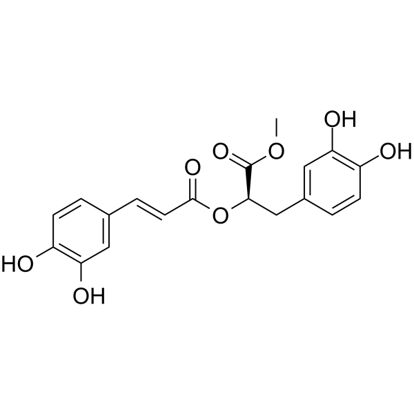
Methyl rosmarinate

Based on 1 Customer Validation
Methyl rosmarinate is an orally active hydroxycinnamic acid. Methyl rosmarinate exhibits an IC50 of 24.70 μM and a Ki of 15.29 μM against PTP1B, an IC50 of 41.46 μg/mL against BChE, a Ki of 0.61 mM against mushroom tyrosinase, and an IC50 of 2.50 μM against SARS-CoV-2 3CLpro. Methyl rosmarinate downregulates the phosphorylation levels of ERK, JNK, p38, Smad2 and Smad3. Methyl rosmarinate activates erythrocyte BPGM and promotes the production of 2,3-BPG. Methyl rosmarinate induces apoptosis of fibroblasts. Methyl rosmarinate prolongs the survival time of hypoxic mice. Methyl rosmarinate improves insulin sensitivity. Methyl rosmarinate binds to SARS-CoV-2 3CLpro and inhibits viral replication. Methyl rosmarinate induces glioblastoma cell death. Methyl rosmarinate activates the TGR5/AMPK axis and reduces the levels of ROS and MDA. Methyl rosmarinate shows inhibitory activity against MMP-1. Methyl rosmarinate can be used in research related to pulmonary fibrosis, hypoxia-induced injury, type 2 diabetes, Alzheimer's disease, hyperpigmentation disorders, COVID-19, glioblastoma and myocardial ischemia-reperfusion injury.
Nur für Forschungszwecke. Wir verkaufen nicht an Patienten.
- Reinheit : 99.46%
- CAS. Nr.: 99353-00-1
- Formel: C19H18O8
- Molecular Weight:374.34
-
Speicherung:
4°C, protect from light
* In solvent : -80°C, 6 months; -20°C, 1 month (protect from light)

To place orders, for customer services and technical support, please contact: MedChemExpress USA
Tel: 609-228-6898 E-mail: [email protected] [email protected]


-
 Biologische Aktivität
-
Chemical Information
-
Lösungsmittel & Löslichkeit
- Protokoll
- Reinheit & Dokumentation
- Verweise
-
 Help & FAQs


-
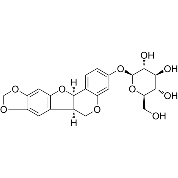
HY-N0616
TrifolirhizinPterocarpan Flavonoid -
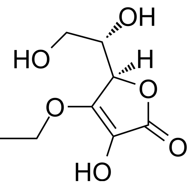
HY-W003607
3-O-Ethyl-L-ascorbic acidVitamin C Derivative -
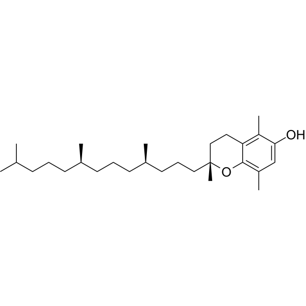
HY-113068
(rel)-β-Tocopherolβ-Tocopherol Isomer -
HY-N1238
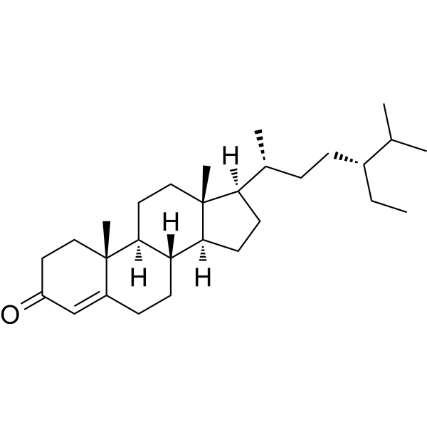
β-SitostenoneAnti-Tumor Agent -
HY-B1461
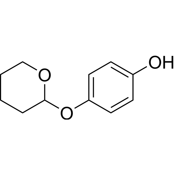
DeoxyarbutinTyrosinase Inhibitor -
HY-P3452
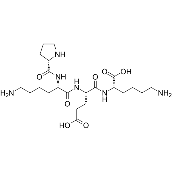
Tetrapeptide-30Skin-Brightening Peptide -
HY-N0192
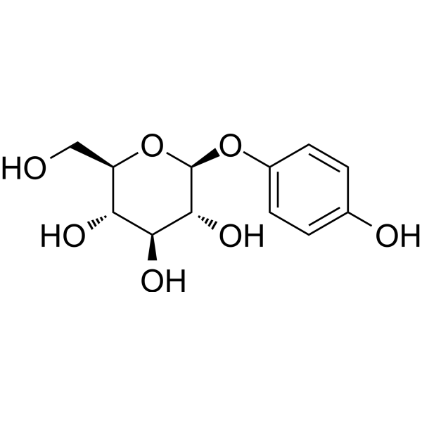
ArbutinEndogenous Metabolite -

HY-N3266R
Methyl rosmarinate (Standard)Tyrosinase Inhibitor -
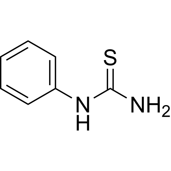
HY-Y0351
PhenylthioureaPhenoloxidase Inhibitor -
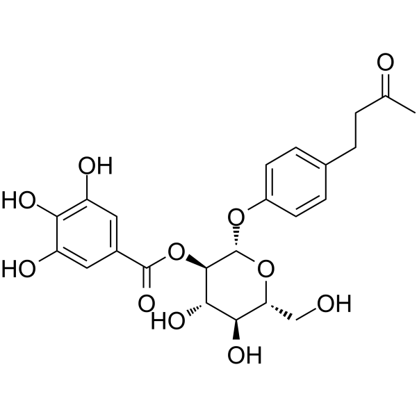
HY-N6244
IsolindleyinTyrosinase Inhibitor -
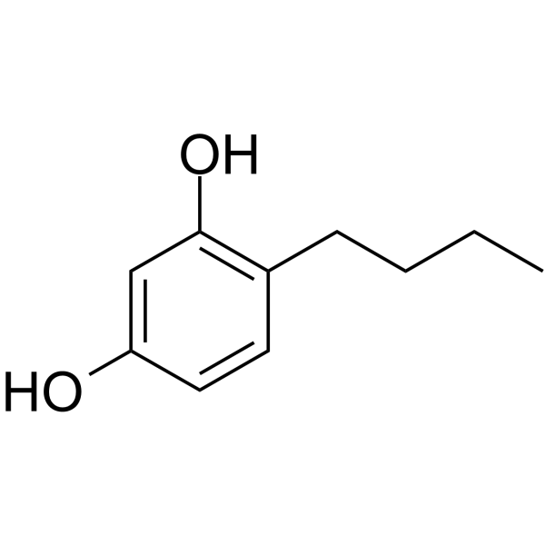
HY-107369
4-ButylresorcinolTyrosinase Inhibitor -
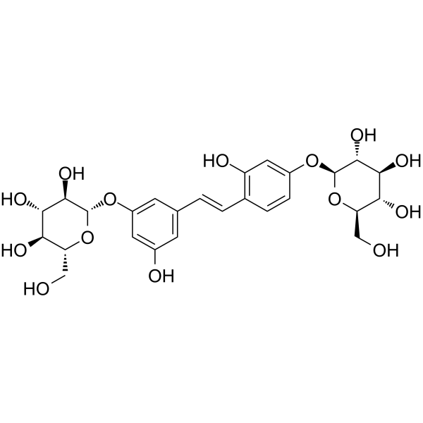
HY-N0619
Mulberroside ATyrosinase Inhibitor
Alle AMPK Isoform-spezifische Produkte anzeigen
More

Biologische Aktivität
Beschreibung
IC50 & Target
IC50: 0.28 mM (mushroom tyrosinase), a -glucosidase[1]
Cellular Effect
|
Cell Line

|
Type | Value | Description | References |
|---|---|---|---|---|
| MOLM-13 | IC50 |
8.4 μM
Compound: 20
|
Antiproliferative activity against human MOLM13 cells by Cell-Titer Glo assay
Antiproliferative activity against human MOLM13 cells by Cell-Titer Glo assay
|
[PMID: 30370766] |
| MOLM-14 | IC50 |
>10 μM
Compound: 20
|
Antiproliferative activity against human MOLM14 cells by Cell-Titer Glo assay
Antiproliferative activity against human MOLM14 cells by Cell-Titer Glo assay
|
[PMID: 30370766] |
| MV4-11 | IC50 |
>10 μM
Compound: 20
|
Antiproliferative activity against human MV4-11 cells by Cell-Titer Glo assay
Antiproliferative activity against human MV4-11 cells by Cell-Titer Glo assay
|
[PMID: 30370766] |
| RAW264.7 | IC50 |
11.3 μM
Compound: 26
|
Antiinflammatory activity against LPS-stimulated mouse RAW264.7 cells assessed as decrease in PGE2 production preincubated for 1 hr followed by LPS stimulation and measured after 24 hrs by ELISA
Antiinflammatory activity against LPS-stimulated mouse RAW264.7 cells assessed as decrease in PGE2 production preincubated for 1 hr followed by LPS stimulation and measured after 24 hrs by ELISA
|
[PMID: 31747281] |
| Vero C1008 | CC50 |
>1000 μM
Compound: 4
|
Cytotoxicity against African green monkey Vero E6 cells assessed as reduction in cell viability incubated for 48 hrs by MTT assay
Cytotoxicity against African green monkey Vero E6 cells assessed as reduction in cell viability incubated for 48 hrs by MTT assay
|
[PMID: 37267066] |
In Vitro
Methyl rosmarinate (10-160 μM; 48 h) exhibits cytotoxicity against L929 cells, with an IC50 of 76.27 μM[1].
Methyl rosmarinate (10-40 μM; 48 h) reduces extracellular matrix protein accumulation in TGF-β1-stimulated L929 cells[1].
Methyl rosmarinate (20-40 μM; 12-72 h) inhibits the proliferation and migration of TGF-β1-stimulated mouse fibroblast L929 cells and increases the cellular apoptosis rate[1].
Methyl rosmarinate (20-40 μM; 48 h) upregulates the expression levels of pro-apoptotic proteins Bax, cleaved caspase 3, and cleaved caspase 9, downregulates the expression level of anti-apoptotic protein Bcl-2, inhibits the phosphorylation of TGF-β1/Smad and MAPK signaling pathways, and alleviates the fibrotic response in TGF-β1-stimulated mouse fibroblast L929 cells[1].
Methyl rosmarinate competitively and reversibly inhibits the activity of recombinant human PTP1B enzyme, with an IC50 of 24.7 μM and a Ki of 15.29 μM[3].
Methyl rosmarinate (6.25-25 μM; 24 h) acts as an insulin sensitizer, enhancing insulin-stimulated glucose uptake and glycogen synthesis in fully differentiated C2C12 myotubes. It activates the insulin signaling pathway and increases insulin-stimulated phosphorylation levels of IRS-1 and Akt, without altering the protein expression level of PTP1B[3].
Methyl rosmarinate inhibits butyrylcholinesterase (BChE) with an IC50 of 41.46 µg/mL, but exerts no significant inhibitory effect on acetylcholinesterase (AChE)[4].
Methyl rosmarinate (0.01-0.4 mM; 2 min preincubation, 5 min reaction) inhibits the diphenolase activity of mushroom tyrosinase with a Ki of 0.61 mM, and inhibits yeast α-glucosidase activity[5].
Methyl rosmarinate acts as an allosteric inhibitor of purified SARS-CoV-2 3CLpro, with an IC50 of 2.5 μM, a Ki of 1.27 μM, and a Kd of 5.93 μM[6].
Methyl rosmarinate (1-200 μM; 52 h total) inhibits the replication of SARS-CoV-2 replicons in Huh7 cells, with an EC50 of 18.91 μM, and only exhibits moderate cytotoxicity at the concentration of 200 μM[6].
Methyl rosmarinate (5-60 μM; 72 h) reduces the viability of human glioblastoma U87 (IC50 = 9.8 μM) and T98 (IC50 = 13 μM) cell lines in a dose-dependent manner[7].
Methyl rosmarinate (9.8-19.6 μM in U87 cells; 13-26 μM in T98 cells; 72 h) induces subG0 and S phase arrest in U87 cells, and subG0 and G2/M phase arrest in T98 cells[7].
Methyl rosmarinate (9.8-19.6 μM in U87 cells; 13-26 μM in T98 cells; 48 h) inhibits the migration of U87 and T98 cell lines in a dose-dependent manner[7].
Methyl rosmarinate inhibits purified human MMP-1 with an IC50 of 14.7 μM[9].
MedChemExpress (MCE) has not independently confirmed the accuracy of these methods. They are for reference only. Further protocols information, click here.
-
Cell Line:mouse fibroblast L929 cells
-
Concentration:0, 10, 20, 40, 80, and 160 μM
-
Incubation Time:48 h
-
Result:Exhibited cytotoxicity toward L929 cells, with an IC50 value of 76.27 μM.
Reduced cell viability to 92.53% of the control at 80 μM.
Reduced cell viability to 48.82% of the control at 160 μM.
-
Cell Line:TGF-β1-stimulated mouse fibroblast L929 cells
-
Concentration:10, 20, 40 μM
-
Incubation Time:48 h (following 72 h of TGF-β1 stimulation)
-
Result:Dose-dependently reduced the elevated expression of collagen-I, collagen-III, vimentin, α-SMA, and snail proteins induced by TGF-β1 stimulation.
Caused significant, concentration-dependent decreases in the expression of all these fibrosis-associated proteins relative to TGF-β1-only treated cells.
-
Cell Line:TGF-β1-stimulated mouse fibroblast L929 cells
-
Concentration:20, 40 μM
-
Incubation Time:12, 24, 48, 72 h (co-treated with TGF-β1)
-
Result:Significantly inhibited the increased proliferation of L929 cells induced by TGF-β1 at all measured time points.
Showed statistically significant inhibitory effects at both 20 μM and 40 μM concentrations relative to TGF-β1-only treated cells.
-
Cell Line:TGF-β1-stimulated mouse fibroblast L929 cells
-
Concentration:20, 40 μM
-
Incubation Time:24, 48 h (following TGF-β1 induction)
-
Result:Significantly reduced the migration rate of TGF-β1-stimulated L929 cells at both 24 and 48 h.
Showed a stronger inhibitory effect at 40 μM concentration relative to the 20 μM concentration.
-
Cell Line:TGF-β1-stimulated mouse fibroblast L929 cells
-
Concentration:20, 40 μM
-
Incubation Time:48 h (following TGF-β1 induction)
-
Result:Significantly increased the apoptosis rate of TGF-β1-stimulated L929 cells.
Increased apoptosis rate to 5.49% at 20 μM relative to 3.53% in TGF-β1-only treated cells.
Increased apoptosis rate to 10.32% at 40 μM relative to 3.53% in TGF-β1-only treated cells.
-
Cell Line:TGF-β1-stimulated mouse fibroblast L929 cells
-
Concentration:20, 40 μM
-
Incubation Time:48 h (following TGF-β1 induction)
-
Result:Increased expression of pro-apoptotic proteins Bax, cleaved caspase 3, and cleaved caspase 9 in a dose-dependent manner relative to TGF-β1-only treated cells.
Decreased expression of anti-apoptotic protein Bcl-2 in a dose-dependent manner relative to TGF-β1-only treated cells.\nDose-dependently reduced the phosphorylation ratios of p-Smad2/Smad2, p-Smad3/Smad3, p-ERK/ERK, p-JNK/JNK, and p-p38/p38 in TGF-β1-stimulated L929 cells.
Caused statistically significant decreases in phosphorylation ratios at both 20 and 40 μM relative to TGF-β1-only treated cells.
-
Cell Line:fully differentiated C2C12 myotubes
-
Concentration:6.25, 12.5 and 25 μM
-
Incubation Time:24 h
-
Result:Did not alter PTP1B protein expression.
Dose-dependently enhanced insulin-stimulated phosphorylation of IRS-1 and Akt, with significant increases observed at 12.5 and 25 μM relative to the insulin-only group.
-
Cell Line:human glioblastoma U87, T98 cell lines
-
Concentration:5, 15, 30, 45, 60 μM
-
Incubation Time:72 h
-
Result:Reduced cell viability in a dose-dependent manner in both cell lines.
Reached a half maximal inhibitory concentration (IC50) of 9.8 μM in U87 cells and 13 μM in T98 cells.
Induced morphological changes including shrinking and death at higher concentrations.
-
Cell Line:human glioblastoma U87, T98 cell lines
-
Concentration:9.8, 19.6 μM (U87 cells); 13, 26 μM (T98 cells)
-
Incubation Time:72 h
-
Result:Increased subG0/G1 phase from 1.9945% to 16.331% and S phase from 7.318% to 15.249% in U87 cells.
Increased subG0/G1 phase from 0.835% to 5.179% and G2/M phase from 4.502 to 16.551 in T98 cells.
Caused statistically significant changes in both cell lines.
In Vivo
Methyl rosmarinate (25-75 mg/kg, i.p.; once daily for 3 consecutive days) prolongs the survival time of mice under normobaric closed hypoxia. It protects mice from damage caused by high-altitude field hypoxia by reducing inflammatory factors, improving tissue oxidative stress, alleviating tissue pathological damage and relieving tissue hypoxia. Additionally, it enhances the glycolysis pathway activity of red blood cells in Mus musculus exposed to high-altitude field hypoxia, activates BPGM, and increases the level of 2,3-BPG, thereby improving the oxygen release capacity of red blood cells[2].
Methyl rosmarinate (6.25-25 mg/kg; oral gavage; daily; 7 weeks) improves insulin sensitivity, restores glucose and lipid homeostasis, protects skeletal muscle and organ function, and enhances β-cell function in type 2 diabetic mice induced by high-fat diet/Streptozotocin (HY-13753)[3].
Methyl rosmarinate (50-200 mg/kg; i.p.; single administration 3 hours before ligation) dose-dependently improves cardiac function, and alleviates myocardial injury, oxidative stress and mitochondrial damage in Mus musculus (mouse) models of myocardial ischemia-reperfusion injury by activating the TGR5/AMPK signaling axis[8].
MedChemExpress (MCE) has not independently confirmed the accuracy of these methods. They are for reference only.
-
Animal Model:C57BL/6 (male, bleomycin-induced pulmonary fibrosis model)[1]
-
Dosage:20 mg/kg
-
Administration:p.o.; daily; day 3 to day 28
-
Result:Significantly improved mouse body weight and reduced lung fibrosis as visualized by small animal MicroCT.
Improved lung structure, reduced inflammatory cell infiltration, and decreased collagen deposition compared to the bleomycin-only group.
Reduced Ashcroft fibrosis score from ~5.2 in the bleomycin-only group to ~1.8.
Reduced collagen deposition area from ~50% in the bleomycin-only group to ~45%.
Significantly reduced lung tissue levels of fibrosis-associated proteins collagen-I and collagen-III compared to the bleomycin-only group.
-
Animal Model:BALB/c (male and female, 18-22 g)[2]
-
Dosage:25 mg/kg; 50 mg/kg; 75 mg/kg
-
Administration:i.p.; daily; 3 days
-
Result:Prolonged survival time by 20.15% (31.89 minutes) at 25 mg/kg vs.
blank control.
Prolonged survival time by 24.35% (33.00 minutes) at 50 mg/kg vs.
blank control.
Prolonged survival time by 24.19% (32.96 minutes) at 75 mg/kg vs.
blank control.
Showed statistically significant survival time increases across all dose groups vs.
blank control.
-
Animal Model:C57BL/6 J (male, 6 weeks old, high-fat diet + streptozotocin-induced type 2 diabetes)[3]
-
Dosage:6.25 mg/kg; 25 mg/kg
-
Administration:i.g.; daily; 7 weeks
-
Result:Reversed decreased body weight and elevated fasting blood glucose levels, with effects comparable to metformin.
Reduced weekly food intake and decreased glycosylated serum protein (GSP) levels in a dose-dependent manner.
Improved glucose clearance during GTT and insulin sensitivity during ITT, as evidenced by reduced area under the curve (AUC) values compared to vehicle-treated T2D mice.
Increased fasting serum insulin (FINS) levels; the 25 mg/kg dose significantly reduced the homeostasis model assessment of insulin resistance (HOMA-IR) index and improved the homeostasis model assessment of β-cell function (HOMA-β) index.
Improved lipid profiles by reducing total cholesterol (T-CHO), triglycerides (TG), and low-density lipoprotein cholesterol (LDL-C) levels, while increasing high-density lipoprotein cholesterol (HDL-C) levels.
Reversed T2D-induced loss of skeletal muscle mass (improved indexes of extensor digitorum longus, tibialis anterior, soleus, gastrocnemius, and quadriceps muscles) and protected against skeletal muscle fiber atrophy.
Elevated glycogen synthesis in skeletal muscle, restored insulin-stimulated phosphorylation of IRS-1 and Akt in skeletal muscle, and downregulated PTP1B protein expression in skeletal muscle (the 25 mg/kg dose returned PTP1B expression to normal levels).
The 25 mg/kg dose reduced liver and kidney indexes, attenuated liver steatosis and renal tubule vacuolation, and decreased alanine aminotransferase (ALT) and aspartate aminotransferase (AST) levels, with no observed toxicity in normal mice.
-
Animal Model:C57BL/6 J (male, 6-8 weeks old, 20-22 g, myocardial ischemia-reperfusion injury model via left anterior descending coronary artery ligation for 30 minutes followed by reperfusion; some groups received tail vein injection of AV-TGR5, AV-sh-TGR5, or negative control AAV9 3 weeks prior to injury)[8]
-
Dosage:50 mg/kg; 100 mg/kg; 200 mg/kg
-
Administration:i.p.; single dose 3 h pre-ligation
-
Result:Increased LVDP, +dp/dtmax, and −dp/dtmax, and decreased LVEDP dose-dependently compared to MIRI controls.
Reduced serum CK-MB and LDH levels, and reduced myocardial infarct size dose-dependently compared to MIRI controls.
Preserved myocardial fiber structure, reduced inflammatory cell infiltration, hemorrhage, edema, and necrosis; reduced myocardial fibrosis area and cardiomyocyte apoptosis rate dose-dependently compared to MIRI controls.
Reduced cleaved caspase 3 and Bax protein levels, and increased Bcl-2 protein levels compared to MIRI controls.
Reduced myocardial ROS, serum MDA, and serum LPO levels, and increased serum SOD levels dose-dependently compared to MIRI controls.
Reduced mitochondrial damage rate, improved mitochondrial structure, and reduced mitochondrial swelling and vacuolar degeneration dose-dependently compared to MIRI controls.
Increased myocardial TGR5 and p-AMPK protein levels dose-dependently compared to MIRI controls.
When combined with 100 mg/kg methyl rosmarinate, TGR5 overexpression further enhanced cardiac function, reduced myocardial injury markers, infarct size, pathological damage, oxidative stress, and mitochondrial damage, and further increased TGR5 and p-AMPK levels compared to methyl rosmarinate alone.
TGR5 knockdown reversed the beneficial effects of methyl rosmarinate, reducing cardiac function, increasing injury markers, pathological damage, oxidative stress, and mitochondrial damage, and decreasing TGR5 and p-AMPK levels compared to methyl rosmarinate alone.


Chemical Information
-
CAS. Nr. 99353-00-1
-
Appearance Solid
-
Molecular Weight 374.34
-
Formel C19H18O8
-
Color White to light yellow
-
SMILES
OC1=C(O)C=CC(/C=C/C(O[C@@H](C(OC)=O)CC2=CC(O)=C(O)C=C2)=O)=C1
-
Structure Classification
-
Initial Source
-
Versand
Room temperature in continental US; may vary elsewhere.
-
Speicherung
4°C, protect from light
* In solvent : -80°C, 6 months; -20°C, 1 month (protect from light)


Lösungsmittel & Löslichkeit
In Vitro:
DMSO : 100 mg/mL (267.14 mM; Need ultrasonic; Hygroscopic DMSO has a significant impact on the solubility of product, please use newly opened DMSO)
Please refer to the solubility information to select the appropriate solvent. Once prepared, please aliquot and store the solution to prevent product inactivation from repeated freeze-thaw cycles.
Storage method and period of stock solution: -80°C, 6 months; -20°C, 1 month (protect from light). When stored at -80°C, please use it within 6 months. When stored at -20°C, please use it within 1 month.
Please refer to the solubility information to select the appropriate solvent. Once prepared, please aliquot and store the solution to prevent product inactivation from repeated freeze-thaw cycles.
Storage method and period of stock solution: -80°C, 6 months; -20°C, 1 month (protect from light). When stored at -80°C, please use it within 6 months. When stored at -20°C, please use it within 1 month.
Konzentration (Stammlösung) × Volumen (Stammlösung) = Konzentration (Ziellösung) × Volumen (Ziellösung)
In Vivo:
Select the appropriate dissolution method based on your experimental animal and administration route.
- For the following dissolution methods, please ensure to first prepare a clear stock solution using an In Vitro approach and then sequentially add co-solvents:
- To ensure reliable experimental results, the clarified stock solution can be appropriately stored based on storage conditions. As for the working solution for In Vivo experiments, it is recommended to prepare freshly and use it on the same day.
- The percentages shown for the solvents indicate their volumetric ratio in the final prepared solution. If precipitation or phase separation occurs during preparation, heat and/or sonication can be used to aid dissolution.
Add each solvent one by one: 10% DMSO 40% PEG300 5% Tween-80 45% Saline
Solubility: ≥ 2.5 mg/mL (6.68 mM); Clear solution
This protocol yields a clear solution of ≥ 2.5 mg/mL (saturation unknown).
Taking 1 mL working solution as an example, add 100 μL DMSO stock solution (25.0 mg/mL) to 400 μL PEG300, and mix evenly; then add 50 μL Tween-80 and mix evenly; then add 450 μL Saline to adjust the volume to 1 mL.
Preparation of Saline: Dissolve 0.9 g sodium chloride in ddH₂O and dilute to 100 mL to obtain a clear Saline solution.
Add each solvent one by one: 10% DMSO 90% (20% SBE-β-CD in Saline)
Solubility: ≥ 2.5 mg/mL (6.68 mM); Clear solution
This protocol yields a clear solution of ≥ 2.5 mg/mL (saturation unknown).
Taking 1 mL working solution as an example, add 100 μL DMSO stock solution (25.0 mg/mL) to 900 μL 20% SBE-β-CD in Saline, and mix evenly.
Preparation of 20% SBE-β-CD in Saline (4°C, storage for one week): 2 g SBE-β-CD powder is dissolved in 10 mL Saline, completely dissolve until clear.
In Vivo Dissolution Calculator
Please enter the basic information of animal experiments:
-
-
-
-
Recommended: Prepare an additional quantity of animals to account for potential losses during experiments.
Please enter your animal formula composition:
-
%DMSO +
Recommended: Keep the proportion of DMSO in working solution below 2% if your animal is weak.
-
%+
-
+%Tween-80 + +
-
%Saline +
The co-solvents required include: DMSO, . All of co-solvents are available by MedChemExpress (MCE). , Tween 80. All of co-solvents are available by MedChemExpress (MCE).
Working solution concentration: 0.22 mg/mL
Method for preparing stock solution: mg drug dissolved in μL DMSO. Stock solution concentration: mg/mL. * In solvent : -80°C, 6 months; -20°C, 1 month (protect from light)
1. Take μL DMSO stock solution;
2. Add μL .
μL , mix evenly;
3. Then add μL Tween 80, mix evenly;
4. Then add μL
Please ensure that the stock solution in the first step is dissolved to a clear state, and add co-solvents in sequence. You can use ultrasonic heating (ultrasonic cleaner, recommended frequency 20-40 kHz), vortexing, etc. to assist dissolution.

Protokoll
-
Western Blot
Western blotting (WB) is a commonly used experimental method in molecular biology, biochemistry, and immunogenetics for identifying and quantifying target proteins. It combines gel electrophoresis with immunoassay, enabling researchers to analyze protein expression, post-translational modifications, and molecular weight.
-
Collagen: Sirius Red Staining
Sirius Red or picrosirius red staining is a histochemical method for visualizing collagen-rich extracellular matrix in tissue sections, and collagen fibers are detected as red-stained structures under bright-field microscopy with enhanced birefringence under polarized light. Picrosirius red is useful for assessing total collagen organization, distribution, and fibrosis burden, but polarized color should not be interpreted as a definitive collagen type I versus type III readout because color is affected by fiber orientation, thickness, and packing.
-
Mitochondrial membrane-potential fluorescent assay
Mitochondrial membrane potential fluorescent assays estimate ΔΨm in living cells using lipophilic cationic dyes such as TMRM, TMRE, rhodamine 123, and JC-1, which accumulate in mitochondria according to membrane polarization; loss of signal after FCCP or CCCP treatment is interpreted as mitochondrial depolarization. TMRM/TMRE and rhodamine 123 are commonly used for semi-quantitative live-cell microscopy or flow cytometry, while JC-1 can report a shift from red aggregate fluorescence to green monomer fluorescence during depolarization; interpretation requires controls because dye concentration, quenching mode, cell type, dye efflux, and mitochondrial mass can affect fluorescence independently of ΔΨm.
-
Mammalian live/dead viability and cytotoxicity staining
Live/dead viability and cytotoxicity staining assays are based on the simultaneous detection of intracellular esterase activity in metabolically active (viable) cells and membrane integrity loss in non-viable cells. In commonly used dual-staining approaches, membrane-permeant fluorogenic substrates are converted by intracellular esterases into fluorescent products in live cells, while impermeant DNA-binding dyes selectively enter cells with compromised plasma membranes and label nucleic acids in dead or dying cells, enabling discrimination between viable and non-viable populations by fluorescence microscopy or flow cytometry.
-
Annexin V plus membrane-impermeant dye apoptosis staining
Annexin V-based apoptosis assays rely on the detection of phosphatidylserine (PS) externalization from the inner leaflet of the plasma membrane to the outer leaflet, an early biochemical hallmark of apoptosis. Fluorescently labeled Annexin V binds PS in a calcium-dependent manner, enabling identification of early apoptotic cells by flow cytometry or fluorescence microscopy. When combined with a membrane-impermeant DNA-binding dye (e. g. , propidium iodide), this approach allows discrimination between viable (Annexin V−/dye−), early apoptotic (Annexin V+/dye−), and late apoptotic or necrotic (Annexin V+/dye+) cell populations by assessing membrane integrity and PS exposure.
-
ROS/oxidative-stress fluorescent staining
ROS/oxidative-stress fluorescent staining uses cell-permeant fluorogenic probes that become fluorescent after oxidation inside cells or tissues; commonly used examples include DCFH-DA/DCFDA for broad cellular oxidant detection, DHE for superoxide-related signal detection, MitoSOX for mitochondrial superoxide-related signal detection, and CellROX probes for oxidative-stress-associated fluorescence readouts. The assay detects probe oxidation rather than a single ROS species unless the probe and analysis method have been chemically validated for that species. DCFH-DA enters cells, is deacetylated by intracellular esterases to DCFH, and produces fluorescent DCF after oxidation, so the readout is used as an operational measure of total cellular oxidative stress rather than a species-specific ROS measurement. DHE and MitoSOX can report superoxide-related oxidation, but red fluorescence alone can include non-specific ethidium-like oxidation products; HPLC or optimized spectral approaches are
-
Connective Tissue: Masson's Trichrome/Collagen Trichrome Staining
Masson’s Trichrome (collagen/trichrome staining) is a histological technique that differentially stains tissue compartments using sequential acidic dyes to distinguish collagen from muscle and cytoplasmic components based on dye affinity and tissue permeability differences, enabling visualization of fibrosis and connective tissue architecture in histological sections. The classical formulation typically uses Weigert's iron hematoxylin for nuclear staining, Biebrich scarlet-acid fuchsin for cytoplasm and muscle, and aniline blue (or light green variants) for collagen, producing a characteristic blue/green collagen signal contrasted against red cytoplasm and dark nuclei. The staining principle relies on selective displacement of smaller dye molecules by larger anionic dyes in collagen-rich regions under controlled acidified conditions, which enhances collagen-specific dye retention. This property makes the method widely used for fibrosis assessment in organs such as heart, liver, lung, a
-
Research Protocol for Metabolic Diseases
AMP-activated protein kinase, AMPK, is a conserved cellular energy sensor that responds to reduced cellular energy status and coordinates metabolism by increasing ATP-generating catabolic pathways while suppressing ATP-consuming anabolic processes. In metabolic disease research, the AMPK pathway is experimentally relevant because it regulates hepatic lipid synthesis, fatty acid oxidation, glucose production, skeletal-muscle glucose disposal, mTORC1-linked biosynthesis, autophagy, mitochondrial homeostasis, and whole-body energy balance. The central pathway logic is that energy stress, metformin, exercise-like stimulation, or direct AMPK activators increase AMPKα Thr172 phosphorylation and downstream substrate phosphorylation, including ACC and RAPTOR. Phosphorylation of ACC suppresses lipogenesis and supports fatty acid oxidation, whereas phosphorylation of RAPTOR suppresses mTORC1 signaling and links cellular energy status to growth and protein synthesis control. The pathway is linked
-
Alzheimer’s Disease Modeling
Alzheimer’s Disease (AD) is a neurodegenerative disorder characterized by a progressive decline in cognitive functions and loss of specific types of neurons and synapses. Alzheimer's symptoms can be simulated in mice by injecting drugs (such as Aβ) or genetically modified.
-
Kinase activity and phosphorylation assays
Kinase activity assays measure the ability of kinases to transfer phosphate groups from ATP to specific substrates, while phosphorylation assays detect the presence and levels of phosphorylated proteins. Common methods include radiolabeled ATP incorporation (e. g. ,), ADP release detection via bioluminescence (e. g. ,[3]), enzyme-linked immunosorbent assays (ELISA) for phospho-specific epitopes (e. g. ,[6]), and microtiter-based formats for high-throughput screening (e. g. ,[8]). The ADP-Glo assay quantifies kinase activity by measuring ADP produced during phosphorylation using a luciferase-based system. Radiometric assays involve autoradiography or scintillation counting after incorporation of 32P-labeled ATP into substrate proteins. ELISA-based approaches rely on phospho-specific antibodies to detect activated kinases in cell lysates or purified samples.
-
Fibrosis/Collagen Morphometry
Fibrosis and collagen morphometry is based on the quantitative visualization of fibrillar collagen deposition in tissue sections using histochemical stains such as Sirius Red (Picrosirius Red) or Masson's trichrome, followed by image-based or polarization-enhanced analysis to estimate collagen proportional area as a surrogate of extracellular matrix accumulation during fibrotic remodeling. Sirius Red combined with polarized light microscopy enhances detection of collagen fibers due to birefringence properties, enabling more specific visualization of collagen type I and III fibrils compared to conventional bright-field histology, while whole-section or region-restricted digital morphometry reduces field-selection bias in fibrosis assessment. Alternative quantitative approaches include second harmonic generation (SHG) and two-photon excited fluorescence microscopy, which enable label-free detection of fibrillar collagen and have been validated against histological staining and biochemica
-
Apoptosis
Apoptosis, also called programmed cell death, is generally characterized by distinct morphological characteristics.
-
TUNEL staining for apoptotic DNA fragmentation
TUNEL staining detects DNA strand breaks by using terminal deoxynucleotidyl transferase to add labeled nucleotides to exposed 3′-OH DNA termini, generating either microscopic staining in fixed cells or tissue sections, or fluorescence/cytometric signal in cell suspensions. TUNEL positivity reflects DNA fragmentation but should not be interpreted alone as definitive apoptosis, because TUNEL can also label necrotic, autolytic, mechanically damaged, or DNA-repair-associated DNA breaks.
-
Cell Cytotoxicity Assay
Cytotoxicity assays are usually based on the assessment of cell membrane damage, which can also be indirectly detected by measuring cell viability. Detection methods include MTT assay, CKK-8 assay, LDH assay and ATP assay, etc.
-
Research Protocol for Endocrine Diseases
Endocrine diseases often arise from disrupted hormone production, hormone signaling, or target-tissue responsiveness; for diabetes-focused endocrine disease models, insulin signaling regulates glucose uptake, hepatic glucose output, lipid metabolism, and β-cell compensation. Type 2 diabetes develops through interacting defects in insulin resistance, β-cell dysfunction, adipose inflammation, hepatic glucose overproduction, altered incretin signaling, and ectopic lipid metabolism. A major unresolved question is whether endocrine dysfunction is driven primarily by target-tissue insulin resistance, intrinsic β-cell failure, immune/inflammatory stress, or combined multi-organ failure that differs by disease stage.
-
Mitochondrial membrane-potential and mitochondrial mass staining
Mitochondrial membrane potential staining measures the electrochemical polarization across the mitochondrial inner membrane in live cells using lipophilic cationic fluorescent probes; early rhodamine-based work showed that selective mitochondrial dye accumulation is lost when the mitochondrial transmembrane potential is dissipated. JC-1 reports mitochondrial polarization by shifting from green monomer fluorescence to red J-aggregate fluorescence as dye concentration increases within energized mitochondria; therefore, the red/green fluorescence ratio is used as a relative readout of mitochondrial membrane potential. TMRE or TMRM staining provides a single-channel relative readout because these cationic rhodamine esters accumulate in polarized mitochondria, and lower fluorescence indicates reduced mitochondrial polarization when acquisition and dye-loading conditions are controlled. Mitochondrial mass staining is commonly performed with MitoTracker Green FM or related MitoTracker dyes as
-
Apoptosis Solutions
Apoptosis is a regulated, generally non-lytic cell-death pathway that removes unwanted, damaged, infected, or abnormal cells through coordinated morphological changes, caspase activation, DNA fragmentation, and membrane remodeling. The intrinsic apoptosis pathway is controlled mainly by mitochondrial outer membrane permeabilization, BCL-2 family proteins, cytochrome c release, apoptosome formation, caspase-9 activation, and downstream executioner caspase-3/7 activation. The extrinsic apoptosis pathway is initiated by death receptors such as Fas, TNFR, and TRAIL receptors, which recruit adaptor proteins and activate caspase-8 before engaging executioner caspases or mitochondrial amplification through BID cleavage. Apoptosis is linked to many phenotypes, including cancer cell killing, tissue homeostasis, immune regulation, neurodegeneration, infection response, and treatment-induced cytotoxicity; unresolved questions include how apoptosis interacts with necroptosis, pyroptosis, ferroptos
-
Protocol for Kinase activity and phosphorylation assays
Kinase activity assays measure transfer of phosphate from ATP to a protein or peptide substrate, generating phosphorylated substrate, ADP, or incorporated radiolabeled phosphate as the readout; phosphorylation assays measure site-specific phosphorylation in cells or tissues as a proxy for kinase-pathway activation, inhibition, or substrate regulation. Phosphorylation can be detected by phospho-specific Western blot, immunoprecipitation kinase assay, phospho-immunofluorescence, phospho-flow cytometry, luminescent ADP detection, radiolabeled ATP incorporation, or reporter-based pathway assays, and these readouts can be applied to cancer cells, primary neurons, mouse tumors, organoids, inflammatory macrophages, ferroptosis studies, and mitophagy studies when the kinase target is biologically relevant.


Reinheit & Dokumentation
-
Data Sheet (300 KB)
-
SDS (394 KB)
- English - EN (394 KB)
- Français - FR (394 KB)
- Deutsch - DE (394 KB)
- Norwegian - NO (394 KB)
- Español - ES (394 KB)
- Swedish - SV (394 KB)
- Italian - IT (394 KB)
- Korean - KR (394 KB)
- Portuguese - PT (394 KB)
-
Handling Instructions (2659 KB)
Verweise
[2]. Guo Q, et al. Study on the Protective Effect of Methyl Rosmarinate on Hypoxic Mice and Their Erythrocytes. Drug Des Devel Ther. 2025;19:3179-3192. Published 2025 Apr 24. [Content Brief]
[3]. Xie B, et al. Bioactivity-based molecular networking-guided identification of methyl rosmarinate from Orthosiphon aristatus as a PTP1B inhibitor that improves insulin sensitivity in diabetic mice. Phytomedicine. 2025;148:157444. [Content Brief]
[4]. Senol Deniz FS, et al. Outlining In Vitro and In Silico Cholinesterase Inhibitory Activity of Twenty-Four Natural Products of Various Chemical Classes: Smilagenin, Kokusaginine, and Methyl Rosmarinate as Emboldening Inhibitors. Molecules. 2021;26(7):2024. Published 2021 Apr 1. [Content Brief]
[5]. Lin L, et al. Comparative evaluation of rosmarinic acid, methyl rosmarinate and pedalitin isolated from Rabdosia serra (MAXIM.) HARA as inhibitors of tyrosinase and α-glucosidase. Food Chem. 2011;129(3):884-889. [Content Brief]
[6]. Li H, et al. Methyl rosmarinate is an allosteric inhibitor of SARS-CoV-2 3 CL protease as a potential candidate against SARS-cov-2 infection. Antiviral Res. 2024;224:105841. [Content Brief]
[7]. Benekou MV, et al.Antineoplastic Activity of Methyl rosmarinate in Glioblastoma Cells. Curr Issues Mol Biol. 2025 Mar 10;47(3):180. [Content Brief]
[8]. Zhou W, et al. Methyl rosmarinate alleviates myocardial ischemia-reperfusion injury in mice via triggering TGR5/AMPK signaling axis. Naunyn Schmiedebergs Arch Pharmacol. 2026;399(4):5267-5284. [Content Brief]
[9]. Yuan H, et al. Synthesis of derivatives of methyl rosmarinate and their inhibitory activities against matrix metalloproteinase-1 (MMP-1). Eur J Med Chem. 2013;62:148-157. [Content Brief]


Complete Stock Solution Preparation Table
Please refer to the solubility information to select the appropriate solvent. Once prepared, please aliquot and store the solution to prevent product inactivation from repeated freeze-thaw cycles.
Storage method and period of stock solution: -80°C, 6 months; -20°C, 1 month (protect from light). When stored at -80°C, please use it within 6 months. When stored at -20°C, please use it within 1 month.
| Optional Solvent | Concentration Solvent Mass | 1 mg | 5 mg | 10 mg | 25 mg |
|---|---|---|---|---|---|
| DMSO | 1 mM | 2.6714 mL | 13.3568 mL | 26.7137 mL | 66.7842 mL |
| 5 mM | 0.5343 mL | 2.6714 mL | 5.3427 mL | 13.3568 mL | |
| 10 mM | 0.2671 mL | 1.3357 mL | 2.6714 mL | 6.6784 mL | |
| 15 mM | 0.1781 mL | 0.8905 mL | 1.7809 mL | 4.4523 mL | |
| 20 mM | 0.1336 mL | 0.6678 mL | 1.3357 mL | 3.3392 mL | |
| 25 mM | 0.1069 mL | 0.5343 mL | 1.0685 mL | 2.6714 mL | |
| 30 mM | 0.0890 mL | 0.4452 mL | 0.8905 mL | 2.2261 mL | |
| 40 mM | 0.0668 mL | 0.3339 mL | 0.6678 mL | 1.6696 mL | |
| 50 mM | 0.0534 mL | 0.2671 mL | 0.5343 mL | 1.3357 mL | |
| 60 mM | 0.0445 mL | 0.2226 mL | 0.4452 mL | 1.1131 mL | |
| 80 mM | 0.0334 mL | 0.1670 mL | 0.3339 mL | 0.8348 mL | |
| 100 mM | 0.0267 mL | 0.1336 mL | 0.2671 mL | 0.6678 mL |


Methyl rosmarinate Related Classifications
HY-N3266 Related Classifications
 Powered by Bioz
Keywords
- Methyl rosmarinate
- 99353-00-1
- Tyrosinase
- Phosphatase
- Cholinesterase (ChE)
- SARS-CoV
- PERK
- JNK
- p38 MAPK
- TGF-beta/Smad
- Apoptosis
- Reactive Oxygen Species (ROS)
- AMPK
- MMP
- SARS-CoV-2 3CLpro
- human glioblastoma T98 cells
- human BChE
- human PTP1B
- TGF-β1/Smad signaling pathways
- human glioblastoma U87 cells
- mushroom tyrosinase
- MAPK signaling pathways
- C2C12 myotubes
- mouse fibroblast L929 cells
- pulmonary fibrosis
- hypoxia-induced injury
- type 2 diabetes
- Alzheimer's disease
- hyperpigmentation disorders
- coronavirus disease 2019 (COVID-19)
- glioblastoma
- myocardial ischemia-reperfusion injury
- Inhibitor
- inhibitor
- inhibit