



Myrislignan
Based on 1 publication(s) in Google Scholar
Myrislignan is a PI3K/AKT/NF-κB inhibitor that can cross the blood-brain barrier. Myrislignan exerts anticancer activity by inducing apoptosis and ferroptosis. Myrislignan inhibits the replication and invasion of Toxoplasma gondii; it induces reactive oxygen species (ROS) imbalance, autophagy, and the death of Toxoplasma gondii tachyzoites. Myrislignan inhibits mitochondrial function and ERK1/2 phosphorylation to improve ovariectomy-induced osteoporosis. Myrislignan can be used in studies related to gastric cancer, glioblastoma, osteoporosis, and toxoplasmosis.
For research use only. We do not sell to patients.
- Purity : 99.53%
- CAS No.: 171485-39-5
- Formula: C21H26O6
- Molecular Weight:374.43
-
Storage:
4°C, protect from light
* In solvent : -80°C, 6 months; -20°C, 1 month (protect from light)

To place orders, for customer services and technical support, please contact: MedChemExpress USA
Tel: 609-228-6898 E-mail: [email protected] [email protected]


-
 Biological Activity
-
Chemical Information
-
Solvent & Solubility
- Protocols
- Purity & Documentation
- References
-
 Help & FAQs


-

HY-10227
BortezomibProteasome Inhibitor -
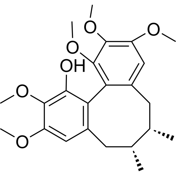
HY-N0859
SchisanhenolAntioxidant Agent -
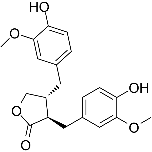
HY-N3312
MatairesinolInflammatory Pathway Inhibitor, Oxidative Stress Inhibitor, Cancer Pathway Inhibitor -
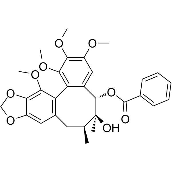
HY-N0694
Schisantherin ANF-κB Inhibitor -

HY-N0859R
Schisanhenol (Standard)Analytical Standard -

HY-N3312R
Matairesinol (Standard)Anti-allergic -

HY-N0694R
Schisantherin A (Standard)NF-κB Inhibitor
Publications Citing Use of MedChemExpress (MCE) Myrislignan
More

Biological Activity
Description
In Vitro
Myrislignan (5-30 μg/mL; 48 h) dose-dependently inhibits the activity of the NF-κB signaling pathway in human glioblastoma cell lines U87 and U251[2].
Myrislignan (5-60 μg/mL; 24-48 h) inhibits the growth of human glioblastoma cell lines U87 and U251, as well as human low-grade glioma cell lines SHG44, HS683, and SW1088 in a dose- and time-dependent manner, while a low dose (30 μg/mL) shows no toxicity to the human normal astrocyte cell line NHA[2].
Myrislignan (5-30 μg/mL; 24 h) inhibits the migration and invasion abilities of human glioblastoma cell lines U87 and U251 in a dose-dependent manner[2].
Myrislignan (5-30 μg/mL; 48 h) dose-dependently impairs the wound healing capacity of human glioblastoma cell lines U87 and U251[2].
Myrislignan (5-15 μg/mL) downregulates the protein level of SLC7A11 in the human glioblastoma cell line U87 in a dose-dependent manner, without altering the expression levels of Nrf2, TFR1 or GPX4[2].
Myrislignan (30 μM; 60 min-7 days) inhibits osteoclastogenesis in primary mouse bone marrow macrophages by suppressing ERK phosphorylation and downstream protein expression; this effect is partially reversed by the ERK agonist LM22B-10 (HY-104047) at a concentration of 30 μM[3].
Myrislignan (32-70 mg/mL; 16 h) induces autophagosome formation in tachyzoites of the RH strain of Toxoplasma gondii[4].
Myrislignan (32-70 mg/mL; 16-24 h) induces the formation of autophagosome-like structures in tachyzoites of Toxoplasma gondii RH strain within infected Vero cells, followed by subsequent degeneration after treatment at 32 or 70 mg/mL for 16 or 24 h[4].
Myrislignan (32-70 mg/mL; 24 h) alters the gene expression of tachyzoites of the RH strain of Toxoplasma gondii; differentially expressed genes (DEGs) are enriched in redox processes and the oxidative phosphorylation pathway[4].
Myrislignan (32-70 mg/mL; 8-24 h) induces a statistically significant time-dependent increase in ROS activity in tachyzoites of the RH strain of Toxoplasma gondii[4].
Myrislignan (0-80 μM; 48-96 h) exerts no effect on the viability of primary mouse bone marrow macrophages and does not induce their apoptosis even at concentrations up to 80 μM and treatment durations as long as 96 h[3].
Myrislignan (15-30 μM; 2 days) impairs the bone resorption function of mature osteoclasts derived from primary mouse bone marrow macrophages[3].
Myrislignan (15-30 μM; 7 days) dose-dependently inhibits the expression of osteoclast-specific genes in RANKL-treated primary mouse bone marrow macrophages[3].
Myrislignan (15-30 μM; 7 days) inhibits the formation of podosome belts-a key cytoskeletal feature of mature osteoclasts-in primary mouse bone marrow macrophages treated with RANKL for 7 days[3].
MedChemExpress (MCE) has not independently confirmed the accuracy of these methods. They are for reference only.
-
Cell Line:human gastric cancer SGC-7901 cells
-
Concentration:25; 50; 100; 200 μM
-
Incubation Time:48 h; 72 h
-
Result:Significantly inhibited SGC-7901 cell viability in a dose-dependent manner at 50, 100, and 200 μM after 48 h and 72 h.
Showed no significant difference in cell viability at 25 μM compared with control groups.
-
Cell Line:human gastric cancer SGC-7901 cells
-
Concentration:25; 50; 100; 200 μM
-
Incubation Time:48 h
-
Result:Increased the apoptosis rate of SGC-7901 cells, with a significant difference between the 200 μM group and the 100 μM group.
-
Cell Line:human gastric cancer SGC-7901 cells
-
Concentration:25; 50; 100; 200 μM
-
Incubation Time:48 h
-
Result:Downregulated the expression levels of PI3K and AKT proteins, and upregulated the expression levels of BAX, Caspase-3, and Caspase-9 proteins in a dose-dependent manner.
Showed relative PI3K expression of approximately 0.6, relative AKT expression of approximately 0.6, relative Caspase-9 expression of approximately 1.0, relative Caspase-3 expression of approximately 1.1, and relative BAX expression of approximately 1.1 at 200 μM.
-
Cell Line:human glioblastoma cell lines U87, U251; human low-grade glioma cell lines SHG44, HS683, SW1088; human normal astrocyte cell line NHA
-
Concentration:5, 10 and 15 μg/mL (U87); 10, 20 and 30 μg/mL (U251); 30, 40, 50 and 60 μg/mL (NHA)
-
Incubation Time:24 h, 48 h (U87, U251); null (NHA)
-
Result:Significantly inhibited the growth of U87 and U251 cells in a time- and dose-dependent manner.
Suppressed the growth of SHG44, HS683, and SW1088 cells.
Did not affect apoptosis or ferroptosis-associated protein levels in NHA cells at 30 μg/mL.
Inhibited NHA growth and caused lipid peroxidation at 40, 50, and 60 μg/mL.
-
Cell Line:human glioblastoma cell lines U87, U251
-
Concentration:5, 10 and 15 μg/mL (U87); 10, 20 and 30 μg/mL (U251)
-
Incubation Time:24 h
-
Result:Significantly reduced the number of migrated U87 and U251 cells in a dose-dependent manner.
Caused statistically significant decreases at all tested concentrations compared to untreated controls.
-
Cell Line:human glioblastoma cell lines U87, U251
-
Concentration:5, 10 and 15 μg/mL (U87); 10, 20 and 30 μg/mL (U251)
-
Incubation Time:48 h
-
Result:Significantly reduced the wound healing percentage of U87 and U251 cells in a dose-dependent manner.
Caused statistically significant decreases at all tested concentrations compared to untreated controls.
In Vivo
MedChemExpress (MCE) has not independently confirmed the accuracy of these methods. They are for reference only.
-
Animal Model:Nude mice (male, 5 weeks old, intracranial xenograft model via stereotactic implantation of U87 cells)[2]
-
Dosage:5 mg/kg
-
Administration:i.p.; every 3 days; 21 days
-
Result:Improved mouse survival compared to the control group.
Reduced tumor weight.
Decreased tumor tissue levels of phosphorylated p65 (p-p65), Slug, and SLC7A11.
Increased tumor tissue levels of E-cadherin.
-
Animal Model:C57BL/6J (11-weeks-old female; osteoporosis induced via bilateral ovariectomy)[3]
-
Dosage:15 mg/kg; 30 mg/kg
-
Administration:i.p.; daily; 6 weeks
-
Result:Increased bone volume/total volume (BV/TV) and trabecular number (Tb.N), and decreased trabecular separation (Tb.Sp) in the 30 mg/kg group compared to the ovariectomy-only group.
Showed intermediate improvements in BV/TV, Tb.N, and Tb.Sp in the 15 mg/kg group compared to the ovariectomy-only group.
Reduced bone surface/trabecular surface (BS/TS) and the number of TRAP-positive osteoclasts per trabecular surface (N.Oc/BS) in both dose groups compared to the ovariectomy-only group, with greater effects at 30 mg/kg.
Reduced the ratio of phosphorylated ERK (p-ERK) to total ERK in both dose groups compared to the ovariectomy-only group, with a more significant reduction at 30 mg/kg.
- Western Blot
- Mitochondrial membrane-potential fluorescent assay
- Annexin V plus membrane-impermeant dye apoptosis staining
- ROS/oxidative-stress fluorescent staining
- Research Protocol for Inflammation-related Diseases
- Kinase activity and phosphorylation assays
- Detection of Apoptosis
- TUNEL staining for apoptotic DNA fragmentation
- Autophagy
- Mitochondrial membrane-potential and mitochondrial mass staining
- Apoptosis Solutions
- Cell invasion
- Ferroptosis Solutions
- Lysosome and acidic-vesicle live-cell staining
- Protocol for Kinase activity and phosphorylation assays
- Macroautophagy Solutions


Chemical Information
-
CAS No. 171485-39-5
-
Appearance Solid
-
Molecular Weight 374.43
-
Formula C21H26O6
-
Color White to off-white
-
SMILES
C[C@@H]([C@@H](C1=CC(OC)=C(O)C=C1)O)OC(C(OC)=CC(CC=C)=C2)=C2OC
-
Structure Classification
-
Initial Source
-
Shipping
Room temperature in continental US; may vary elsewhere.
-
Storage
4°C, protect from light
* In solvent : -80°C, 6 months; -20°C, 1 month (protect from light)


Publications (1)
-
Journal Impact Factor
-
Most Recent


Solvent & Solubility
In Vitro:
DMSO : 100 mg/mL (267.07 mM; Need ultrasonic; Hygroscopic DMSO has a significant impact on the solubility of product, please use newly opened DMSO)
Please refer to the solubility information to select the appropriate solvent. Once prepared, please aliquot and store the solution to prevent product inactivation from repeated freeze-thaw cycles.
Storage method and period of stock solution: -80°C, 6 months; -20°C, 1 month (protect from light). When stored at -80°C, please use it within 6 months. When stored at -20°C, please use it within 1 month.
Please refer to the solubility information to select the appropriate solvent. Once prepared, please aliquot and store the solution to prevent product inactivation from repeated freeze-thaw cycles.
Storage method and period of stock solution: -80°C, 6 months; -20°C, 1 month (protect from light). When stored at -80°C, please use it within 6 months. When stored at -20°C, please use it within 1 month.
Concentration (start) × Volume (start) = Concentration (final) × Volume (final)
In Vivo:
Select the appropriate dissolution method based on your experimental animal and administration route.
- For the following dissolution methods, please ensure to first prepare a clear stock solution using an In Vitro approach and then sequentially add co-solvents:
- To ensure reliable experimental results, the clarified stock solution can be appropriately stored based on storage conditions. As for the working solution for In Vivo experiments, it is recommended to prepare freshly and use it on the same day.
- The percentages shown for the solvents indicate their volumetric ratio in the final prepared solution. If precipitation or phase separation occurs during preparation, heat and/or sonication can be used to aid dissolution.
Add each solvent one by one: 10% DMSO 40% PEG300 5% Tween-80 45% Saline
Solubility: ≥ 2.5 mg/mL (6.68 mM); Clear solution
This protocol yields a clear solution of ≥ 2.5 mg/mL (saturation unknown).
Taking 1 mL working solution as an example, add 100 μL DMSO stock solution (25.0 mg/mL) to 400 μL PEG300, and mix evenly; then add 50 μL Tween-80 and mix evenly; then add 450 μL Saline to adjust the volume to 1 mL.
Preparation of Saline: Dissolve 0.9 g sodium chloride in ddH₂O and dilute to 100 mL to obtain a clear Saline solution.
Add each solvent one by one: 10% DMSO 90% (20% SBE-β-CD in Saline)
Solubility: ≥ 2.5 mg/mL (6.68 mM); Clear solution
This protocol yields a clear solution of ≥ 2.5 mg/mL (saturation unknown).
Taking 1 mL working solution as an example, add 100 μL DMSO stock solution (25.0 mg/mL) to 900 μL 20% SBE-β-CD in Saline, and mix evenly.
Preparation of 20% SBE-β-CD in Saline (4°C, storage for one week): 2 g SBE-β-CD powder is dissolved in 10 mL Saline, completely dissolve until clear.
In Vivo Dissolution Calculator
Please enter the basic information of animal experiments:
-
-
-
-
Recommended: Prepare an additional quantity of animals to account for potential losses during experiments.
Please enter your animal formula composition:
-
%DMSO +
Recommended: Keep the proportion of DMSO in working solution below 2% if your animal is weak.
-
%+
-
+%Tween-80 + +
-
%Saline +
The co-solvents required include: DMSO, . All of co-solvents are available by MedChemExpress (MCE). , Tween 80. All of co-solvents are available by MedChemExpress (MCE).
Working solution concentration: 0.22 mg/mL
Method for preparing stock solution: mg drug dissolved in μL DMSO. Stock solution concentration: mg/mL. * In solvent : -80°C, 6 months; -20°C, 1 month (protect from light)
1. Take μL DMSO stock solution;
2. Add μL .
μL , mix evenly;
3. Then add μL Tween 80, mix evenly;
4. Then add μL
Please ensure that the stock solution in the first step is dissolved to a clear state, and add co-solvents in sequence. You can use ultrasonic heating (ultrasonic cleaner, recommended frequency 20-40 kHz), vortexing, etc. to assist dissolution.


Purity & Documentation
-
Data Sheet (298 KB)
-
SDS (393 KB)
- English - EN (393 KB)
- Français - FR (393 KB)
- Deutsch - DE (393 KB)
- Norwegian - NO (393 KB)
- Español - ES (393 KB)
- Swedish - SV (393 KB)
- Italian - IT (393 KB)
- Korean - KR (393 KB)
- Portuguese - PT (393 KB)
-
Handling Instructions (2659 KB)
References
[1].
Zhou YJ, et al. Myrislignan Induces Apoptosis in Gastric Cancer Cell Line Through PI3K/AKT Signaling Pathway. Sichuan Da Xue Xue Bao Yi Xue Ban. 2023 Jan;54(1):136-141. Chinese.
[Content Brief]
[2]. Zhou Y, et al. NF-B Inhibitor Myrislignan Induces Ferroptosis of Glioblastoma Cells via Regulating Epithelial-Mesenchymal Transformation in a Slug-Dependent Manner. Oxidative medicine and cellular longevity. 2023;2023:7098313. [Content Brief]
[3]. Yang T, et al. Myrislignan targets extracellular signal-regulated kinase (ERK) and modulates mitochondrial function to dampen osteoclastogenesis and ovariectomy-induced osteoporosis. Journal of translational medicine. 2023 Nov 22;21(1):839. [Content Brief]
[4].
Zhang J, et al. Myrislignan Induces Redox Imbalance and Activates Autophagy in Toxoplasma gondii. Front Cell Infect Microbiol. 2021 Sep 3;11:730222.
[Content Brief]


Complete Stock Solution Preparation Table
Please refer to the solubility information to select the appropriate solvent. Once prepared, please aliquot and store the solution to prevent product inactivation from repeated freeze-thaw cycles.
Storage method and period of stock solution: -80°C, 6 months; -20°C, 1 month (protect from light). When stored at -80°C, please use it within 6 months. When stored at -20°C, please use it within 1 month.
| Optional Solvent | Concentration Solvent Mass | 1 mg | 5 mg | 10 mg | 25 mg |
|---|---|---|---|---|---|
| DMSO | 1 mM | 2.6707 mL | 13.3536 mL | 26.7073 mL | 66.7682 mL |
| 5 mM | 0.5341 mL | 2.6707 mL | 5.3415 mL | 13.3536 mL | |
| 10 mM | 0.2671 mL | 1.3354 mL | 2.6707 mL | 6.6768 mL | |
| 15 mM | 0.1780 mL | 0.8902 mL | 1.7805 mL | 4.4512 mL | |
| 20 mM | 0.1335 mL | 0.6677 mL | 1.3354 mL | 3.3384 mL | |
| 25 mM | 0.1068 mL | 0.5341 mL | 1.0683 mL | 2.6707 mL | |
| 30 mM | 0.0890 mL | 0.4451 mL | 0.8902 mL | 2.2256 mL | |
| 40 mM | 0.0668 mL | 0.3338 mL | 0.6677 mL | 1.6692 mL | |
| 50 mM | 0.0534 mL | 0.2671 mL | 0.5341 mL | 1.3354 mL | |
| 60 mM | 0.0445 mL | 0.2226 mL | 0.4451 mL | 1.1128 mL | |
| 80 mM | 0.0334 mL | 0.1669 mL | 0.3338 mL | 0.8346 mL | |
| 100 mM | 0.0267 mL | 0.1335 mL | 0.2671 mL | 0.6677 mL |


Myrislignan Related Classifications
HY-N0608 Related Classifications
 Powered by Bioz