



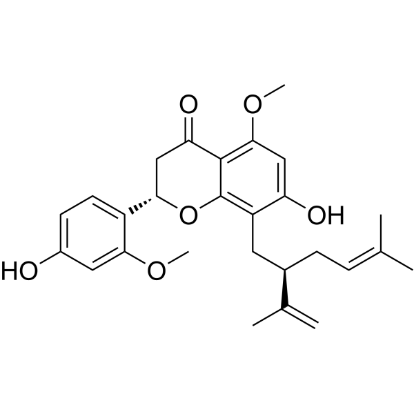
(2S)-2'-Methoxykurarinone

Based on 1 Customer Validation
(2S)-2'-Methoxykurarinone (2'-O-Methylkurarinone) is a flavanone natural product found in the roots of Sophora flavescens, possessing anti-sepsis, inhibitory effects on osteoclast differentiation and bone resorption, anti-inflammatory, and anti-Plasmodium activities. (2S)-2'-Methoxykurarinone exhibits an EC50 = 2.4 μM against the FCR-3 strain of Plasmodium falciparum. In sepsis models, (2S)-2'-Methoxykurarinone modulates immune cell function and inhibits inflammation and oxidative stress. In bone-related models, (2S)-2'-Methoxykurarinone blocks osteoclast differentiation and bone resorption, and inhibits RANKL-mediated Akt, p38, and JNK signaling pathways. In skin inflammation models, (2S)-2'-Methoxykurarinone inhibits NF-κB pathway activation, upregulates HO-1 expression, and suppresses CCL27 chemokine production in HaCaT cells. (2S)-2'-Methoxykurarinone is useful for research on osteoporosis, sepsis, inflammatory skin diseases (such as atopic dermatitis and allergic contact dermatitis), and malaria.
For research use only. We do not sell to patients.
- Purity : 98.86%
- CAS No.: 270249-38-2
- Formula: C27H32O6
- Molecular Weight:452.54
-
Storage:
4°C, protect from light
* In solvent : -80°C, 6 months; -20°C, 1 month (protect from light)

To place orders, for customer services and technical support, please contact: MedChemExpress USA
Tel: 609-228-6898 E-mail: [email protected] [email protected]


-
 Biological Activity
-
Chemical Information
-
Solvent & Solubility
- Protocols
- Purity & Documentation
- References
-
 Help & FAQs


All Parasite Isoforms
More

Biological Activity
Description
|
NF-κB |
HO-1 |
Plasmodium |
JNK |
Akt |
p38 MAP kinase |
Cellular Effect
|
Cell Line

|
Type | Value | Description | References |
|---|---|---|---|---|
| HL-60 | IC50 |
13.7 μM
Compound: 1
|
Cytotoxicity against human HL60 cells after 96 hrs by MTT assay
Cytotoxicity against human HL60 cells after 96 hrs by MTT assay
|
[PMID: 10843587] |
In Vitro
(2S)-2'-Methoxykurarinone (MK) (5-20 μM; 4-7 d) inhibits IL-1-induced osteoclast formation in bone marrow cell-osteoblast co-culture systems; it inhibits IL-1-induced osteoclast formation in calvarial bone organ cultures; and it inhibits RANKL-induced differentiation of bone marrow macrophages into osteoclasts[1].
(2S)-2'-Methoxykurarinone (20 μM; 1 h pre-incubation, followed by 24 h culture with RANKL) inhibits the bone resorption activity of mature osteoclasts[1].
(2S)-2'-Methoxykurarinone (20 μM; 1 h pretreatment, followed by RANKL stimulation for 0-48 h) inhibits the mRNA expression of c-Fos, NFATc1, OSCAR, and TRAP in RANKL-induced bone marrow macrophages[1].
(2S)-2'-Methoxykurarinone (20 μM; 1 h pretreatment, followed by RANKL stimulation for 0-48 h or 0-30 min) inhibits RANKL-induced activation of Akt, p38, and JNK signaling pathways, and suppresses c-Fos and NFATc1 protein expression in bone marrow macrophages[1].
(2S)-2'-Methoxykurarinone (MOK) (5-40 μM; 18-24 h) dose-dependently inhibits the expression of CTACK/CCL27 mRNA and protein in TNF-α- and IL-1β-induced human HaCaT keratinocytes[3].
(2S)-2'-Methoxykurarinone (5-40 μM; 18 h) dose-dependently inhibits the formation of NF-κB-DNA complexes in TNF-α- and IL-1β-induced human HaCaT keratinocytes; it inhibits NF-κB binding activity and nuclear translocation, and the phosphorylation of IκBα; it induces HO-1 mRNA and protein expression[3].
(2S)-2'-Methoxykurarinone (compound 1) (72 h) exhibits moderate antimalarial activity against the Plasmodium falciparum FCR-3 strain, with an EC50 of 2.4 μM[4].
MedChemExpress (MCE) has not independently confirmed the accuracy of these methods. They are for reference only. Further protocols information, click here.
In Vivo
MedChemExpress (MCE) has not independently confirmed the accuracy of these methods. They are for reference only.


Chemical Information
-
CAS No. 270249-38-2
-
Appearance Solid
-
Molecular Weight 452.54
-
Formula C27H32O6
-
Color Light yellow to brown
-
SMILES
O=C1C[C@@H](C2=CC=C(O)C=C2OC)OC3=C(C[C@H](C(C)=C)C/C=C(C)\C)C(O)=CC(OC)=C13
-
Synonyms
2'-O-Methylkurarinone
-
Structure Classification
-
Initial Source
-
Shipping
Room temperature in continental US; may vary elsewhere.
-
Storage
4°C, protect from light
* In solvent : -80°C, 6 months; -20°C, 1 month (protect from light)


Solvent & Solubility
In Vitro:
DMSO : 25 mg/mL (55.24 mM; Need ultrasonic; Hygroscopic DMSO has a significant impact on the solubility of product, please use newly opened DMSO)
Please refer to the solubility information to select the appropriate solvent. Once prepared, please aliquot and store the solution to prevent product inactivation from repeated freeze-thaw cycles.
Storage method and period of stock solution: -80°C, 6 months; -20°C, 1 month (protect from light). When stored at -80°C, please use it within 6 months. When stored at -20°C, please use it within 1 month.
Please refer to the solubility information to select the appropriate solvent. Once prepared, please aliquot and store the solution to prevent product inactivation from repeated freeze-thaw cycles.
Storage method and period of stock solution: -80°C, 6 months; -20°C, 1 month (protect from light). When stored at -80°C, please use it within 6 months. When stored at -20°C, please use it within 1 month.
Concentration (start) × Volume (start) = Concentration (final) × Volume (final)
In Vivo:
Select the appropriate dissolution method based on your experimental animal and administration route.
- For the following dissolution methods, please ensure to first prepare a clear stock solution using an In Vitro approach and then sequentially add co-solvents:
- To ensure reliable experimental results, the clarified stock solution can be appropriately stored based on storage conditions. As for the working solution for In Vivo experiments, it is recommended to prepare freshly and use it on the same day.
- The percentages shown for the solvents indicate their volumetric ratio in the final prepared solution. If precipitation or phase separation occurs during preparation, heat and/or sonication can be used to aid dissolution.
Add each solvent one by one: 10% DMSO 40% PEG300 5% Tween-80 45% Saline
Solubility: ≥ 2.5 mg/mL (5.52 mM); Clear solution
This protocol yields a clear solution of ≥ 2.5 mg/mL (saturation unknown).
Taking 1 mL working solution as an example, add 100 μL DMSO stock solution (25.0 mg/mL) to 400 μL PEG300, and mix evenly; then add 50 μL Tween-80 and mix evenly; then add 450 μL Saline to adjust the volume to 1 mL.
Preparation of Saline: Dissolve 0.9 g sodium chloride in ddH₂O and dilute to 100 mL to obtain a clear Saline solution.
Add each solvent one by one: 10% DMSO 90% (20% SBE-β-CD in Saline)
Solubility: 2.5 mg/mL (5.52 mM); Clear solution; Need ultrasonic
This protocol yields a clear solution of 2.5 mg/mL.
Taking 1 mL working solution as an example, add 100 μL DMSO stock solution (25.0 mg/mL) to 900 μL 20% SBE-β-CD in Saline, and mix evenly.
Preparation of 20% SBE-β-CD in Saline (4°C, storage for one week): 2 g SBE-β-CD powder is dissolved in 10 mL Saline, completely dissolve until clear.
In Vivo Dissolution Calculator
Please enter the basic information of animal experiments:
-
-
-
-
Recommended: Prepare an additional quantity of animals to account for potential losses during experiments.
Please enter your animal formula composition:
-
%DMSO +
Recommended: Keep the proportion of DMSO in working solution below 2% if your animal is weak.
-
%+
-
+%Tween-80 + +
-
%Saline +
The co-solvents required include: DMSO, . All of co-solvents are available by MedChemExpress (MCE). , Tween 80. All of co-solvents are available by MedChemExpress (MCE).
Working solution concentration: 0.22 mg/mL
Method for preparing stock solution: mg drug dissolved in μL DMSO. Stock solution concentration: mg/mL. * In solvent : -80°C, 6 months; -20°C, 1 month (protect from light)
1. Take μL DMSO stock solution;
2. Add μL .
μL , mix evenly;
3. Then add μL Tween 80, mix evenly;
4. Then add μL
Please ensure that the stock solution in the first step is dissolved to a clear state, and add co-solvents in sequence. You can use ultrasonic heating (ultrasonic cleaner, recommended frequency 20-40 kHz), vortexing, etc. to assist dissolution.

Protocols
-
RNA extraction experimental
By lysing cells, releasing RNA, and removing impurities such as proteins and DNA, high-purity RNA products are finally obtained. The commonly used traditional method is the guanidine isothiocyanate/phenol/chloroform method (Trizol), which is suitable for a variety of animal materials including animal tissues, microorganisms, cultured cells, etc., and most plant materials.
-
LPS-Induced Endotoxemia/Systemic Inflammation
Lipopolysaccharide (LPS)-induced endotoxemia is a widely used in vivo model of acute systemic inflammation in which LPS, a Gram-negative bacterial endotoxin, activates innate immune signaling primarily through TLR4, leading to rapid and transient induction of pro-inflammatory cytokines such as TNF-α, IL-6, and IL-1β in circulation and tissues. This cytokine surge is commonly used as a measurable readout of systemic inflammatory activation and immune dysregulation, and is typically assessed within hours after intraperitoneal LPS administration in mouse models of endotoxemia. The model captures key features of systemic inflammatory response syndrome, including cytokine release, immune cell activation, and downstream tissue responses, and has been used to evaluate anti-inflammatory interventions such as cytokine modulation, lipid mediators, and immune cell-targeting therapies.
-
Imiquimod-Induced Psoriasiform Dermatitis
Imiquimod (IMQ)-induced psoriasiform dermatitis is a widely used murine model in which topical application of IMQ, a Toll-like receptor 7 (TLR7) agonist, triggers innate immune activation in the skin and induces a psoriasis-like inflammatory cascade characterized by epidermal hyperplasia, immune cell infiltration, and cytokine production dominated by the IL-23/IL-17 axis. This inflammatory response is mediated through activation of dendritic cells and downstream induction of IL-23, IL-17A, IL-22, and related pro-inflammatory mediators, recapitulating key features of human plaque psoriasis and enabling mechanistic and therapeutic studies. The model is commonly induced using Aldara (5% IMQ cream) applied topically to murine skin, resulting in rapid onset of erythema, scaling, and thickening that can be quantified as disease severity indices and validated histologically.
-
ROS/oxidative-stress fluorescent staining
ROS/oxidative-stress fluorescent staining uses cell-permeant fluorogenic probes that become fluorescent after oxidation inside cells or tissues; commonly used examples include DCFH-DA/DCFDA for broad cellular oxidant detection, DHE for superoxide-related signal detection, MitoSOX for mitochondrial superoxide-related signal detection, and CellROX probes for oxidative-stress-associated fluorescence readouts. The assay detects probe oxidation rather than a single ROS species unless the probe and analysis method have been chemically validated for that species. DCFH-DA enters cells, is deacetylated by intracellular esterases to DCFH, and produces fluorescent DCF after oxidation, so the readout is used as an operational measure of total cellular oxidative stress rather than a species-specific ROS measurement. DHE and MitoSOX can report superoxide-related oxidation, but red fluorescence alone can include non-specific ethidium-like oxidation products; HPLC or optimized spectral approaches are
-
TPA/Croton Oil Ear Edema and Dermatitis
The TPA (12-O-tetradecanoylphorbol-13-acetate) and croton oil-induced mouse ear edema model is a well-established acute cutaneous inflammation system used to evaluate topical anti-inflammatory activity by measuring edema formation, neutrophil infiltration, vascular permeability, and cytokine-mediated skin responses in vivo. The inflammatory response is triggered by topical application of phorbol esters (TPA) or croton oil constituents, leading to rapid activation of protein kinase C signaling, leukocyte recruitment, and increased vascular permeability, which can be quantified by ear thickness, weight, dye extravasation, and biochemical markers such as myeloperoxidase (MPO) activity and pro-inflammatory mediators in ear tissue homogenates. This model is widely used for screening anti-inflammatory agents, where reductions in edema and inflammatory biomarkers reflect suppression of acute dermal inflammation and immune cell infiltration. Histological evaluation typically confirms epidermal
-
Contact Hypersensitivity Dermatitis
Contact hypersensitivity (CHS) dermatitis is a T cell-mediated delayed-type (Type IV) immune reaction in which low-molecular-weight haptens applied to the skin bind host proteins to form complete antigens, triggering sensitization followed by a secondary inflammatory response upon re-exposure (elicitation phase), which is commonly quantified by ear swelling as a readout of skin inflammation in murine models. This model is widely used to study allergic contact dermatitis because it is antigen-specific, reproducible, and reflects key immunological events including dendritic cell activation, T cell priming in draining lymph nodes, and effector T cell-driven tissue inflammation. DNFB- and oxazolone-induced CHS models are standard systems for evaluating both acute and chronic T cell-dependent skin inflammation and for testing immunomodulatory interventions.
-
Research Protocol for Inflammation-related Diseases
The NLRP3 inflammasome is a cytosolic innate immune signaling platform that integrates priming signals and danger-signal activation to promote caspase-1 activation, maturation of IL-1β and IL-18, and gasdermin D-mediated pyroptotic cell death. The core experimental logic is to determine whether inflammatory disease phenotypes are driven by increased NLRP3 expression, ASC-containing inflammasome assembly, caspase-1 cleavage, GSDMD cleavage, and extracellular release of IL-1β/IL-18 rather than by nonspecific cell injury alone. The pathway is strongly linked to inflammation-related disease phenotypes because monosodium urate crystals activate NALP3/NLRP3 inflammasome signaling in gout-like crystal inflammation, cholesterol crystals activate NLRP3 inflammasomes in atherogenesis models, and DSS-induced intestinal inflammation has been reported to involve NLRP3 inflammasome activity. However, experimental colitis studies also show context-dependent protective effects of NLRP3 inflammasome co
-
Osteoclast differentiation from monocyte/macrophage precursors
Osteoclast differentiation is an in vitro induction assay in which monocyte/macrophage-lineage precursors are exposed to macrophage colony-stimulating factor (M-CSF) and receptor activator of NF-κB ligand (RANKL), generating multinucleated osteoclasts that are commonly identified by tartrate-resistant acid phosphatase (TRAP) staining and functionally confirmed by resorption pits on dentin, bone, or mineralized substrates. M-CSF supports survival and expansion of osteoclast precursors, while RANKL binding to RANK drives osteoclast commitment, fusion, maturation, and resorptive function; osteoprotegerin inhibits this pathway by binding RANKL and preventing RANK activation. The main readouts are the number of TRAP-positive multinucleated cells, formation of F-actin rings, and resorbed surface area; TRAP-positive multinucleated cells indicate osteoclast differentiation, whereas pit formation on dentin, bone, or mineralized coating indicates functional bone-resorbing activity.


Purity & Documentation
-
Data Sheet (290 KB)
-
SDS (252 KB)
- English - EN (252 KB)
- Français - FR (252 KB)
- Deutsch - DE (252 KB)
- Norwegian - NO (252 KB)
- Español - ES (252 KB)
- Swedish - SV (252 KB)
- Italian - IT (252 KB)
- Korean - KR (252 KB)
- Portuguese - PT (252 KB)
-
Handling Instructions (2659 KB)
References


Complete Stock Solution Preparation Table
Please refer to the solubility information to select the appropriate solvent. Once prepared, please aliquot and store the solution to prevent product inactivation from repeated freeze-thaw cycles.
Storage method and period of stock solution: -80°C, 6 months; -20°C, 1 month (protect from light). When stored at -80°C, please use it within 6 months. When stored at -20°C, please use it within 1 month.
| Optional Solvent | Concentration Solvent Mass | 1 mg | 5 mg | 10 mg | 25 mg |
|---|---|---|---|---|---|
| DMSO | 1 mM | 2.2097 mL | 11.0487 mL | 22.0975 mL | 55.2437 mL |
| 5 mM | 0.4419 mL | 2.2097 mL | 4.4195 mL | 11.0487 mL | |
| 10 mM | 0.2210 mL | 1.1049 mL | 2.2097 mL | 5.5244 mL | |
| 15 mM | 0.1473 mL | 0.7366 mL | 1.4732 mL | 3.6829 mL | |
| 20 mM | 0.1105 mL | 0.5524 mL | 1.1049 mL | 2.7622 mL | |
| 25 mM | 0.0884 mL | 0.4419 mL | 0.8839 mL | 2.2097 mL | |
| 30 mM | 0.0737 mL | 0.3683 mL | 0.7366 mL | 1.8415 mL | |
| 40 mM | 0.0552 mL | 0.2762 mL | 0.5524 mL | 1.3811 mL | |
| 50 mM | 0.0442 mL | 0.2210 mL | 0.4419 mL | 1.1049 mL |


(2S)-2'-Methoxykurarinone Related Classifications
HY-N1746 Related Classifications
 Powered by Bioz