



Borrelidin
Based on 1 publication(s) in Google Scholar
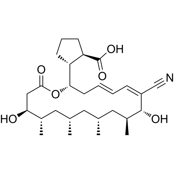
Borrelidin (Treponemycin) is a bacterial and eukaryal threonyl-tRNA synthetase inhibitor which is a nitrile-containing macrolide antibiotic isolated from Streptomyces rochei. Borrelidin is an inhibitor of Cdc28/Cln2 of the budding yeast, with an IC50 of 24 μM. Borrelidin is a potent angiogenesis inhibitor, with an IC50 of 0.8 nM. Borrelidin induces apoptosis in the tube-forming cells. Borrelidin has strong antimalarial activities, with IC50s of 1.9 nM and 1.8 nM against K1 and FCR3 strains of Plasmodium falciparum, respectively.
For research use only. We do not sell to patients.
- Purity : 99.71%
- CAS No.: 7184-60-3
- Formula: C28H43NO6
- Molecular Weight:489.64
-
Storage:
-20°C, sealed storage, away from moisture
* In solvent : -80°C, 6 months; -20°C, 1 month (sealed storage, away from moisture)

To place orders, for customer services and technical support, please contact: MedChemExpress USA
Tel: 609-228-6898 E-mail: [email protected] [email protected]


-
 Biological Activity
-
Chemical Information
-
Solvent & Solubility
- Protocols
- Purity & Documentation
- References
-
 Help & FAQs


-
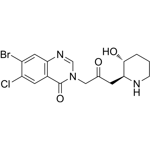
HY-N1584
HalofuginoneNatural Product -
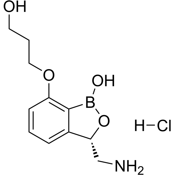
HY-12479A
Epetraborole hydrochlorideLeuRS Inhibitor -
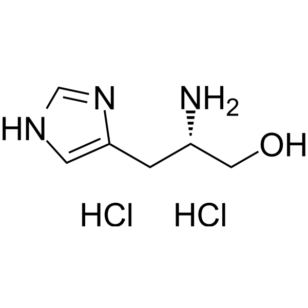
HY-W014233
L-Histidinol dihydrochlorideHistidyl-tRNA Synthetase Inhibitor -
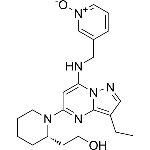
HY-10492
DinaciclibCDK2/5/9 Inhibitor -
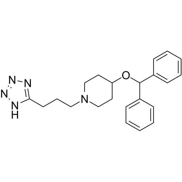
HY-108259
HQL-79Prostaglandin D Synthase Inhibitor -
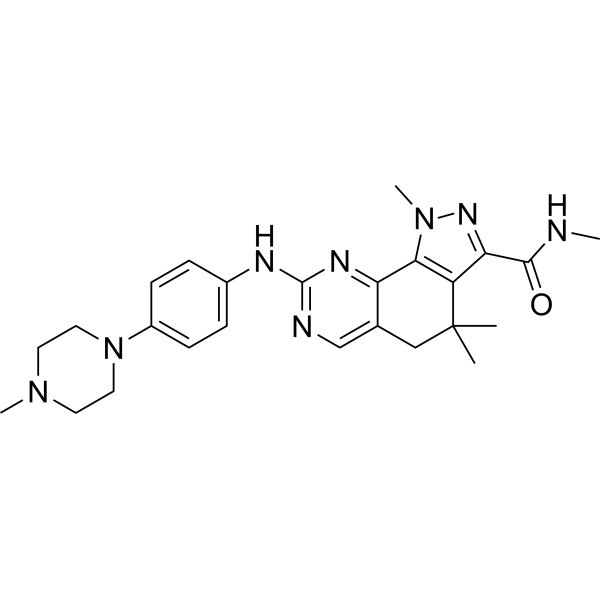
HY-10424
MilciclibCDK/TRK Inhibitor -
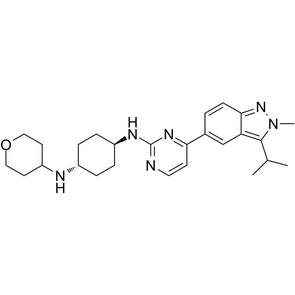
HY-12293
LY2857785CDK9/8/7 Inhibitor -
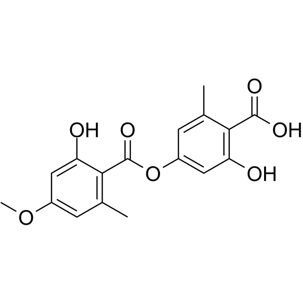
HY-121362
Evernic AcidTrxR1 Inhibitor -
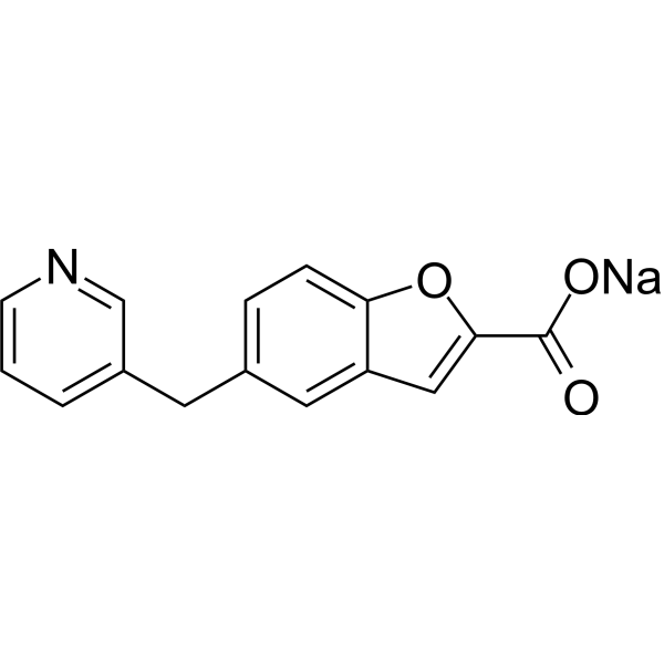
HY-106080A
Furegrelate sodiumThromboxane Synthase Inhibitor -

HY-108259R
HQL-79 (Standard)Reference Standard -

HY-106080AR
Furegrelate sodium (Standard)Thromboxane Synthase Inhibitor -

HY-10492R
Dinaciclib (Standard)Reference Standard
Publications Citing Use of MedChemExpress (MCE) Borrelidin
MoreAll Parasite Isoforms
MoreAll Antibiotic Isoforms
More

Biological Activity
Description
IC50 & Target
|
Plasmodium |
Macrolide |
Cellular Effect
|
Cell Line

|
Type | Value | Description | References |
|---|---|---|---|---|
| HEK-293T | IC50 |
13.97 μg/mL
Compound: Borrelidin
|
Cytotoxicity against HEK293T cells assessed as growth inhibition after 48 hrs by MTT assay
Cytotoxicity against HEK293T cells assessed as growth inhibition after 48 hrs by MTT assay
|
[PMID: 30442507] |
| HT-29 | IC50 |
0.92 μM
Compound: 1
|
Growth inhibition of human HT-29 cells
Growth inhibition of human HT-29 cells
|
[PMID: 25393949] |
| HUVEC | IC50 |
0.5 nM
Compound: 1, Borrelidin
|
Growth inhibition in HUVEC
Growth inhibition in HUVEC
|
[PMID: 16962775] |
| MDA-MB-361 | IC50 |
0.42 μM
Compound: 1
|
Growth inhibition of human MDA-MB-361 cells
Growth inhibition of human MDA-MB-361 cells
|
[PMID: 25393949] |
| NCI-N87 | IC50 |
0.05 μM
Compound: 1
|
Growth inhibition of human NCI-N87 cells
Growth inhibition of human NCI-N87 cells
|
[PMID: 25393949] |
In Vitro


Chemical Information
-
CAS No. 7184-60-3
-
Appearance Solid
-
Molecular Weight 489.64
-
Formula C28H43NO6
-
Color White to off-white
-
SMILES
O=C([C@H]1[C@H]([C@H](C/C=C/C=C(C#N)\[C@H](O)[C@@H](C)C[C@H](C)C[C@H](C)C[C@H](C)[C@@H](O)C2)OC2=O)CCC1)O
-
Synonyms
Treponemycin
-
Structure Classification
-
Initial Source
Streptomyces rochei
-
Shipping
Room temperature in continental US; may vary elsewhere.
-
Storage
-20°C, sealed storage, away from moisture
* In solvent : -80°C, 6 months; -20°C, 1 month (sealed storage, away from moisture)


Publications (1)
-
Journal Impact Factor
-
Most Recent
-
Cell Metab
2023 Dec 5;35(12):2216-2230.e8. PMID: 37979583


Solvent & Solubility
In Vitro:
Ethanol : ≥ 100 mg/mL (204.23 mM)
* "≥" means soluble, but saturation unknown.
Please refer to the solubility information to select the appropriate solvent. Once prepared, please aliquot and store the solution to prevent product inactivation from repeated freeze-thaw cycles.
Storage method and period of stock solution: -80°C, 6 months; -20°C, 1 month (sealed storage, away from moisture). When stored at -80°C, please use it within 6 months. When stored at -20°C, please use it within 1 month.
Please refer to the solubility information to select the appropriate solvent. Once prepared, please aliquot and store the solution to prevent product inactivation from repeated freeze-thaw cycles.
Storage method and period of stock solution: -80°C, 6 months; -20°C, 1 month (sealed storage, away from moisture). When stored at -80°C, please use it within 6 months. When stored at -20°C, please use it within 1 month.
Concentration (start) × Volume (start) = Concentration (final) × Volume (final)
In Vivo:
Select the appropriate dissolution method based on your experimental animal and administration route.
- For the following dissolution methods, please ensure to first prepare a clear stock solution using an In Vitro approach and then sequentially add co-solvents:
- To ensure reliable experimental results, the clarified stock solution can be appropriately stored based on storage conditions. As for the working solution for In Vivo experiments, it is recommended to prepare freshly and use it on the same day.
- The percentages shown for the solvents indicate their volumetric ratio in the final prepared solution. If precipitation or phase separation occurs during preparation, heat and/or sonication can be used to aid dissolution.
Add each solvent one by one: 10% EtOH 40% PEG300 5% Tween-80 45% Saline
Solubility: ≥ 2.5 mg/mL (5.11 mM); Clear solution
This protocol yields a clear solution of ≥ 2.5 mg/mL (saturation unknown).
Taking 1 mL working solution as an example, add 100 μL EtOH stock solution (25.0 mg/mL) to 400 μL PEG300, and mix evenly; then add 50 μL Tween-80 and mix evenly; then add 450 μL Saline to adjust the volume to 1 mL.
Preparation of Saline: Dissolve 0.9 g sodium chloride in ddH₂O and dilute to 100 mL to obtain a clear Saline solution.
Add each solvent one by one: 10% EtOH 90% Corn Oil
Solubility: ≥ 2.5 mg/mL (5.11 mM); Clear solution
This protocol yields a clear solution of ≥ 2.5 mg/mL (saturation unknown). If the continuous dosing period exceeds half a month, please choose this protocol carefully.
Taking 1 mL working solution as an example, add 100 μL EtOH stock solution (25.0 mg/mL) to 900 μL Corn oil, and mix evenly.
In Vivo Dissolution Calculator
Please enter the basic information of animal experiments:
-
-
-
-
Recommended: Prepare an additional quantity of animals to account for potential losses during experiments.
Working solution concentration: 0.22 mg/mL

Protocols
-
Human pluripotent stem cell endothelial-cell differentiation
Human pluripotent stem cell endothelial differentiation is based on stepwise developmental patterning: early activation of WNT/GSK3β inhibition promotes mesodermal or vascular progenitor entry, followed by endothelial specification using VEGF-related signaling, BMP4, FGF2, Notch modulation, or cAMP depending on the published protocol. Endothelial differentiation is read out by acquisition of CD31, CD34, VE-cadherin/CD144, KDR/VEGFR2, vWF, Tie2, NOS3, acetylated LDL uptake, tube/network formation, barrier function, and in vivo vessel-forming capacity where tested.
-
Apoptosis
Apoptosis, also called programmed cell death, is generally characterized by distinct morphological characteristics.
-
TUNEL staining for apoptotic DNA fragmentation
TUNEL staining detects DNA strand breaks by using terminal deoxynucleotidyl transferase to add labeled nucleotides to exposed 3′-OH DNA termini, generating either microscopic staining in fixed cells or tissue sections, or fluorescence/cytometric signal in cell suspensions. TUNEL positivity reflects DNA fragmentation but should not be interpreted alone as definitive apoptosis, because TUNEL can also label necrotic, autolytic, mechanically damaged, or DNA-repair-associated DNA breaks.
-
Cell Cytotoxicity Assay
Cytotoxicity assays are usually based on the assessment of cell membrane damage, which can also be indirectly detected by measuring cell viability. Detection methods include MTT assay, CKK-8 assay, LDH assay and ATP assay, etc.
-
Vascular/Branching Fractal Analysis
Vascular/branching fractal analysis quantifies the geometric complexity of vessel trees or vascular networks from segmented 2D images, commonly by converting vessels into binary and/or skeletonized maps and estimating fractal dimension using box-counting or related approaches. Fractal dimension is interpreted as an image-derived readout of vascular branching complexity, space filling, or density, and has been applied to retinal photographs, fluorescein angiography, OCT angiography, capillary perfusion maps, and in vitro Matrigel angiogenesis networks. The assay readout is generated from vessel-positive pixels after image preprocessing, vessel segmentation, binarization, and optional skeletonization; reported outputs include fractal dimension, vessel density, branchpoint density, endpoint density, vessel length density, tortuosity, and generation-based branching metrics when VESGEN-style analysis is used. The biological interpretation is limited to quantitative vascular patterning and s
-
Annexin V plus membrane-impermeant dye apoptosis staining
Annexin V-based apoptosis assays rely on the detection of phosphatidylserine (PS) externalization from the inner leaflet of the plasma membrane to the outer leaflet, an early biochemical hallmark of apoptosis. Fluorescently labeled Annexin V binds PS in a calcium-dependent manner, enabling identification of early apoptotic cells by flow cytometry or fluorescence microscopy. When combined with a membrane-impermeant DNA-binding dye (e. g. , propidium iodide), this approach allows discrimination between viable (Annexin V−/dye−), early apoptotic (Annexin V+/dye−), and late apoptotic or necrotic (Annexin V+/dye+) cell populations by assessing membrane integrity and PS exposure.
-
Endothelial Tube Formation Assay
Endothelial tube formation assay evaluates the ability of endothelial cells to attach, migrate, align, and organize into capillary-like networks when cultured on gelled basement membrane extract or Matrigel; the readout is the morphology and quantity of tube-like networks, which reflects an in vitro endothelial morphogenesis step related to angiogenesis. Basement membrane extract/Matrigel provides laminin-rich extracellular matrix cues that support endothelial differentiation into capillary-like structures, but it can contain biologically active growth factors, so growth-factor-reduced matrix is preferred when testing defined angiogenic stimulators or inhibitors.
-
Apoptosis Solutions
Apoptosis is a regulated, generally non-lytic cell-death pathway that removes unwanted, damaged, infected, or abnormal cells through coordinated morphological changes, caspase activation, DNA fragmentation, and membrane remodeling. The intrinsic apoptosis pathway is controlled mainly by mitochondrial outer membrane permeabilization, BCL-2 family proteins, cytochrome c release, apoptosome formation, caspase-9 activation, and downstream executioner caspase-3/7 activation. The extrinsic apoptosis pathway is initiated by death receptors such as Fas, TNFR, and TRAIL receptors, which recruit adaptor proteins and activate caspase-8 before engaging executioner caspases or mitochondrial amplification through BID cleavage. Apoptosis is linked to many phenotypes, including cancer cell killing, tissue homeostasis, immune regulation, neurodegeneration, infection response, and treatment-induced cytotoxicity; unresolved questions include how apoptosis interacts with necroptosis, pyroptosis, ferroptos
-
Endothelial Cell Migration/Angiogenic Sprouting Assay
Endothelial cell migration and angiogenic sprouting assays are in vitro (and partially ex vivo-adapted) functional models that quantify the ability of endothelial cells to undergo coordinated migration, extracellular matrix invasion, and multicellular organization into capillary-like sprouts in response to pro-angiogenic stimuli such as VEGF, bFGF, or conditioned microenvironments. These assays are used to model early angiogenic events including tip-cell formation, directional migration, and lumen-like sprout extension, which collectively reflect angiogenic activation and vascular morphogenesis processes observed in vivo.


Purity & Documentation
-
Data Sheet (278 KB)
-
SDS (396 KB)
- English - EN (396 KB)
- Français - FR (396 KB)
- Deutsch - DE (396 KB)
- Norwegian - NO (396 KB)
- Español - ES (396 KB)
- Swedish - SV (396 KB)
- Italian - IT (396 KB)
- Korean - KR (396 KB)
- Portuguese - PT (396 KB)
-
Handling Instructions (2659 KB)
References
[1]. Habibi D, et al. Borrelidin, a small molecule nitrile-containing macrolide inhibitor of threonyl-tRNA synthetase, is a potent inducer of apoptosis in acute lymphoblastic leukemia. Invest New Drugs. 2012 Aug;30(4):1361-70. [Content Brief]
[2]. Tsuchiya E, et al. Borrelidin inhibits a cyclin-dependent kinase (CDK), Cdc28/Cln2, of Saccharomyces cerevisiae. J Antibiot (Tokyo). 2001 Jan;54(1):84-90. [Content Brief]
[3]. Wakabayashi T, et al. Borrelidin is an angiogenesis inhibitor; disruption of angiogenic capillary vessels in a rat aorta matrix culture model. J Antibiot (Tokyo). 1997 Aug;50(8):671-6. [Content Brief]
[4]. Otoguro K, et al. In vitro and in vivo antimalarial activities of a non-glycosidic 18-membered macrolide antibiotic, borrelidin, against drug-resistant strains of Plasmodia. J Antibiot (Tokyo). 2003 Aug;56(8):727-9. [Content Brief]


Complete Stock Solution Preparation Table
Please refer to the solubility information to select the appropriate solvent. Once prepared, please aliquot and store the solution to prevent product inactivation from repeated freeze-thaw cycles.
Storage method and period of stock solution: -80°C, 6 months; -20°C, 1 month (sealed storage, away from moisture). When stored at -80°C, please use it within 6 months. When stored at -20°C, please use it within 1 month.
| Optional Solvent | Concentration Solvent Mass | 1 mg | 5 mg | 10 mg | 25 mg |
|---|---|---|---|---|---|
| Ethanol | 1 mM | 2.0423 mL | 10.2116 mL | 20.4232 mL | 51.0579 mL |
| 5 mM | 0.4085 mL | 2.0423 mL | 4.0846 mL | 10.2116 mL | |
| 10 mM | 0.2042 mL | 1.0212 mL | 2.0423 mL | 5.1058 mL | |
| 15 mM | 0.1362 mL | 0.6808 mL | 1.3615 mL | 3.4039 mL | |
| 20 mM | 0.1021 mL | 0.5106 mL | 1.0212 mL | 2.5529 mL | |
| 25 mM | 0.0817 mL | 0.4085 mL | 0.8169 mL | 2.0423 mL | |
| 30 mM | 0.0681 mL | 0.3404 mL | 0.6808 mL | 1.7019 mL | |
| 40 mM | 0.0511 mL | 0.2553 mL | 0.5106 mL | 1.2764 mL | |
| 50 mM | 0.0408 mL | 0.2042 mL | 0.4085 mL | 1.0212 mL | |
| 60 mM | 0.0340 mL | 0.1702 mL | 0.3404 mL | 0.8510 mL | |
| 80 mM | 0.0255 mL | 0.1276 mL | 0.2553 mL | 0.6382 mL | |
| 100 mM | 0.0204 mL | 0.1021 mL | 0.2042 mL | 0.5106 mL |


Borrelidin Related Classifications
HY-N6742 Related Classifications
 Powered by Bioz