



Lazertinib
Based on 4 publication(s) in Google Scholar
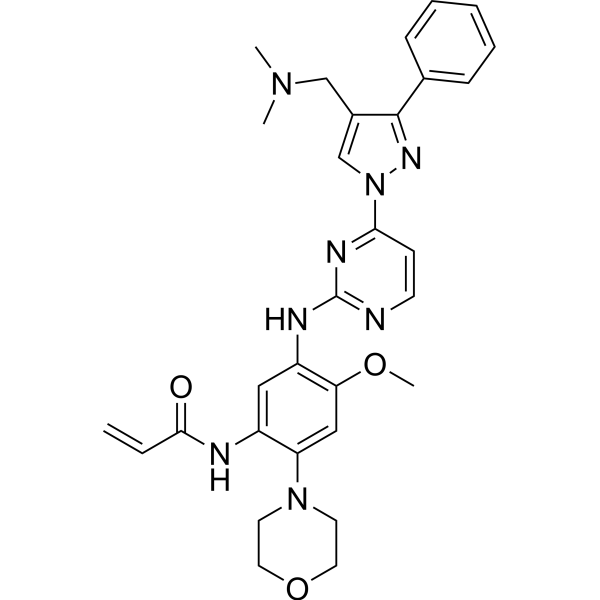
Lazertinib (YH25448; GNS-1480) is an orally active, blood-brain barrier permeable third-generation EGFR tyrosine kinase inhibitor, as well as an ABCB1/ABCG2 inhibitor and a TRPA1 activator. Lazertinib exhibits IC50 values of 0.4 mM and 0.2 mM against human ABCB1 and ABCG2, respectively. By inhibiting mutant EGFR signaling, EGFR phosphorylation and the downstream ERK/AKT pathway, as well as upregulating surface expression of EGFR/MET, Lazertinib induces cell cycle arrest, apoptosis, spontaneous calcium responses, hyperexcitability of dorsal root ganglion (DRG) neurons, and TRPA1-dependent pain-like behaviors. Lazertinib competitively binds to the substrate-binding sites of ABCB1/ABCG2, stimulates their ATPase activity without altering their expression or plasma membrane localization, thereby enhancing ADCC activity, acting as a chemosensitizer, and reversing ABCB1-mediated multidrug resistance. It exerts antitumor activity as a single agent or in combination with other drugs. Lazertinib is applicable to research related to non-small cell lung cancer, multidrug-resistant cancers, and paresthesia.
For research use only. We do not sell to patients.
- Purity : 99.87%
- CAS No.: 1903008-80-9
- Formula: C30H34N8O3
- Molecular Weight:554.64
-
Storage:Powder -20°C, 3 years , 4°C, 2 years ; In solvent -80°C, 2 years , -20°C, 1 year

To place orders, for customer services and technical support, please contact: MedChemExpress USA
Tel: 609-228-6898 E-mail: [email protected] [email protected]


-
 Biological Activity
-
Chemical Information
-
Solvent & Solubility
- Protocols
- Purity & Documentation
- References
-
 Help & FAQs


-
HY-15772
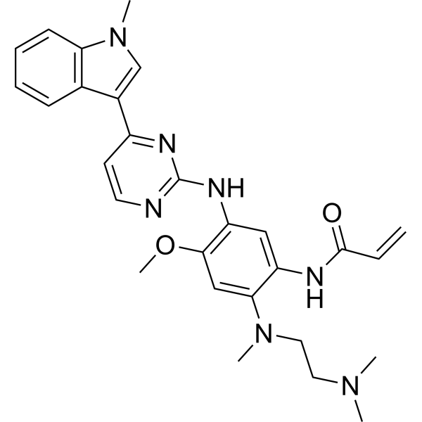
OsimertinibMutant-Selective EGFR Inhibitor -
HY-114277
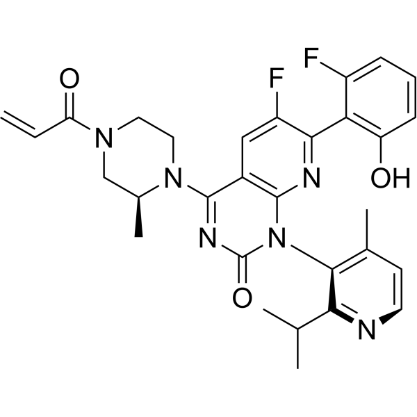
SotorasibKRAS G12C Inhibitor -

HY-P9977
AmivantamabEGFR-MET Dual Antibody -
HY-130149
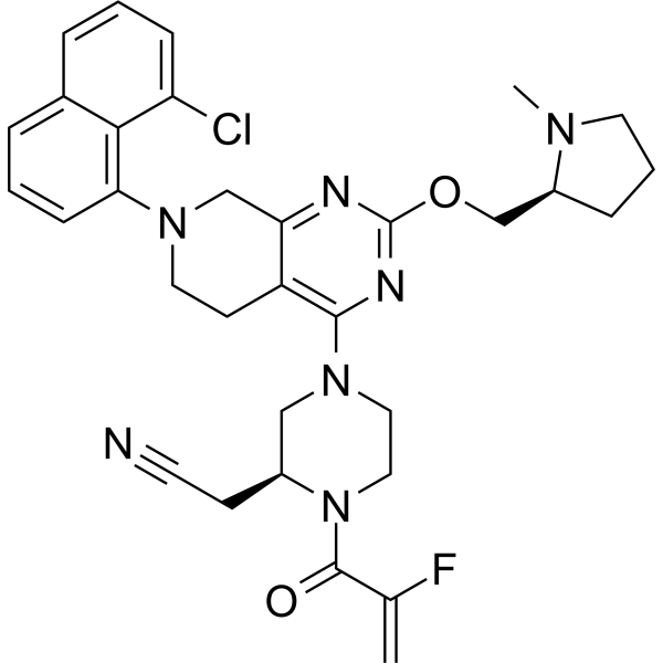
AdagrasibKRAS G12C Inhibitor -
HY-12215
LorlatinibALK/ROS1 Inhibitor -
HY-16069
TucatinibHER2 Inhibitor -
HY-10820
Pemetrexed -
HY-50898
LapatinibEGFR/HER2 Inhibitor -
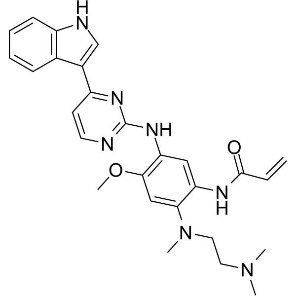
HY-B0793
AZ-5104EGFR Inhibitor -
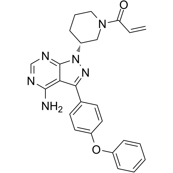
HY-10997
IbrutinibBtk Inhibitor -
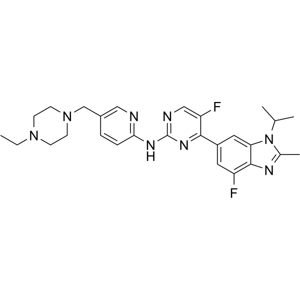
HY-16297A
AbemaciclibCDK4/6 Inhibitor -

HY-15141
StaurosporinePKC/PKA Inhibitor
Publications Citing Use of MedChemExpress (MCE) Lazertinib
MoreAll EGFR Isoforms
More

Biological Activity
Description
IC50 & Target
[1]|
EGFRT790M |
TRPA1 |
Cellular Effect
|
Cell Line

|
Type | Value | Description | References |
|---|---|---|---|---|
| A-431 | IC50 |
3.6 μM
Compound: 5; YH25448
|
Antiproliferative activity against human A-431 cells harboring wild type EGFR assessed as inhibition of cell growth by MTT assay
Antiproliferative activity against human A-431 cells harboring wild type EGFR assessed as inhibition of cell growth by MTT assay
|
[PMID: 31869655] |
| BaF3 | EC50 |
>100 nM
Compound: YH25448
|
Antiproliferative activity against mouse BAF3 cells harboring HER2 wild type after 72 hrs by resazurin dye-based fluorescence assay
Antiproliferative activity against mouse BAF3 cells harboring HER2 wild type after 72 hrs by resazurin dye-based fluorescence assay
|
[PMID: 36518696] |
| BaF3 | EC50 |
4.8 nM
Compound: YH25448
|
Antiproliferative activity against mouse BAF3 cells harboring EGFR L858R mutant after 72 hrs by resazurin dye-based fluorescence assay
Antiproliferative activity against mouse BAF3 cells harboring EGFR L858R mutant after 72 hrs by resazurin dye-based fluorescence assay
|
[PMID: 36518696] |
| BaF3 | EC50 |
7.4 nM
Compound: YH25448
|
Antiproliferative activity against mouse BAF3 cells harboring EGFR L858R/T790M mutant after 72 hrs by resazurin dye-based fluorescence assay
Antiproliferative activity against mouse BAF3 cells harboring EGFR L858R/T790M mutant after 72 hrs by resazurin dye-based fluorescence assay
|
[PMID: 36518696] |
| NCI-H1975 | IC50 |
<1 μM
Compound: 5; YH25448
|
Antiproliferative activity against human NCI-H1975 cells expressing EGFR L858R/T790M mutant assessed as inhibition of cell growth by MTT assay
Antiproliferative activity against human NCI-H1975 cells expressing EGFR L858R/T790M mutant assessed as inhibition of cell growth by MTT assay
|
[PMID: 31869655] |
| U-87MG ATCC | IC50 |
33.2 μM
Compound: 5; YH25448
|
Antiproliferative activity against human U-87MG cells harboring wild type EGFR assessed as inhibition of cell growth by MTT assay
Antiproliferative activity against human U-87MG cells harboring wild type EGFR assessed as inhibition of cell growth by MTT assay
|
[PMID: 31869655] |
In Vitro
Lazertinib (0.1-1000 nM; 3 d) inhibits the viability of Ba/F3 cells expressing various rare EGFR mutations, with IC50 values ranging from 3.5 nM to 108.3 nM[1].
Lazertinib (10 nM (YUO-139 PDOs); 100 nM (YU-1092 PDCs); 72 h) upregulates EGFR expression on the surface of EGFRG719S-mutant YUO-139 patient-derived organoids and EGFRL861Q-mutant YU-1092 patient-derived cells after 72 h of treatment[1].
Lazertinib (0.001-10 μM; 72 h) inhibits the viability of EGFR-mutant non-small cell lung cancer (NSCLC) cell lines after 72 h of incubation[2].
Lazertinib (0.01-3.0 μM; 7 d, medium changed every 72 h) inhibits the long-term proliferation of PC-9 EGFR-mutant NSCLC cells in a concentration-dependent manner within 7 days, but fails to achieve complete growth inhibition at the tested concentrations[2].
Lazertinib (100 nM; 4 h; 72 h) inhibits the phosphorylation of EGFR, ERK and AKT in PC-9 and HCC4011 EGFR-mutant NSCLC cells, but its inhibitory effects on ERK and AKT disappear by 72 h[2].
Lazertinib (100 nM; 48 h) upregulates the expression of the anti-apoptotic protein MCL-1 in PC-9, HCC4011 and H1975 EGFR-mutant non-small cell lung cancer cells[2].
Lazertinib (100 μM; 72 h) exhibits equivalent cytotoxicity in drug-sensitive parental cancer cell lines (HepG2, KB, S1, HEK293/Vector) and their multidrug-resistant (MDR) sublines with overexpressed ABCB1 or ABCG2. It reduces the IC50 values of Vincristine (HY-N0488A), Doxorubicin (HY-15142A), Mitoxantrone (HY-13502), and Topotecan (HY-13768), respectively, but shows no significant effect on Cisplatin (HY-17394)[3].
Lazertinib (0.1-1 μM; 24 h) does not induce neurite fragmentation in primary cultured dorsal root ganglion (DRG) neurons from the L3, L4 and L5 segments of adult mice[4].
MedChemExpress (MCE) has not independently confirmed the accuracy of these methods. They are for reference only.
-
Cell Line:Ba/F3 cells expressing EGFR G719S, S768I, G719A/S768I, L861Q, G719S/S768I, and S768I/L858R uncommon mutations
-
Concentration:0.1-1000 nM
-
Incubation Time:3 d
-
Result:Inhibited cell viability with an IC50 of 3.5 nM in EGFR G719S Ba/F3 cells. Inhibited cell viability with an IC50 of 108.3 nM in EGFR S768I Ba/F3 cells. Inhibited cell viability with an IC50 of 36.8 nM in EGFR G719A/S768I Ba/F3 cells. Inhibited cell viability with an IC50 of 13.9 nM in EGFR L861Q Ba/F3 cells. Inhibited cell viability with an IC50 of 21.9 nM in EGFR G719S/S768I Ba/F3 cells. Inhibited cell viability with an IC50 of 7.2 nM in EGFR S768I/L858R Ba/F3 cells.
-
Cell Line:PC-9, HCC4011, H1975, PC-9GXR, KPP-03, HCC827, HCC4006 (EGFR-mutant non-small cell lung cancer cell lines)
-
Concentration:0.001-10 μM
-
Incubation Time:72 h
-
Result:Inhibited cell viability in all seven cell lines, with IC50 values: PC-9: 0.103 μM; HCC4011: 0.161 μM; H1975: 4.074 μM; PC-9GXR: 1.379 μM; KPP-03: >10.0 μM; HCC827: <0.001 μM; HCC4006: 0.002 μM. Showed highest sensitivity in HCC827 and HCC4006, and lowest sensitivity in KPP-03.
-
Cell Line:ABCB1-overexpressing MDR cells (KBv200, HepG2/adr, HEK293/ABCB1), ABCG2-overexpressing MDR cells (S1-MI-80, HEK293/ABCG2), and their parental sensitive cells
-
Concentration:0.0625-0.25 μM
-
Incubation Time:3 d
-
Result:Reduced the IC50 of vincristine (HY-N0488A) by up to 25.21-fold, doxorubicin (HY-15142A) by up to 14.82-fold, and paclitaxel by up to 15.90-fold in ABCB1-overexpressing cells. Reduced the IC50 of mitoxantrone (HY-13502) by up to 8.97-fold and topotecan (HY-13768) by up to 11.94-fold in ABCG2-overexpressing cells. Did not significantly alter the IC50 values of substrate drugs in parental sensitive cells, or the IC50 of non-substrate cisplatin (HY-17394) in either sensitive or MDR cells.
In Vivo
Mesylate of Lazertinib (10 mg/kg; p.o.; once daily; for 19 consecutive days) fails to inhibit tumor growth in the gefitinib (HY-50895)-resistant non-small cell lung cancer EGFRL861Q YU-1092 xenograft model[1].
Mesylate of Lazertinib (10 mg/kg; p.o.; once daily; administered until day 29) induces initial tumor regression but fails to achieve durable control in the EGFR-TKI-resistant non-small cell lung cancer YHIM-1008 EGFRG719C/S768I xenograft model[1].
Mesylate of Lazertinib (3 mg/kg; daily; for 31 consecutive days) exerts transient anti-tumor activity in PC-9 CDX tumors, with the tumors exhibiting regrowth within 3 weeks after the initiation of treatment[2].
Mesylate of Lazertinib (10 mg/kg; p.o.; once every 3 days; 6 doses total) fails to inhibit the growth of ABCB1-overexpressing HepG2/adr xenografts. However, when combined with Doxorubicin (HY-15142A), it effectively reverses ABCB1-mediated multidrug resistance, significantly reduces tumor volume and weight, with no observed toxicity[3].
Mesylate of Lazertinib (1 mM, 20 μL; plantar injection; single administration) induces TRPA1-dependent pain-like behavior in male C57BL/6J mice, with an average licking and biting duration of 58.91 seconds within 15 minutes[4].
MedChemExpress (MCE) has not independently confirmed the accuracy of these methods. They are for reference only.
-
Animal Model:nu/nu mice (6-week-old female)[1]
-
Dosage:10 mg/kg
-
Administration:p.o.; daily; 29 days
-
Result:Induced a 141% tumor growth inhibition (TGI). Showed less durable responses than the lazertinib-amivantamab combination, with tumor regrowth observed in a subset of treated mice after treatment discontinuation.
-
Animal Model:Athymic nude mice (4-6 weeks old, 16-20 g, subcutaneous inoculation with ABCB1-overexpressing HepG2/adr cells)[3]
-
Dosage:10 mg/kg (monotherapy); 10 mg/kg (combination with doxorubicin)
-
Administration:p.o.; q3d; 6 doses
-
Result:Did not reduce tumor volume or weight relative to saline control (monotherapy). Produced a remarkable decrease in tumor volume and tumor weight relative to saline group and doxorubicin-alone group (combination with doxorubicin). Showed no obvious changes in mouse body weight, indicating good tolerability (monotherapy and combination groups).
-
Animal Model:C57BL/6J mice (5- to 6-week-old, male)[4]
-
Dosage:1 mM, 20 μL
-
Administration:intraplantar; single dose
-
Result:Increased average licking/biting time to 58.91 seconds over 15 minutes, compared to 6.832 seconds in vehicle-treated mice. Reduced total licking/biting time to 46.11 seconds when preinjected with TRPA1 antagonist HC-030031, compared to 105.6 seconds in vehicle-preinjected group.
- Western Blot
- RNA extraction experimental
- Annexin V plus membrane-impermeant dye apoptosis staining
- BrdU Incorporation Assay
- Protocol for Cell Cycle
- Primary Dorsal Root Ganglion Sensory Neuron Culture
- Kinase activity and phosphorylation assays
- Detection of Apoptosis
- TUNEL staining for apoptotic DNA fragmentation
- Flow cytometric DNA-content cell-cycle staining
- Apoptosis Solutions
- Protocol for Kinase activity and phosphorylation assays
- Subcutaneous Cell-Line-Derived Xenograft
Clinical Trial
| NCT Number | Sponsor | Condition | Start Date |
Phase
|
|---|---|---|---|---|
| NCT01329991 | Plexxikon| | 2011-05 | PHASE1 |


Chemical Information
-
CAS No. 1903008-80-9
-
Appearance Solid
-
Molecular Weight 554.64
-
Formula C30H34N8O3
-
Color Light yellow to yellow
-
SMILES
C=CC(NC1=CC(NC2=NC=CC(N3N=C(C4=CC=CC=C4)C(CN(C)C)=C3)=N2)=C(OC)C=C1N5CCOCC5)=O
-
Synonyms
YH25448; GNS-1480
-
Shipping
Room temperature in continental US; may vary elsewhere.
-
Storage
Powder -20°C 3 years 4°C 2 years In solvent -80°C 2 years -20°C 1 year


Publications (4)
-
Journal Impact Factor
-
Most Recent
-
Cancer Res
Enozertinib is a Selective, Brain-penetrant EGFR inhibitor for Treating Non-small Cell Lung Cancers with EGFR Exon 20 and Atypical Mutations. [Abstract]2025 Nov 6. PMID: 41196054 -
Drug Discov Ther
Solid phase extraction and high-performance liquid chromatographic determination of lazertinib in human plasma. [Abstract]2025 Oct 17. PMID: 41110965 -
-


Solvent & Solubility
In Vitro:
DMSO : 3.33 mg/mL (6.00 mM; ultrasonic and warming and heat to 60°C; Hygroscopic DMSO has a significant impact on the solubility of product, please use newly opened DMSO)
Please refer to the solubility information to select the appropriate solvent. Once prepared, please aliquot and store the solution to prevent product inactivation from repeated freeze-thaw cycles.
Storage method and period of stock solution: -80°C, 2 years; -20°C, 1 year. When stored at -80°C, please use it within 2 years. When stored at -20°C, please use it within 1 year.
Please refer to the solubility information to select the appropriate solvent. Once prepared, please aliquot and store the solution to prevent product inactivation from repeated freeze-thaw cycles.
Storage method and period of stock solution: -80°C, 2 years; -20°C, 1 year. When stored at -80°C, please use it within 2 years. When stored at -20°C, please use it within 1 year.
Concentration (start) × Volume (start) = Concentration (final) × Volume (final)
In Vivo:
Select the appropriate dissolution method based on your experimental animal and administration route.
- For the following dissolution methods, please ensure to first prepare a clear stock solution using an In Vitro approach and then sequentially add co-solvents:
- To ensure reliable experimental results, the clarified stock solution can be appropriately stored based on storage conditions. As for the working solution for In Vivo experiments, it is recommended to prepare freshly and use it on the same day.
- The percentages shown for the solvents indicate their volumetric ratio in the final prepared solution. If precipitation or phase separation occurs during preparation, heat and/or sonication can be used to aid dissolution.
Add each solvent one by one: 10% DMSO 40% PEG300 5% Tween-80 45% Saline
Solubility: ≥ 0.9 mg/mL (1.62 mM); Clear solution
This protocol yields a clear solution of ≥ 0.9 mg/mL (saturation unknown).
Taking 1 mL working solution as an example, add 100 μL DMSO stock solution (9.0 mg/mL) to 400 μL PEG300, and mix evenly; then add 50 μL Tween-80 and mix evenly; then add 450 μL Saline to adjust the volume to 1 mL.
Preparation of Saline: Dissolve 0.9 g sodium chloride in ddH₂O and dilute to 100 mL to obtain a clear Saline solution.
Add each solvent one by one: 10% DMSO 90% (20% SBE-β-CD in Saline)
Solubility: ≥ 0.9 mg/mL (1.62 mM); Clear solution
This protocol yields a clear solution of ≥ 0.9 mg/mL (saturation unknown).
Taking 1 mL working solution as an example, add 100 μL DMSO stock solution (9.0 mg/mL) to 900 μL 20% SBE-β-CD in Saline, and mix evenly.
Preparation of 20% SBE-β-CD in Saline (4°C, storage for one week): 2 g SBE-β-CD powder is dissolved in 10 mL Saline, completely dissolve until clear.
In Vivo Dissolution Calculator
Please enter the basic information of animal experiments:
-
-
-
-
Recommended: Prepare an additional quantity of animals to account for potential losses during experiments.
Please enter your animal formula composition:
-
%DMSO +
Recommended: Keep the proportion of DMSO in working solution below 2% if your animal is weak.
-
%+
-
+%Tween-80 + +
-
%Saline +
The co-solvents required include: DMSO, . All of co-solvents are available by MedChemExpress (MCE). , Tween 80. All of co-solvents are available by MedChemExpress (MCE).
Working solution concentration: 0.22 mg/mL
Method for preparing stock solution: mg drug dissolved in μL DMSO. Stock solution concentration: mg/mL.
1. Take μL DMSO stock solution;
2. Add μL .
μL , mix evenly;
3. Then add μL Tween 80, mix evenly;
4. Then add μL
Please ensure that the stock solution in the first step is dissolved to a clear state, and add co-solvents in sequence. You can use ultrasonic heating (ultrasonic cleaner, recommended frequency 20-40 kHz), vortexing, etc. to assist dissolution.


Purity & Documentation
-
Data Sheet (288 KB)
-
SDS (643 KB)
- English - EN (643 KB)
- Français - FR (643 KB)
- Deutsch - DE (643 KB)
- Norwegian - NO (643 KB)
- Español - ES (643 KB)
- Swedish - SV (643 KB)
- Italian - IT (643 KB)
- Korean - KR (643 KB)
- Portuguese - PT (643 KB)
-
Handling Instructions (2659 KB)
References
[1]. Oh SY, et al. The potential of lazertinib and amivantamab combination therapy as a treatment strategy for uncommon EGFR-mutated NSCLC. Cell Rep Med. 2025;6(2):101929. [Content Brief]
[2]. Matsui Y, et al. Initial AXL and MCL-1 inhibition contributes to abolishing lazertinib tolerance in EGFR-mutant lung cancer cells. Cancer Sci. 2024;115(10):3333-3345. [Content Brief]
[4]. Kim H, et al. EGFR Tyrosine Kinase Inhibitor Lazertinib Activates a Subset of Mouse Sensory Neurons Via TRPA1. J Pain. 2024;25(5):104435. [Content Brief]


Complete Stock Solution Preparation Table
Please refer to the solubility information to select the appropriate solvent. Once prepared, please aliquot and store the solution to prevent product inactivation from repeated freeze-thaw cycles.
Storage method and period of stock solution: -80°C, 2 years; -20°C, 1 year. When stored at -80°C, please use it within 2 years. When stored at -20°C, please use it within 1 year.
| Optional Solvent | Concentration Solvent Mass | 1 mg | 5 mg | 10 mg | 25 mg |
|---|---|---|---|---|---|
| DMSO | 1 mM | 1.8030 mL | 9.0149 mL | 18.0297 mL | 45.0743 mL |
| 5 mM | 0.3606 mL | 1.8030 mL | 3.6059 mL | 9.0149 mL |


Lazertinib Related Classifications
HY-109061 Related Classifications
 Powered by Bioz