



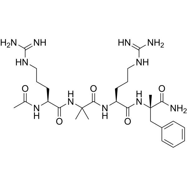
Cenupatide

Based on 1 Customer Validation
Cenupatide (UPARANT) is a uPAR and FPR antagonist with anti-angiogenic, anti-inflammatory, and vascular barrier regulatory activities, as well as high stability and resistance to enzymatic degradation in blood/plasma. Cenupatide blocks the uPAR-FPR interaction, reduces VEGF-induced phosphorylation levels of AKT, VEGFR-2, STAT3, JNK, p38 MAPK, ERK1/2, and NF-κB p65, and inhibits αvβ3 integrin activation. Cenupatide suppresses endothelial cell migration, invasion, tube formation, and angiogenic signaling pathways, restores tight junctions and blood-retinal barrier integrity, reduces pro-inflammatory marker levels, and inhibits apoptosis. Cenupatide reduces retinal neovascularization, renal fibrosis, and vascular leakage, and restores visual function in preclinical models. Cenupatide is applicable to research related to retinopathy, diabetic complications, ocular diseases, cancer, and inflammatory diseases.
For research use only. We do not sell to patients.
- Purity : 99.94%
- CAS No.: 1006388-38-0
- Formula: C28H47N11O5
- Molecular Weight:617.74
-
Storage:
Sealed storage, away from moisture.
Powder -80°C, 2 years , -20°C, 1 year* In solvent : -80°C, 6 months; -20°C, 1 month (sealed storage, away from moisture)

To place orders, for customer services and technical support, please contact: MedChemExpress USA
Tel: 609-228-6898 E-mail: [email protected] [email protected]


-
 Biological Activity
-
Chemical Information
-
Solvent & Solubility
- Protocols
- Purity & Documentation
- References
-
 Help & FAQs


All VEGFR Isoforms
More

Biological Activity
Description
|
uPAR |
FPR |
ERK1 |
ERK2 |
Akt |
VEGFR2 |
STAT3 |
JNK |
p38 MAPK |
NF-κB |
p65 |
αvβ3 |
In Vitro
Cenupatide (UPARANT) (1-100 nM; 24-72 h) does not affect viability or proliferation of primary human retinal endothelial cells, either alone or when co-treated with VEGF-A[1].
Cenupatide (10-100 nM; 24 h) significantly reduces VEGF-A-induced migration of primary human retinal endothelial cells by ~30% after 24 hours, while having no effect on migration alone[1].
Cenupatide (10 nM; 24 h) reduces VEGF-A-induced invasion of primary human retinal endothelial cells by 40% after 24 hours of incubation[1].
Cenupatide (10 nM; 6 h) reduces VEGF-A-stimulated tube formation of primary human retinal endothelial cells by 50.4% in branching points after 6 hours of incubation[1].
Cenupatide (10-100 nM; 24-72 h) completely prevents VEGF-A-induced loss of transendothelial electrical resistance in confluent primary human retinal endothelial cells over 72 hours, while 1 nM provides partial protection[1].
Cenupatide (10 nM; 72 h) prevents VEGF-A-induced reduction in tight junction protein expression in primary human retinal endothelial cells after 72 hours of incubation[1].
Cenupatide (10 nM; 30 min preincubation prior to 5-60 min VEGF-A stimulation) inhibits VEGF-A-induced VEGFR-2 phosphorylation in primary human retinal endothelial cells by 37.5% at 5 minutes and restores levels to control by 30 minutes, without altering total VEGFR-2 protein or mRNA expression[1].
Cenupatide (10 nM; 24 h for protein analysis; 30 min preincubation prior to 1-4 h VEGF-A stimulation for mRNA analysis) reduces VEGF-A-induced autocrine VEGF-A protein expression by 64% after 24 hours and reduces VEGF-A mRNA expression by up to 77% after 4 hours in primary human retinal endothelial cells[1].
Cenupatide (10 nM; 30 min preincubation prior to 5-60 min VEGF-A stimulation) inhibits VEGF-A-induced phosphorylation of AKT, JNK, p38, and ERK 1/2 in primary human retinal endothelial cells, completely preventing JNK and p38 activation at 5 minutes and returning all four protein phosphorylation levels to near baseline by 30 to 60 minutes[1].
Cenupatide (10 nM; 30 min preincubation prior to 5-60 min VEGF-A stimulation for STAT3 analysis; 30 min preincubation prior to 1-4 h VEGF-A stimulation for HIF-1α analysis) prevents VEGF-A-induced STAT3 phosphorylation and increases HIF-1α mRNA expression in primary human retinal endothelial cells, maintaining STAT3 activation near control levels and restoring HIF-1α mRNA to control levels by 1 hour[1].
Cenupatide (0.001-1 nM; 24 h) dose-dependently inhibits PDR vitreous fluid-induced sprouting in HUVEC spheroids, with maximum inhibition of ~72% at 0.1 nM[2].
Cenupatide (10 nM; 30 minutes) at 10 nM prevents VEGF-induced cytoskeletal reorganization and αvβ3 integrin redistribution to focal adhesions in HUVEC[8].
Cenupatide (10 nM; 4 hours) at 10 nmol/L inhibits VEGF adhesion to vitronectin, both in basal conditions and in the presence of VEGF[8].
Cenupatide (10 aM-10 μM; 4 hours) inhibits VEGF-dependent migration of HUVEC in a dose-dependent manner, with activity starting at femtomolar concentrations[8].
Cenupatide (10 fM-10 μM; 6 hours) potently inhibits VEGF-dependent tube formation in HUVEC, with a 100-fold lower IC50 than RERF and 75% inhibition at 10 nM[8].
Cenupatide (0.15 mM; 60 min) is not inhibited in binding or internalization by PDR vitreous fluid in cultured HUVECs[2].
Cenupatide shows high resistance to chymotrypsin digestion (no detectable breakdown over 60 minutes) and reduced susceptibility to trypsin digestion compared to unmodified peptides, with a trypsin T1/2 of 11.4 minutes at a 10:1 peptide:enzyme ratio with 0.6 mg/L trypsin[8].
Cenupatide (100 nM; 60 minutes) competes with fMLF for binding to FPR on HUVEC, effectively reducing fluorescent fMLF receptor binding when used at 100 nM[8].
Cenupatide (100 nM; 30 minutes) inhibits agonist-dependent FPR internalization in HUVEC when used at 100 nM, while causing minimal internalization of its own fluorescently labeled form[8].
Cenupatide (10 nM; 5-60 minutes) at 10 nM inhibits VEGF-induced phosphorylation of Akt and ERK1/2 in HUVEC[8].
Cenupatide (10 nM,-10 μM; 24-72 hours) at concentrations up to 10 μM does not affect the viability or proliferation of VEGF-stimulated HUVEC over 72 hours[8].
MedChemExpress (MCE) has not independently confirmed the accuracy of these methods. They are for reference only. Further protocols information, click here.
-
Cell Line:Primary human retinal endothelial cells
-
Concentration:1 nM; 10 nM; 100 nM
-
Incubation Time:24 h; 48 h; 72 h
-
Result:Whether used alone or in combination with VEGF-A, did not affect the viability or proliferation of primary human retinal endothelial cells.
-
Cell Line:Primary human retinal endothelial cells
-
Concentration:10 nM; 100 nM
-
Incubation Time:24 h
-
Result:Significantly reduced VEGF-A-induced migration of primary human retinal endothelial cells, with migration decreasing by approximately 30% after 24 hours, whereas it had no effect on cell migration when administered alone.
-
Cell Line:Primary human retinal endothelial cells
-
Concentration:10 nM
-
Incubation Time:24 h
-
Result:Reduced the invasion rate of VEGF-A-induced primary human retinal endothelial cells by 40% after 24 hours of incubation.
-
Cell Line:Primary human retinal endothelial cells
-
Concentration:10 nM
-
Incubation Time:72 h; 30 min preincubation prior to 5-60 min VEGF-A stimulation
-
Result:Inhibited the VEGF-A-induced downregulation of tight junction protein expression in primary human retinal endothelial cells after 72 hours of incubation.
Inhibited VEGF-A-induced VEGFR-2 phosphorylation: the inhibition rate reached 37.5% at 5 minutes, and phosphorylation levels returned to those of the control group by 30 minutes, without altering total VEGFR-2 protein or mRNA expression.
Inhibited VEGF-A-induced phosphorylation of AKT, JNK, p38, and ERK 1/2; completely blocked JNK and p38 activation at 5 minutes; and restored the phosphorylation levels of these four proteins to near-baseline levels between 30 and 60 minutes.
-
Cell Line:Human umbilical vein endothelial cells (HUVEC)
-
Concentration:10 aM-10 μM
-
Incubation Time:4 hours
-
Result:Inhibited VEGF-directed HUVEC migration in a dose-dependent manner, with inhibition starting in the low femtomolar range and plateauing in the nanomolar range.
Reduced VEGF-dependent migration by over 60% at the highest tested concentrations.
-
Cell Line:Human umbilical vein endothelial cells (HUVEC)
-
Concentration:10 nM
-
Incubation Time:5, 10, 15, 60 minutes
-
Result:Consistently decreased VEGF-induced, time-dependent phosphorylation of Akt and ERK1/2 in HUVEC.
-
Cell Line:Human umbilical vein endothelial cells (HUVEC)
-
Concentration:10 nM-10 μM
-
Incubation Time:24, 48, 72 hours
-
Result:Did not modify VEGF-induced HUVEC growth over 72 hours, as measured by MTS absorbance and xCELLigence cell index, at both 10 nmol/L and 10 μmol/L.
Parmacokinetics
In Vivo
Cenupatide (7 mg/kg; s.c.; three times weekly) prevents diabetic retinal complications and reduces ocular vascular leakage in genetically modified fat rats[3].
Cenupatide (75-150 μg/kg; subcutaneous; single dose) dose-dependently inhibits VEGF-induced angiogenesis in a murine Matrigel sponge assay, reducing vascularization by up to 72% at the 150 μg/kg dose[8].
Cenupatide (5 μg per pellet; subcorneal; single sustained-release dose) completely prevents VEGF-induced corneal neovascularization in rabbits, reducing angiogenic activity by 75% at the 5 μg per pellet dose[8].
Cenupatide (7 mg/kg; s.c.; three times a week) prevents diabetic retinal complications and restores blood-retinal barrier function in genetically diabetic Torii rats[9].
Cenupatide (7.6 g/L; intravitreal; 3 injections on days 4, 8, 12; 1 μL per injection) normalizes iris macro- and microvascular density, FPR1 expression, inflammatory and extracellular matrix degradation marker levels, and CREB/NFκB phosphorylation to non-induced control levels in a mouse model of rubeosis iridis[4].
Cenupatide (15.2 mg/kg; subcutaneous; daily; 5 days from day 4 through day 8) normalizes iris vascular density, FPR1 expression, and inflammatory and extracellular matrix degradation marker levels to non-induced control levels in a mouse model of rubeosis iridis[4].
Cenupatide (1-8 mg/kg; s.c.; daily; 5 days) significantly ameliorates Streptozotocin (HY-13753)-induced diabetic nephropathy in male Sprague-Dawley rats by normalizing uPA activity, αvβ3 integrin/Rac-1 signaling, renal fibrosis, vascular permeability, inflammatory markers, renal function parameters, and glomerular morphology[5].
Cenupatide (4-20 mg/kg; s.c.; daily for 3 days; daily for 5 days; daily for 7 days; single dose) restores retinal ERG function, BRB integrity, and normalizes inflammatory/proangiogenic marker levels in rats with streptozotocin-induced diabetic retinopathy, with ERG recovery lasting ~2 weeks post-treatment; higher and lower/different duration doses lack efficacy or serve safety/Pharmacokinetic profiling[6].
Cenupatide (20 mg/kg; s.c.; single dose) rapidly distributes to plasma, eye, and retina in healthy rats, with detectable levels persisting for at least 24 hours post-dose[6].
Cenupatide (6-24 mg/kg; i.p.; single dose) with single intraperitoneal administration at 12 and 24 mg/kg significantly reduces Carrageenan (HY-125474)-induced paw oedema in male CD-1 mice in both early and late inflammatory phases, while the 6 mg/kg dose shows no significant effect[10].
Cenupatide (12-24 mg/kg; i.p.; 5 total doses) with repeated intraperitoneal administration at 12 and 24 mg/kg dose-dependently abolishes the late phase of carrageenan-induced paw oedema in male CD-1 mice[10].
Cenupatide (7 mg/kg; s.c.; 3 times a week; 19 weeks) prevents retinal impairment in spontaneously diabetic Torii rats, maintaining ERG function, BRB integrity, and retinal cell viability, while reducing proangiogenic and inflammatory marker levels, with a key effect of normalizing uPAR/FPR expression and downstream transcription factor activation in this type 2 diabetic retinopathy model[7].
Cenupatide (8 mg/kg; s.c.; daily; 5 days) reduces activation of proinflammatory transcription factors in streptozotocin-induced diabetic rats with type 1 diabetic retinopathy, without altering uPAR/FPR system expression[7].
Cenupatide (12-24 mg/kg; i.p.; single administration) reduces inflammatory mediator overproduction in rodent models of acute inflammation[9].
Cenupatide (8 mg/kg; s.c.; daily; 5 days) restores renal vascular integrity and modulates aquaporin-2 expression in streptozotocin-induced diabetic nephropathy in Sprague-Dawley rats[9].
Cenupatide (16 mg/kg; s.c.; daily; postnatal day 10 to postnatal day 30) limits retinal cell apoptosis in a rodent model of retinitis pigmentosa[9].
Cenupatide (6-24 mg/kg; i.p.; single dose) with single intraperitoneal administration at 12 and 24 mg/kg significantly reduces carrageenan-induced paw oedema, as well as iNOS, COX-2, and NOx levels in male Wistar rats, while the 6 mg/kg dose shows no significant effect[10].
Cenupatide (6-24 mg/kg; i.p.; single dose) with single intraperitoneal administration at 12 and 24 mg/kg significantly reduces inflammatory cell influx and NOx production in Zymosan (Zymosan A) (HY-W250113)-induced peritonitis in male CD-1 mice, while the 6 mg/kg dose only significantly reduces NOx levels[10].
MedChemExpress (MCE) has not independently confirmed the accuracy of these methods. They are for reference only.
-
Animal Model:C57BL/6 (mixed gender, postnatal day 7 pups, oxygen-induced retinopathy model)[2]
-
Dosage:0.0015 μg/μL; 0.015 μg/μL; 0.15 μg/μL; 1.5 μg/μL
-
Administration:intravitreal; at postnatal day 12 and 15
-
Result:Showed no effect on neovascular tuft area, VEGF levels, VEGFR-2 phosphorylation, HIF-1α accumulation, or STAT3 phosphorylation at 0.0015 μg/μL.
Reduced neovascular tuft area by ~62% (P < 0.001), reduced VEGFR-2 phosphorylation by ~32% (P < 0.01), and showed no effect on VEGF levels, HIF-1α accumulation, or STAT3 phosphorylation at 0.015 μg/μL.
Reduced neovascular tuft area by ~75% (P < 0.001), reduced VEGF levels by ~49% (P < 0.01), reduced VEGFR-2 phosphorylation by ~52% (P < 0.001), reduced HIF-1α accumulation by ~50% (P < 0.01), and reduced STAT3 phosphorylation by ~49% (P < 0.01) at 0.15 μg/μL.
Reduced neovascular tuft area by ~85% (P < 0.0001), reduced VEGF levels by ~63% (P < 0.001), reduced VEGFR-2 phosphorylation by ~78% (P < 0.001), reduced HIF-1α accumulation by ~82% (P < 0.001), reduced STAT3 phosphorylation by ~84% (P < 0.001), restored occludin and albumin levels to control values to reduce blood-retinal barrier leakage, increased a-wave, b-wave, and sum oscillatory potential amplitudes in electroretinography to improve visual function, reduced TNF-α mRNA by ~38% (P < 0.01), IL-1β mRNA by ~48% (P < 0.001), IL-6 mRNA by ~66% (P < 0.01), iNOS mRNA by ~50% (P < 0.05), ICAM-1 mRNA by ~41% (P < 0.001), and GFAP mRNA by ~83% (P < 0.001), and showed no effect on Bax/Bcl-2 ratio, cytochrome c levels, retinal cell apoptosis, or retinal layer thickness at 1.5 μg/μL.
-
Animal Model:genetically modified fat rats (diabetic retinal complication risk)[3]
-
Dosage:7 mg/kg
-
Administration:s.c.; three times weekly
-
Result:Prevented the onset of diabetic retinal complications by reducing vascular leakage into the eye.
-
Animal Model:BALB/c mice (either sex, 12.5-day-old pups, induced via two self-sealing uveal punctures with a 30G needle immediately posterior to the limbus, repeated every 4 days from experimental day 0 through day 12)[4]
-
Dosage:7.6 g/L
-
Administration:intravitreal; 3 injections (days 4, 8, 12); 1 μL per injection
-
Result:Reduced iris macrovasculature density from a ~20% increase in rubeosis iridis eyes to non-punctured control levels by experimental day 8, with this effect sustained through day 15.
Reduced rubeosis iridis-associated microvascular changes (30% increases in total vasculature, sprouts, and vascular branching) back to control levels.
Normalized rubeosis iridis-induced 3.5-fold overexpression of FPR1 transcript levels to control levels, with no effect on FPR2 or FPR3.
Reduced rubeosis iridis-induced increased phosphorylation of CREB (p=0.002 versus rubeosis iridis) and NFκB (p=0.036 versus rubeosis iridis) back to control levels.
Normalized rubeosis iridis-induced overexpression of extracellular matrix degradation markers (PAI-1, uPA, uPAR) and inflammation markers (IL-1β, IL-6, TGFα, CCL2, CXCR4) at the transcript level to control levels.
Reduced rubeosis iridis-induced elevated IL-6 protein levels back to control levels, and reduced MMP2 protein levels significantly compared to rubeosis iridis eyes (p=0.036).
-
Animal Model:BALB/c mice (either sex, 12.5-day-old pups, induced via two self-sealing uveal punctures with a 30G needle immediately posterior to the limbus, repeated every 4 days from experimental day 0 through day 12)[4]
-
Dosage:15.2 mg/kg
-
Administration:subcutaneous; daily; 5 days (day 4 through day 8)
-
Result:Reduced rubeosis iridis-induced iris vascular density (over 25% increase at day 4) to non-punctured control levels by experimental day 8, with this effect sustained through day 15.
Normalized rubeosis iridis-induced overexpression of extracellular matrix degradation markers (PAI-1, uPA, uPAR) and inflammation markers (IL-1β, IL-6, CCL2, CXCR4) at the transcript level to control levels.
Reduced rubeosis iridis-induced elevated FPR1 transcript levels back to control levels.
-
Animal Model:Sprague-Dawley (male, 150-200 g, streptozotocin-induced diabetic nephropathy)[5]
-
Dosage:1 mg/kg; 8 mg/kg
-
Administration:s.c.; daily; 5 days
-
Result:Showed no significant effects on any measured parameters at 1 mg/kg.
Reduced renal uPA levels by 1.6-fold and renal uPA activity by 1.4-fold at 8 mg/kg.
Reduced plasma uPA levels by 1.2-fold and plasma uPA activity by 1.3-fold at 8 mg/kg.
Reduced glomerular FPR2 transcript levels by 1.3-fold at 8 mg/kg.
Reduced renal αvβ3 integrin levels by 1.6-fold at 8 mg/kg.
Reduced renal β3 integrin phosphorylation by 2.0-fold at 8 mg/kg.
Reduced renal Rac-1 activity by 1.2-fold at 8 mg/kg.
Reduced renal Plg levels by 1.7-fold at 8 mg/kg.
Increased renal plasmin levels by 1.5-fold and renal plasmin activity by 1.7-fold at 8 mg/kg.
Increased renal MMP-2 levels by 2.0-fold, renal MMP-9 levels by 1.7-fold, and renal MMP-2/MMP-9 activity by 1.9-fold at 8 mg/kg.
Increased plasma plasmin activity by 1.3-fold at 8 mg/kg.
Reduced renal fibronectin levels by 1.6-fold, renal collagen I levels by 3.1-fold, and renal collagen IV levels by 1.8-fold at 8 mg/kg.
Reduced glomerular fibrotic areas as measured by Masson's Trichrome staining at 8 mg/kg.
Reduced urine output by 1.7-fold, urine albumin by 1.8-fold, urine creatinine by 1.3-fold, albumin-to-creatinine ratio by 2.3-fold, plasma creatinine by 1.4-fold, creatinine clearance by 1.3-fold, and blood urea nitrogen by 1.7-fold at 8 mg/kg.
Increased renal ZO-1 levels by 1.7-fold and renal occludin levels by 1.8-fold at 8 mg/kg.
Reduced renal VEGF levels by 1.8-fold and Evans blue dye leakage by 1.4-fold at 8 mg/kg.
Increased renal medullary AQP2 levels by 2.8-fold and enhanced apical AQP2 localization in medullary collecting ducts at 8 mg/kg.
Reduced renal iNOS levels by 3.0-fold, renal ICAM-1 levels by 2.5-fold, renal NF-κB p65 phosphorylation by 3.8-fold, renal CREB phosphorylation by 2.4-fold, and renal HIF-1α levels by 3.9-fold at 8 mg/kg.
Reduced glomerular area by 1.1-fold and mesangial area by 1.2-fold at 8 mg/kg.
Attenuated glomerular basement membrane thickening and restored podocyte foot process architecture at 8 mg/kg.
-
Animal Model:Sprague-Dawley (male, 150-200 g, streptozotocin-induced diabetic retinopathy)[6]
-
Dosage:4 mg/kg (ERG); 8 mg/kg (ERG, BRB, markers); 10 mg/kg (safety); 20 mg/kg (Pharmacokinetic)
-
Administration:s.c.; daily for 3 days; daily for 5 days; daily for 7 days; single dose
-
Result:Restored scotopic ERG a-wave and b-wave amplitudes to levels not significantly different from non-diabetic controls.
Recovered b-wave Vmax to 736 μV (vs. 534 μV in untreated diabetic rats).
Recovered retinal sensitivity (k) to -1.38 log cd-s/m2 (vs. -0.3 log cd-s/m2 in untreated diabetic rats).
Maintained ERG recovery for approximately 2 weeks after treatment withdrawal.
Reduced diabetes-induced BRB leakage from ~2.3-fold above control levels to near-control levels.
Reduced retinal mRNA levels of inflammatory markers IL-1β by ~1.5-fold, IL-6 by ~1.3-fold, iNOS by ~1.4-fold, GFAP by ~1.4-fold relative to untreated/vehicle-treated diabetic rats.
Reduced retinal mRNA levels of proangiogenic markers VEGF by ~1.5-fold, IGF-1 by ~1.4-fold, FGF-2 by ~1.7-fold, PDGF-B by ~1.4-fold relative to untreated/vehicle-treated diabetic rats.
Restored ICAM-1 and Ang-2 levels to control levels.
Failed to restore ERG function at doses of 8 mg/kg daily for 3 days and 4 mg/kg daily for 5 days.
Achieved plasma Cmax of 5.9 μg/mL at 2 hours post single 20 mg/kg dose, terminal half-life of 3.0 hours, AUCinf of 34.4 μg-h/mL, with detectable plasma levels at 24 hours post-dose.
Reached concentration of 1.10 μg/g in the eye and 0.16 μg/g in the retina at 2 hours post single 20 mg/kg dose; reached concentration of 0.22 μg/g in the eye and 0.04 μg/g in the retina at 24 hours post-dose.
Showed no significant effects on body weight, hematologic/biochemical parameters, gross organ morphology, or liver/kidney histopathology at 10 mg/kg daily for 7 days.
-
Animal Model:Sprague-Dawley (male, 150-200 g, healthy)[6]
-
Dosage:20 mg/kg
-
Administration:s.c.; single dose
-
Result:Was quantifiable in plasma as early as 0.25 hours post-dose.
Reached peak plasma concentration (Cmax) of 5.9 μg/mL at 2 hours post-dose.
Remained detectable in plasma at 24 hours post-dose.
Reached concentration of 1.10 μg/g in the eye and 0.16 μg/g in the retina at 2 hours post-dose.
Reached concentration of 0.22 μg/g in the eye and 0.04 μg/g in the retina at 24 hours post-dose.
Had a terminal plasma half-life of 3.0 hours and AUCinf of 34.4 μg-h/mL.
-
Animal Model:Spontaneously Diabetic Torii (SDT) rats (male, inbred from Sprague-Dawley rats, spontaneously developed hyperglycemia after 20 weeks of age leading to retinal impairment)[7]
-
Dosage:7 mg/kg
-
Administration:s.c.; 3 times a week; 19 weeks
-
Result:Prevented ERG dysfunction, with a-wave and b-wave amplitudes across specified light intensities remaining not significantly different from non-diabetic Sprague-Dawley controls, while untreated/vehicle-treated rats showed significant amplitude reduction (P < 0.001).
Preserved b-wave Vmax (758.20 μV) and retinal sensitivity k (-1.31 log cd-s/m2), matching control values, whereas untreated/vehicle-treated rats had reduced Vmax and k (P < 0.001).
Prevented retinal vascular leakage, eliminating abnormal fluorescein leakage and vasodilation seen in untreated/vehicle-treated rats.
Prevented the 2.2-fold increase in Evans blue dye leakage (P < 0.001) seen in untreated/vehicle-treated rats, maintaining levels matching controls.
Restored transcript and protein levels of BRB markers (claudin-1, claudin-5, ZO-1) that were reduced by ~2-fold (P < 0.001) in untreated/vehicle-treated rats to levels not significantly different from controls.
Prevented retinal damage, reducing upregulated GFAP transcript (by 1.4-fold, P < 0.05) and protein (by ~2-fold, P < 0.001) levels, and upregulated caspase-3 transcript (by 1.3-fold, P < 0.05) and active caspase-3 protein (by ~1.4-fold, P < 0.01) levels seen in untreated/vehicle-treated rats, restoring values to near control levels.
Prevented upregulation of proangiogenic factors, reducing upregulated VEGF transcript (by ~1.2-fold, P < 0.05) and protein (by ~1.8-fold, P < 0.001) levels, and FGF-2 transcript (by ~2-fold, P < 0.01) and protein (by ~2.6-fold, P < 0.001) levels seen in untreated/vehicle-treated rats, restoring values to near control levels.
Modulated uPAR/FPR system and inflammatory pathways, reducing upregulated uPAR (by 1.7-fold, P < 0.01), FPR1 (by 1.8-fold, P < 0.01), and FPR2 (by 1.9-fold, P < 0.01) transcript and protein levels (no effect on FPR3).
Reduced phosphorylation of STAT3 (Tyr705) by 3.4-fold (P < 0.001) and NF-κB p65 (Ser276) by 1.6-fold (P < 0.001).
Reduced upregulated TNF-α transcript (by 1.4-fold, P < 0.01) and protein (by 1.6-fold, P < 0.01) levels, IL-1β transcript (by 1.7-fold, P < 0.01) and protein (by 2.1-fold, P < 0.001) levels, and IL-6 transcript (by 1.3-fold, P < 0.05) and protein (by 1.6-fold, P < 0.01) levels seen in untreated/vehicle-treated rats.
-
Animal Model:Sprague-Dawley (SD) rats (male, type 1 diabetes induced by single intraperitoneal injection of 65 mg/kg streptozotocin)[7]
-
Dosage:8 mg/kg
-
Administration:s.c.; daily; 5 days
-
Result:Reduced phosphorylation of STAT3 (Tyr705) by 3.0-fold (P < 0.01) and NF-κB p65 (Ser276) by 1.7-fold (P < 0.01) in streptozotocin-induced diabetic rats, which had shown 10.3-fold and 3.3-fold increases in phosphorylation (P < 0.001) compared to non-diabetic controls.
Showed no effect on upregulated uPAR, FPR1, FPR2, or FPR3 transcript and protein levels seen in streptozotocin-induced diabetic rats.
-
Animal Model:C57BL/6J (male, 6-8 weeks old, 23-25 g)[8]
-
Dosage:75 μg/kg; 150 μg/kg
-
Administration:subcutaneous; single dose
-
Result:Reduced hemoglobin content (a marker of vascularization) by 64% at the 75 μg/kg dose relative to VEGF-only controls.
Reduced hemoglobin content (a marker of vascularization) by 72% at the 150 μg/kg dose relative to VEGF-only controls.
-
Animal Model:New Zealand White (female, 2.5-3.0 kg)[8]
-
Dosage:5 μg per pellet
-
Administration:subcorneal; single sustained-release dose
-
Result:Reduced the median angiogenic score from 3.3 (VEGF-alone group) to 0.84, representing a 75% inhibition of VEGF-induced neovascularization.
Did not elicit an inflammatory or angiogenic response when administered alone.
-
Animal Model:CD-1 mice; Wistar rats[9]
-
Dosage:12 mg/kg; 24 mg/kg
-
Administration:i.p.; single administration
-
Result:Reduced inducible nitric oxide synthase (iNOS), cyclo-oxygenase 2 (COX2), and nitric oxide (NO) overproduction in both inflammation models.
-
Animal Model:Torii rats (genetically modified, diabetic)[9]
-
Dosage:7 mg/kg
-
Administration:s.c.; three times a week
-
Result:Prevented onset of diabetes retinal complications by reducing vascular leakage into the eye.
Downregulated transcripts and proteins of blood-retinal barrier markers including claudin-1, claudin-5, and zonula occludens-1, restoring barrier integrity.
-
Animal Model:Sprague-Dawley rats (streptozotocin-induced diabetic)[9]
-
Dosage:8 mg/kg
-
Administration:s.c.; daily; 5 days
-
Result:Restored vascular permeability integrity.
Increased aquaporin-2 expression in the renal medulla.
-
Animal Model:Unspecified rodent strain[9]
-
Dosage:16 mg/kg
-
Administration:s.c.; daily; postnatal day 10 to postnatal day 30
-
Result:Significantly reduced the Bax:Bcl2 ratio and active caspase-3 levels.
Limited retinal cell apoptosis without affecting autophagy.
-
Animal Model:CD-1 (male, 32-35 g, carrageenan-induced paw oedema model)[10]
-
Dosage:6 mg/kg; 12 mg/kg; 24 mg/kg
-
Administration:i.p.; single dose
-
Result:Did not significantly affect paw oedema at 4 hours (265 μL) and 12 hours (261 μL) at 6 mg/kg dose.
Significantly reduced paw oedema to 180 μL at 4 hours and 165 μL at 12 hours at 12 mg/kg dose.
Significantly reduced paw oedema to 137 μL at 4 hours and 140 μL at 12 hours at 24 mg/kg dose.
-
Animal Model:CD-1 (male, 32-35 g, carrageenan-induced paw oedema model)[10]
-
Dosage:12 mg/kg; 24 mg/kg
-
Administration:i.p.; 5 total doses (1 hour pre-carrageenan, then at 12, 24, 36, 48 hours post-carrageenan)
-
Result:Reduced paw oedema to 80 μL at 12 hours post-carrageenan at 12 mg/kg dose.
Reduced paw oedema to 49 μL at 12 hours post-carrageenan at 24 mg/kg dose, with an effect comparable to dexamethasone.
-
Animal Model:Wistar (male, 250-300 g, carrageenan-induced paw oedema model)[10]
-
Dosage:6 mg/kg; 12 mg/kg; 24 mg/kg
-
Administration:i.p.; single dose
-
Result:Did not significantly affect paw oedema at 4 hours (2.25 mL) at 6 mg/kg dose.
Significantly reduced paw oedema to 1.35 mL at 4 hours at 12 mg/kg dose.
Significantly reduced paw oedema to 1.23 mL at 4 hours at 24 mg/kg dose.
Significantly reduced iNOS and COX-2 protein expression and NOx levels in paw exudates at 12 and 24 mg/kg doses.
-
Animal Model:CD-1 (male, 32-35 g, zymosan-induced peritonitis model)[10]
-
Dosage:6 mg/kg; 12 mg/kg; 24 mg/kg
-
Administration:i.p.; single dose
-
Result:Did not significantly reduce peritoneal cell influx (3433 × 104 cells) but significantly reduced NOx levels in peritoneal exudates at 6 mg/kg dose.
Significantly reduced peritoneal cell influx to 2233 × 104 cells and NOx levels in peritoneal exudates at 12 mg/kg dose.
Significantly reduced peritoneal cell influx to 2360 × 104 cells and NOx levels in peritoneal exudates at 24 mg/kg dose.


Chemical Information
-
CAS No. 1006388-38-0
-
Appearance Solid
-
Molecular Weight 617.74
-
Formula C28H47N11O5
-
Color White to off-white
-
Synonyms
UPARANT
-
Sequence
Ac-Arg-{Aib}-Arg-{aMePhe}-NH2
-
Sequence Shortening
Ac-R-{Aib}-R-{aMePhe}-NH2
-
Shipping
Room temperature in continental US; may vary elsewhere.
-
Storage
Sealed storage, away from moisture
Powder -80°C 2 years -20°C 1 year * In solvent : -80°C, 6 months; -20°C, 1 month (sealed storage, away from moisture)


Solvent & Solubility
In Vitro:
DMSO : 100 mg/mL (161.88 mM; Need ultrasonic; Hygroscopic DMSO has a significant impact on the solubility of product, please use newly opened DMSO)
H2O : ≥ 100 mg/mL (161.88 mM)
* "≥" means soluble, but saturation unknown.
Please refer to the solubility information to select the appropriate solvent. Once prepared, please aliquot and store the solution to prevent product inactivation from repeated freeze-thaw cycles.
Storage method and period of stock solution: -80°C, 6 months; -20°C, 1 month (sealed storage, away from moisture). When stored at -80°C, please use it within 6 months. When stored at -20°C, please use it within 1 month.
* Note: If you choose water as the stock solution, please dilute it to the working solution, then filter and sterilize it with a 0.22 μm filter before use.
Please refer to the solubility information to select the appropriate solvent. Once prepared, please aliquot and store the solution to prevent product inactivation from repeated freeze-thaw cycles.
Storage method and period of stock solution: -80°C, 6 months; -20°C, 1 month (sealed storage, away from moisture). When stored at -80°C, please use it within 6 months. When stored at -20°C, please use it within 1 month.
* Note: If you choose water as the stock solution, please dilute it to the working solution, then filter and sterilize it with a 0.22 μm filter before use.
Concentration (start) × Volume (start) = Concentration (final) × Volume (final)

Protocols
-
Western Blot
Western blotting (WB) is a commonly used experimental method in molecular biology, biochemistry, and immunogenetics for identifying and quantifying target proteins. It combines gel electrophoresis with immunoassay, enabling researchers to analyze protein expression, post-translational modifications, and molecular weight.
-
Scratch/Wound-Healing Migration Assay
The scratch/wound-healing migration assay measures collective migration of adherent cells into an experimentally created cell-free gap in a confluent monolayer. The readout is generated by imaging the gap immediately after scratching and at later time points, then quantifying reduction in wound area, wound width, or percentage closure as cells move into the denuded region. Gap closure reflects cell migration but may also include cell proliferation, so interpretation should distinguish migration-focused conditions from proliferation-driven closure when possible, such as by using short assay windows, serum-controlled conditions, cell counting, or proliferation controls reported in published protocols.
-
Collagen: Sirius Red Staining
Sirius Red or picrosirius red staining is a histochemical method for visualizing collagen-rich extracellular matrix in tissue sections, and collagen fibers are detected as red-stained structures under bright-field microscopy with enhanced birefringence under polarized light. Picrosirius red is useful for assessing total collagen organization, distribution, and fibrosis burden, but polarized color should not be interpreted as a definitive collagen type I versus type III readout because color is affected by fiber orientation, thickness, and packing.
-
Annexin V plus membrane-impermeant dye apoptosis staining
Annexin V-based apoptosis assays rely on the detection of phosphatidylserine (PS) externalization from the inner leaflet of the plasma membrane to the outer leaflet, an early biochemical hallmark of apoptosis. Fluorescently labeled Annexin V binds PS in a calcium-dependent manner, enabling identification of early apoptotic cells by flow cytometry or fluorescence microscopy. When combined with a membrane-impermeant DNA-binding dye (e. g. , propidium iodide), this approach allows discrimination between viable (Annexin V−/dye−), early apoptotic (Annexin V+/dye−), and late apoptotic or necrotic (Annexin V+/dye+) cell populations by assessing membrane integrity and PS exposure.
-
Cell migration
Cell migration is a method that plays an important role in wound healing, cell differentiation, embryonic development, etc.
-
Neural Crest/Neuronal Cell Migration Explant Assay
Neural crest (NC) and neuronal cell migration explant assays are in vitro systems in which neural tube-derived tissues are cultured to allow premigratory or newly emigrated neural crest cells to undergo epithelial-to-mesenchymal transition (EMT), migrate away from the explant, and form a measurable radial outgrowth that reflects migratory capacity and environmental responsiveness. These assays typically quantify migration by measuring the expansion of cell outgrowth from neural tube or neural plate border explants over time, often comparing early and later timepoints to derive a migration index such as a radius ratio, which reflects net cell dispersal from the explant core. Neural tube explant cultures preserve key aspects of neural crest behavior, including EMT, migration, and early differentiation, making them suitable for assessing intrinsic migratory ability and extrinsic cue dependence. However, studies emphasize that migratory outgrowth from neural tube explants may include non-n
-
Connective Tissue: Masson's Trichrome/Collagen Trichrome Staining
Masson’s Trichrome (collagen/trichrome staining) is a histological technique that differentially stains tissue compartments using sequential acidic dyes to distinguish collagen from muscle and cytoplasmic components based on dye affinity and tissue permeability differences, enabling visualization of fibrosis and connective tissue architecture in histological sections. The classical formulation typically uses Weigert's iron hematoxylin for nuclear staining, Biebrich scarlet-acid fuchsin for cytoplasm and muscle, and aniline blue (or light green variants) for collagen, producing a characteristic blue/green collagen signal contrasted against red cytoplasm and dark nuclei. The staining principle relies on selective displacement of smaller dye molecules by larger anionic dyes in collagen-rich regions under controlled acidified conditions, which enhances collagen-specific dye retention. This property makes the method widely used for fibrosis assessment in organs such as heart, liver, lung, a
-
Research Protocol for Inflammation-related Diseases
The NLRP3 inflammasome is a cytosolic innate immune signaling platform that integrates priming signals and danger-signal activation to promote caspase-1 activation, maturation of IL-1β and IL-18, and gasdermin D-mediated pyroptotic cell death. The core experimental logic is to determine whether inflammatory disease phenotypes are driven by increased NLRP3 expression, ASC-containing inflammasome assembly, caspase-1 cleavage, GSDMD cleavage, and extracellular release of IL-1β/IL-18 rather than by nonspecific cell injury alone. The pathway is strongly linked to inflammation-related disease phenotypes because monosodium urate crystals activate NALP3/NLRP3 inflammasome signaling in gout-like crystal inflammation, cholesterol crystals activate NLRP3 inflammasomes in atherogenesis models, and DSS-induced intestinal inflammation has been reported to involve NLRP3 inflammasome activity. However, experimental colitis studies also show context-dependent protective effects of NLRP3 inflammasome co
-
Kinase activity and phosphorylation assays
Kinase activity assays measure the ability of kinases to transfer phosphate groups from ATP to specific substrates, while phosphorylation assays detect the presence and levels of phosphorylated proteins. Common methods include radiolabeled ATP incorporation (e. g. ,), ADP release detection via bioluminescence (e. g. ,[3]), enzyme-linked immunosorbent assays (ELISA) for phospho-specific epitopes (e. g. ,[6]), and microtiter-based formats for high-throughput screening (e. g. ,[8]). The ADP-Glo assay quantifies kinase activity by measuring ADP produced during phosphorylation using a luciferase-based system. Radiometric assays involve autoradiography or scintillation counting after incorporation of 32P-labeled ATP into substrate proteins. ELISA-based approaches rely on phospho-specific antibodies to detect activated kinases in cell lysates or purified samples.
-
Fibrosis/Collagen Morphometry
Fibrosis and collagen morphometry is based on the quantitative visualization of fibrillar collagen deposition in tissue sections using histochemical stains such as Sirius Red (Picrosirius Red) or Masson's trichrome, followed by image-based or polarization-enhanced analysis to estimate collagen proportional area as a surrogate of extracellular matrix accumulation during fibrotic remodeling. Sirius Red combined with polarized light microscopy enhances detection of collagen fibers due to birefringence properties, enabling more specific visualization of collagen type I and III fibrils compared to conventional bright-field histology, while whole-section or region-restricted digital morphometry reduces field-selection bias in fibrosis assessment. Alternative quantitative approaches include second harmonic generation (SHG) and two-photon excited fluorescence microscopy, which enable label-free detection of fibrillar collagen and have been validated against histological staining and biochemica
-
Apoptosis
Apoptosis, also called programmed cell death, is generally characterized by distinct morphological characteristics.
-
TUNEL staining for apoptotic DNA fragmentation
TUNEL staining detects DNA strand breaks by using terminal deoxynucleotidyl transferase to add labeled nucleotides to exposed 3′-OH DNA termini, generating either microscopic staining in fixed cells or tissue sections, or fluorescence/cytometric signal in cell suspensions. TUNEL positivity reflects DNA fragmentation but should not be interpreted alone as definitive apoptosis, because TUNEL can also label necrotic, autolytic, mechanically damaged, or DNA-repair-associated DNA breaks.
-
Chemotaxis Gradient Chamber Assay 1
The chemotaxis gradient chamber assay is based on the principle of observing directional cell migration in response to a stable, linear or quasi-linear concentration gradient of a chemoattractant formed between two fluid reservoirs separated by a narrow observation chamber. Cells placed within the chamber respond to the gradient by polarized movement toward higher chemoattractant concentrations, allowing quantification of chemotactic behavior in real time under microscopy. The classic Zigmond chamber design enables simultaneous visualization of gradient formation and individual cell trajectories, making it suitable for studying leukocyte chemotaxis and other motile cell types in vitro.
-
Under-Agarose Cell Migration Assay
The under-agarose cell migration assay is a classical in vitro chemotaxis method designed to evaluate directed cell movement through a semi-solid agarose matrix toward soluble chemoattractant gradients, enabling visualization and quantification of leukocyte or motile cell migration in a confined 2D-like environment. In this system, cells and chemoattractants are placed in separate wells cut into an agarose gel, allowing diffusion-driven gradient formation that guides directional migration, which is typically assessed by measuring migration distance, cell morphology changes, and accumulation toward the chemoattractant source. This assay has been widely used to study neutrophil and leukocyte chemotaxis as a simple alternative to filter-based migration systems and allows direct microscopic observation of migrating cells under near-physiological confinement conditions.
-
Transwell/Boyden Chamber Migration Assay
The Transwell/Boyden chamber migration assay measures cell movement through a porous membrane separating an upper and lower chamber, usually after a chemoattractant gradient is established by placing cells in the upper chamber and chemoattractant-containing medium in the lower chamber. The readout is generated by quantifying cells that traverse the membrane and appear on the lower membrane surface or in the lower chamber, depending on whether the cell type is adherent or non-adherent. This assay reflects chemotactic or haptotactic migration rather than matrix invasion unless an extracellular-matrix barrier is added to the membrane.
-
3D Collagen/Hydrogel Matrix Migration Assay
The 3D collagen/hydrogel matrix migration assay is based on embedding cells within or on top of a fibrillar collagen type I-rich three-dimensional matrix to model in vivo-like extracellular matrix (ECM) architecture, enabling analysis of cell migration through a physically and biochemically relevant scaffold. In contrast to 2D migration systems, cells in 3D matrices interact with fibrillar collagen networks, requiring coordinated adhesion remodeling and proteolytic or non-proteolytic deformation mechanisms to move through confined spaces, thereby providing a more physiologically relevant readout of invasive and migratory behavior in tissue-like environments. Cell movement in 3D collagen matrices is typically quantified by tracking single-cell trajectories, invasion depth, or matrix penetration over time, reflecting combined effects of cytoskeletal dynamics, cell-ECM adhesion turnover, and ECM remodeling. These systems are widely used to study tumor cell invasion and stromal cell motili
-
Transepithelial/transendothelial electrical resistance assay
TEER measures electrical resistance across epithelial or endothelial monolayers cultured on permeable supports, and the readout reflects ionic conductance through the cell barrier, especially the paracellular pathway regulated by junctional integrity. TEER can be measured without destroying the monolayer and is commonly used before or during transport, permeability, barrier-disruption, and barrier-maturation experiments. TEER values are influenced by biological maturation and technical conditions; reported factors include temperature, medium formulation, passage number, electrode geometry, membrane properties, and junctional length during early monolayer maturation. Therefore, TEER should be interpreted with blank-insert subtraction, area normalization, repeated readings, and, when possible, orthogonal barrier readouts such as FITC-dextran flux or tight-junction staining.
-
Apoptosis Solutions
Apoptosis is a regulated, generally non-lytic cell-death pathway that removes unwanted, damaged, infected, or abnormal cells through coordinated morphological changes, caspase activation, DNA fragmentation, and membrane remodeling. The intrinsic apoptosis pathway is controlled mainly by mitochondrial outer membrane permeabilization, BCL-2 family proteins, cytochrome c release, apoptosome formation, caspase-9 activation, and downstream executioner caspase-3/7 activation. The extrinsic apoptosis pathway is initiated by death receptors such as Fas, TNFR, and TRAIL receptors, which recruit adaptor proteins and activate caspase-8 before engaging executioner caspases or mitochondrial amplification through BID cleavage. Apoptosis is linked to many phenotypes, including cancer cell killing, tissue homeostasis, immune regulation, neurodegeneration, infection response, and treatment-induced cytotoxicity; unresolved questions include how apoptosis interacts with necroptosis, pyroptosis, ferroptos
-
Cell invasion
Cell invasion is the ability of cells to migrate from one area to another via the extracellular matrix. Cell invasion is the response of normal and cancer cells to chemical and mechanical stimuli. Before migrating to a new region, the extracellular matrix is degraded by proteases within the cell. Cell invasion often occurs during wound repair, vascularization and inflammation, abnormal tissue invasion, and tumor cell metastasis.
-
Protocol for Kinase activity and phosphorylation assays
Kinase activity assays measure transfer of phosphate from ATP to a protein or peptide substrate, generating phosphorylated substrate, ADP, or incorporated radiolabeled phosphate as the readout; phosphorylation assays measure site-specific phosphorylation in cells or tissues as a proxy for kinase-pathway activation, inhibition, or substrate regulation. Phosphorylation can be detected by phospho-specific Western blot, immunoprecipitation kinase assay, phospho-immunofluorescence, phospho-flow cytometry, luminescent ADP detection, radiolabeled ATP incorporation, or reporter-based pathway assays, and these readouts can be applied to cancer cells, primary neurons, mouse tumors, organoids, inflammatory macrophages, ferroptosis studies, and mitophagy studies when the kinase target is biologically relevant.
-
Cell-Exclusion Zone Migration Assay
The Cell-Exclusion Zone (CEZ) migration assay is an in vitro 2D cell migration method in which a defined cell-free area is created using removable physical barriers such as silicone stoppers, allowing cells to be seeded around the barrier and subsequently migrate into the cleared zone after barrier removal. This approach enables quantification of collective cell migration by monitoring repopulation of the initially cell-free region over time using microscopy-based imaging. Compared with scratch-based wound healing assays, barrier-based exclusion methods are designed to avoid mechanical damage to the extracellular matrix and reduce injury-induced effects on boundary cells, thereby improving interpretability of migration behavior in vitro. The assay readout is typically the progressive reduction in the cell-free area or the number of cells invading the exclusion zone, reflecting coordinated cell motility relevant to physiological processes such as wound healing, epithelial repair, and ca
-
Chemotaxis Gradient Chamber Assay 2
Chemotaxis gradient chamber assays measure directional cell migration in response to a soluble chemical gradient by imaging cells as they move across a defined observation region; the readout is generated from time-lapse cell trajectories, displacement toward the gradient, forward migration index, trajectory plots, rose/polar plots, and statistical tests of non-random directionality. The Dunn chamber is a direct-viewing glass chamber in which cells migrate across a bridge between control and chemoattractant wells, allowing observation of cells in a linear concentration gradient; related direct-viewing formats include the Insall chamber, which supports defined unidirectional gradients and high numerical-aperture microscopy, and the μ-Slide Chemotaxis chamber, which supports long-term live-cell imaging and gradient characterization with fluorescent dye.
-
Endothelial Cell Migration/Angiogenic Sprouting Assay
Endothelial cell migration and angiogenic sprouting assays are in vitro (and partially ex vivo-adapted) functional models that quantify the ability of endothelial cells to undergo coordinated migration, extracellular matrix invasion, and multicellular organization into capillary-like sprouts in response to pro-angiogenic stimuli such as VEGF, bFGF, or conditioned microenvironments. These assays are used to model early angiogenic events including tip-cell formation, directional migration, and lumen-like sprout extension, which collectively reflect angiogenic activation and vascular morphogenesis processes observed in vivo.


Purity & Documentation
-
Data Sheet (327 KB)
-
SDS (254 KB)
- English - EN (254 KB)
- Français - FR (254 KB)
- Deutsch - DE (254 KB)
- Norwegian - NO (254 KB)
- Español - ES (254 KB)
- Swedish - SV (254 KB)
- Italian - IT (254 KB)
- Korean - KR (254 KB)
- Portuguese - PT (254 KB)
-
Handling Instructions (2659 KB)
References
[1]. Motta C, et al. Molecular Mechanisms Mediating Antiangiogenic Action of the Urokinase Receptor-Derived Peptide UPARANT in Human Retinal Endothelial Cells. Investigative ophthalmology & visual science. 2016 Oct 01;57(13):5723-5735. [Content Brief]
[2]. Dal Monte M, et al. Antiangiogenic Effectiveness of the Urokinase Receptor-Derived Peptide UPARANT in a Model of Oxygen-Induced Retinopathy. Investigative ophthalmology & visual science. 2015 Apr;56(4):2392-407. [Content Brief]
[3]. Yuan C, et al. Development of inhibitors for uPAR: blocking the interaction of uPAR with its partners. Drug discovery today. 2021 Apr;26(4):1076-1085. [Content Brief]
[4]. Locri F, et al. UPARANT is an effective antiangiogenic agent in a mouse model of rubeosis iridis. Journal of molecular medicine (Berlin, Germany). 2019 Sep;97(9):1273-1283. [Content Brief]
[5]. Dal Monte M, et al. Inhibiting the urokinase-type plasminogen activator receptor system recovers STZ-induced diabetic nephropathy. Journal of cellular and molecular medicine. 2019 Feb;23(2):1034-1049. [Content Brief]
[6]. Cammalleri M, et al. The Urokinase Receptor-Derived Peptide UPARANT Recovers Dysfunctional Electroretinogram and Blood-Retinal Barrier Leakage in a Rat Model of Diabetes. Investigative ophthalmology & visual science. 2017 Jun 01;58(7):3138-3148. [Content Brief]
[7]. Cammalleri M, et al. Diabetic Retinopathy in the Spontaneously Diabetic Torii Rat: Pathogenetic Mechanisms and Preventive Efficacy of Inhibiting the Urokinase-Type Plasminogen Activator Receptor System. Journal of diabetes research. 2017;2017:2904150. [Content Brief]
[8]. Carriero MV, et al. UPARANT: a urokinase receptor-derived peptide inhibitor of VEGF-driven angiogenesis with enhanced stability and in vitro and in vivo potency. Molecular cancer therapeutics. 2014 May;13(5):1092-104. [Content Brief]
[9]. D'Alonzo D, et al. COVID-19 and pneumonia: a role for the uPA/uPAR system. Drug discovery today. 2020 Aug;25(8):1528-1534. [Content Brief]
[10]. Boccella S, et al. Preclinical evaluation of the urokinase receptor-derived peptide UPARANT as an anti-inflammatory drug. Inflammation research : official journal of the European Histamine Research Society ... [et al.]. 2017 Aug;66(8):701-709. [Content Brief]


Complete Stock Solution Preparation Table
Please refer to the solubility information to select the appropriate solvent. Once prepared, please aliquot and store the solution to prevent product inactivation from repeated freeze-thaw cycles.
Storage method and period of stock solution: -80°C, 6 months; -20°C, 1 month (sealed storage, away from moisture). When stored at -80°C, please use it within 6 months. When stored at -20°C, please use it within 1 month.
| Optional Solvent | Concentration Solvent Mass | 1 mg | 5 mg | 10 mg | 25 mg |
|---|---|---|---|---|---|
| DMSO / H2O | 1 mM | 1.6188 mL | 8.0940 mL | 16.1880 mL | 40.4701 mL |
| 5 mM | 0.3238 mL | 1.6188 mL | 3.2376 mL | 8.0940 mL | |
| 10 mM | 0.1619 mL | 0.8094 mL | 1.6188 mL | 4.0470 mL | |
| 15 mM | 0.1079 mL | 0.5396 mL | 1.0792 mL | 2.6980 mL | |
| 20 mM | 0.0809 mL | 0.4047 mL | 0.8094 mL | 2.0235 mL | |
| 25 mM | 0.0648 mL | 0.3238 mL | 0.6475 mL | 1.6188 mL | |
| 30 mM | 0.0540 mL | 0.2698 mL | 0.5396 mL | 1.3490 mL | |
| 40 mM | 0.0405 mL | 0.2024 mL | 0.4047 mL | 1.0118 mL | |
| 50 mM | 0.0324 mL | 0.1619 mL | 0.3238 mL | 0.8094 mL | |
| 60 mM | 0.0270 mL | 0.1349 mL | 0.2698 mL | 0.6745 mL | |
| 80 mM | 0.0202 mL | 0.1012 mL | 0.2024 mL | 0.5059 mL | |
| 100 mM | 0.0162 mL | 0.0809 mL | 0.1619 mL | 0.4047 mL |
* Note: If you choose water as the stock solution, please dilute it to the working solution, then filter and sterilize it with a 0.22 μm filter before use.


Cenupatide Related Classifications
HY-P1615 Related Classifications
 Powered by Bioz
Keywords
- Cenupatide
- 1006388-38-0
- UPARANT
- Transmembrane Glycoprotein
- Formyl Peptide Receptor (FPR)
- VEGFR
- Akt
- STAT
- JNK
- p38 MAPK
- ERK
- NF-κB
- Apoptosis
- Integrin
- diabetic retinopathy
- uPAR
- retinal neovascularization
- primary human retinal endothelial cells
- HUVEC
- blood-retinal barrier
- rubeosis iridis
- renal fibrosis
- VEGF
- FPR
- Inhibitor
- inhibitor
- inhibit