



Sweroside
Based on 4 publication(s) in Google Scholar
Sweroside is an iridoid glycoside that targets multiple targets, including the Keap1/Nrf2 axis, NLRP3 inflammasome, SIRT1, NF-κB, AMPK/mTOR pathway, and caspase family. Sweroside promotes Nrf2 nuclear translocation by competitively binding to Keap1. Sweroside also inhibits oxidative stress and NLRP3-mediated pyroptosis by activating Nrf2, inhibits NF-κB inflammatory pathway by activating SIRT1, and promotes autophagy and induces caspase-dependent apoptosis via the AMPK/mTOR pathway. Sweroside has antioxidant, anti-inflammatory, anti-apoptotic, and lipid metabolism regulating activities, and can be used in the research of myocardial ischemia-reperfusion injury, leukemia, acute lung injury, non-alcoholic fatty liver disease, and other fields.
For research use only. We do not sell to patients.
- Purity : 99.71%
- CAS No.: 14215-86-2
- Formula: C16H22O9
- Molecular Weight:358.34
-
Storage:
4°C, protect from light
* In solvent : -80°C, 6 months; -20°C, 1 month (protect from light)

To place orders, for customer services and technical support, please contact: MedChemExpress USA
Tel: 609-228-6898 E-mail: [email protected] [email protected]


-
 Biological Activity
-
Chemical Information
-
Solvent & Solubility
- Protocols
- Purity & Documentation
- References
-
 Help & FAQs


-
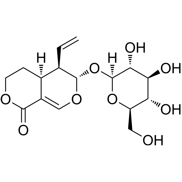
HY-N0494
GentiopicrosideCytochrome P450 Inhibitor -
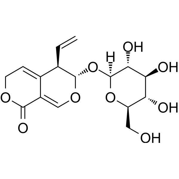
HY-N0807
SwertiamarinNatural Product -

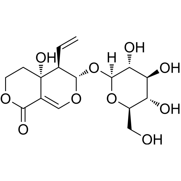
HY-N0806R
Sweroside (Standard)Keap1/Nrf2 Activator -
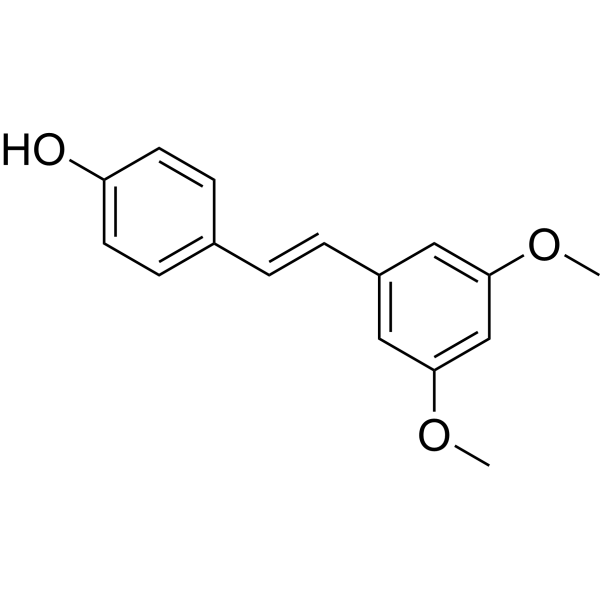
HY-N0828
PterostilbeneAutophagy Inducer -
HY-N0083
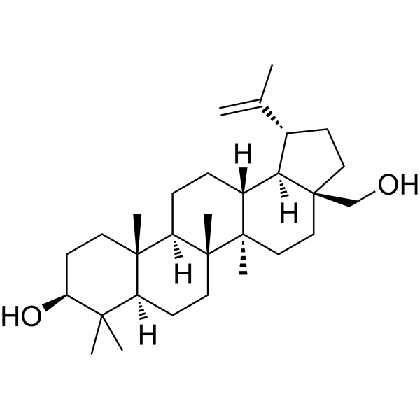
BetulinEndogenous Metabolite -
HY-N0664
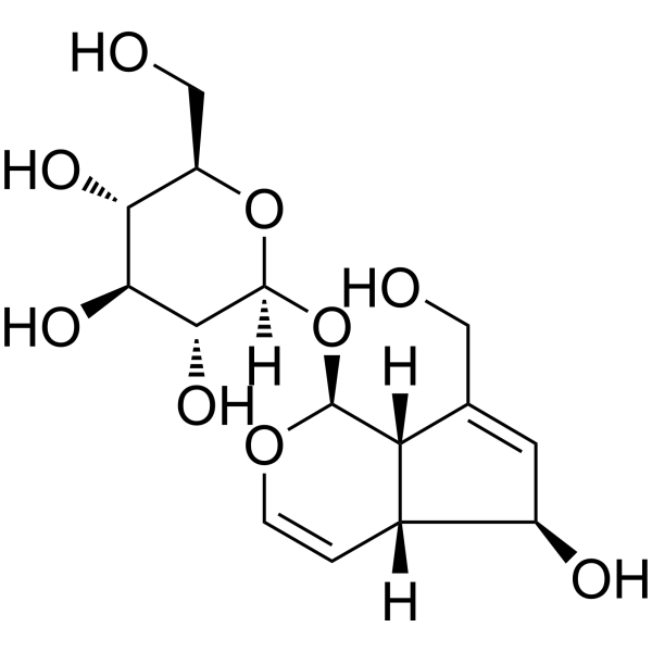
AucubinBacterial Inhibitor -
HY-N0889
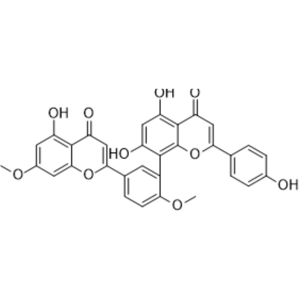
GinkgetinAutophagy Inducer -
HY-N0576
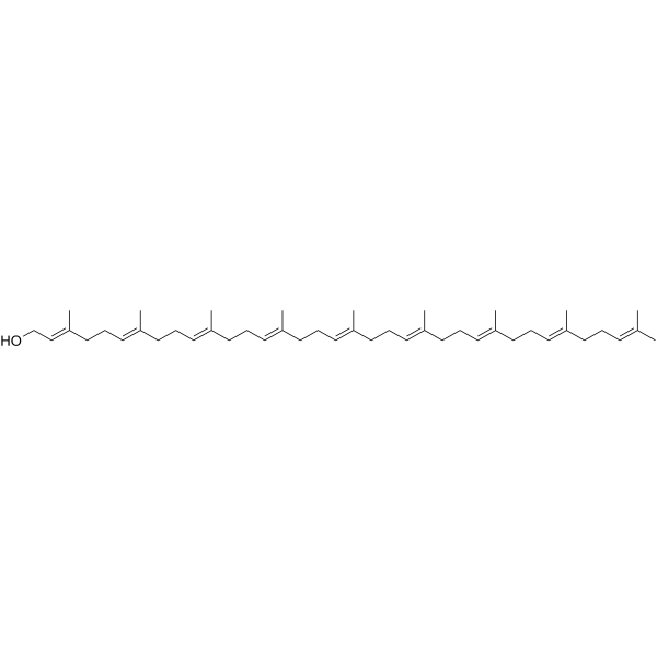
SolanesolAliphatic Terpene Alcohol -

HY-N0828R
Pterostilbene (Standard)Autophagy Inducer -
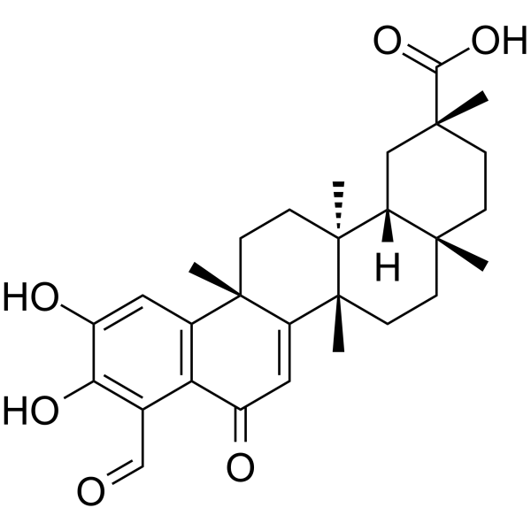
HY-N0587
DemethylzeylasteralApoptosis Inducer -
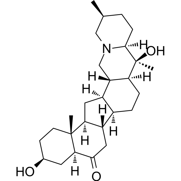
HY-N0213
PeiminineAnti-Inflammatory Agent -

HY-N0664R
Aucubin (Standard)Bacterial Inhibitor
Publications Citing Use of MedChemExpress (MCE) Sweroside
More Customer Validation & Images

-
Cell Proliferation/Viability Assay

-
Cell Imaging/Staining

-
WB

-
IF

-
Histological Imaging/Staining

All AMPK Isoforms
More

Biological Activity
Description
IC50 & Target
|
SIRT1 |
NLRP3 |
Cellular Effect
|
Cell Line

|
Type | Value | Description | References |
|---|---|---|---|---|
| HeLa | IC50 |
>50 μM
Compound: 18
|
Cytotoxicity against human HeLa cells after 48 hrs by MTT assay
Cytotoxicity against human HeLa cells after 48 hrs by MTT assay
|
[PMID: 23002924] |
In Vitro
Sweroside (10-100 μM; pretreatment for 24 h followed by hypoxia/reoxygenation) enhances cell viability and reduced CK-MB and LDH release in H9c2 cells[1].
Sweroside (50 μM; 24 h+hypoxia/reoxygenation treatment) reduces ROS and MDA levels, increases SOD and GSH-Px activities, upregulates HO-1 expression, reduces caspase-1 activity and IL-1β levels, and downregulates NLRP3, ASC, cleaved caspase-1 and IL-1β expression in H9c2 cells[1].
Sweroside (50 μM; 24 h+hypoxia/reoxygenation treatment) inhibits Keap1 expression in H9c2 cells and promotes Nrf2 nuclear translocation[1].
Sweroside (5-320 μM; 24-48 h) reduces cell viability in leukemia cell lines (K562, U937, HL-60, etc.) and has low toxicity to normal cells[2].
Sweroside (20-80 μM; 24 h) induces cell cycle arrest at the S and G2/M phases in HL-60 cells, downregulates Cyclin D1, CDK4, CDC2, upregulates p53, p21, and induces apoptosis, activates caspase-3, -9 and PARP, upregulates Bax, and downregulates Bcl-2[2].
Sweroside reduces LPS-induced inflammation in RAW264.7 cells by activating SIRT1 to mediate NF-κB and FOXO1 pathways[3].
Sweroside (0.1-10 μg/mL; 24 h) reduces lipid accumulation and activates autophagy (upregulation of LC3B-II and downregulation of P62) in palmitic acid-treated primary mouse hepatocytes, acting through the AMPK/mTOR pathway[4].
MedChemExpress (MCE) has not independently confirmed the accuracy of these methods. They are for reference only. Further protocols information, click here.
-
Cell Line:K562, U937, HL-60, NB4, THP-1 (leukemia cells); PBMC, BMC, HL-7702, AD293, BEAS-2B (normal cells)
-
Concentration:5, 10, 20, 40, 80, 160, 320 μM
-
Incubation Time:24 h, 48 h
-
Result:Reduced viability of leukemia cells in a dose-dependent manner (IC50: 156.83 μM for K562, 67.92 μM for U937, 62.58 μM for HL-60, etc.) without significant toxicity to normal cells.
In Vivo
Sweroside (25-100 mg/kg; intraperitoneal injection; daily; 28 days) inhibits tumor growth, prolonged survival, and induces tumor cell apoptosis in a mouse HL-60 xenograft tumor model[2].
Sweroside (15-60 mg/kg; intraperitoneal injection before LPS treatment; single dose) reduces lung wet-to-dry ratio and MPO activity, reduces inflammatory cells and cytokines, activates SIRT1, and inhibits NF-κB in an LPS-induced mouse acute lung injury model[3].
EX-527 (HY-15452) (60 mg/kg Sweroside+10 mg/kg EX-527; intraperitoneal injection before LPS treatment; single dose) can antagonize the in vivo effects of sweroside in the acute lung injury model induced by LPS in mice[3].
Sweroside (120 mg/kg/day; daily; 3 months) reduces hepatic lipid accumulation, activates AMPK/mTOR-mediated autophagy, and improves metabolic indicators in the high-fat diet-induced non-alcoholic fatty liver disease model in C57BL/6J mice[4].
In a high-fat diet-induced non-alcoholic fatty liver disease model in C57BL/6J mice, 3-Methyladenine (HY-19312) (120 mg/kg/day Sweroside for 3 months+30 mg/kg 3-Methyladenine (3-MA) ??3 times a week for 2 weeks; administered for the last 2 weeks) reverses the improvement of hepatic lipid accumulation induced by sweroside[4].
MedChemExpress (MCE) has not independently confirmed the accuracy of these methods. They are for reference only.
-
Animal Model:2-month-old C57BL/6J mice (20-25 g) with LPS-induced acute lung injury (ALI) model (inhaled LPS 5 mg/kg)[3]
-
Dosage:15, 30, 60 mg/kg
-
Administration:Intraperitoneal injection, single dose before LPS inhalation
-
Result:Decreased lung wet-to-dry ratio, inhibited MPO activity, reduced numbers of inflammatory cells (neutrophils, macrophages) in BALF, alleviated lung histopathological damage (alveolar destruction, wall thickening), reduced TNF-α and IL-1β levels, upregulated SIRT1 expression, and downregulated NF-κB activation (p-p65, p-IκBα).
-
Animal Model:4-6-week-old male C57BL/6J mice with high-fat diet (HFD)-induced nonalcoholic fatty liver disease (NAFLD) model (HFD with 60% calories from fat for 3 months)[4]
-
Dosage:120 mg/kg/day
-
Administration:Injection, daily, 3 months
-
Result:Reduced body weight and liver mass, improved glucose and insulin tolerance, decreased serum levels of TG, TC, FFA, ALT, AST, TNF-α, and IL-1β, reduced hepatic lipid accumulation (Oil Red O staining), activated autophagy (upregulated LC3B-II, downregulated P62), and activated AMPK/mTOR pathway (increased p-AMPK, decreased p-mTOR).


Chemical Information
-
CAS No. 14215-86-2
-
Appearance Solid
-
Molecular Weight 358.34
-
Formula C16H22O9
-
Color White to off-white
-
SMILES
C=C[C@@H]1[C@@](CCOC2=O)([H])C2=CO[C@H]1O[C@]([C@@H]([C@@H](O)[C@@H]3O)O)([H])O[C@@H]3CO
-
Structure Classification
-
Shipping
Room temperature in continental US; may vary elsewhere.
-
Storage
4°C, protect from light
* In solvent : -80°C, 6 months; -20°C, 1 month (protect from light)


Publications (4)
-
Journal Impact Factor
-
Most Recent
-
Phytomedicine
Liquiritin targets NF-κB/MAPK signaling to attenuate osteoclastogenesis and triggers apoptosis through PPARγ activation. [Abstract]2025 Dec:149:157604. PMID: 41308395 -
Int Immunopharmacol
Sweroside ameliorates endothelial dysfunction via the KLF2-mediated repression of the FABP4/CCL20 signaling axis. [Abstract]2026 Jul 15:181:116771. PMID: 42068897 -
Int Immunopharmacol
Therapeutic potential of Sweroside in postmenopausal osteoporosis: Inhibition of osteoclast differentiation and promotion of osteoclast apoptosis via NF-κB and MAPK pathways. [Abstract]2025 May 16:155:114630. PMID: 40220621
Sweroside purchased from MedChemExpress. Usage Cited in: Int Immunopharmacol. 2025 May 16:155:114630. [Abstract]
The effect of Sweroside on BMDMs viability was assessed using CCK-8 assays over a range of Sweroside concentrations from 0 to 400 μM.
Sweroside purchased from MedChemExpress. Usage Cited in: Int Immunopharmacol. 2025 May 16:155:114630. [Abstract]
TRAcP staining was performed to evaluate osteoclast differentiation in BMDMs treated with different concentrations of Sweroside (0, 10, 20 and 40 μM).
Sweroside purchased from MedChemExpress. Usage Cited in: Int Immunopharmacol. 2025 May 16:155:114630. [Abstract]
Protein expressions of integrin-β3, CTSK, NFATc1, and c-Fos were evaluated by Western blotting after treatment with RANKL and Sweroside (40 μM) at 0, 1, 3, and 5 days.
Sweroside purchased from MedChemExpress. Usage Cited in: Int Immunopharmacol. 2025 May 16:155:114630. [Abstract]
Immunofluorescence staining and quantitative assessment of the average nuclear p65 fluorescence intensity were conducted to evaluate the localization of p65 in RANKL and Sweroside (40 μM)-treated BMDMs.
Sweroside purchased from MedChemExpress. Usage Cited in: Int Immunopharmacol. 2025 May 16:155:114630. [Abstract]
Histomorphometric differences among Sham, OVX, and OVX + Sweroside (30 mg/kg/day) groups were assessed using H&E and TRAcP staining.
-
Bioorg Chem
Discovery of cyanidin-3-O-galactoside as a novel CNT2 inhibitor for the treatment of hyperuricemia. [Abstract]2025 Jan:154:108108. PMID: 39753042


Solvent & Solubility
In Vitro:
DMSO : 100 mg/mL (279.06 mM; Need ultrasonic; Hygroscopic DMSO has a significant impact on the solubility of product, please use newly opened DMSO)
H2O : 50 mg/mL (139.53 mM; Need ultrasonic)
Please refer to the solubility information to select the appropriate solvent. Once prepared, please aliquot and store the solution to prevent product inactivation from repeated freeze-thaw cycles.
Storage method and period of stock solution: -80°C, 6 months; -20°C, 1 month (protect from light). When stored at -80°C, please use it within 6 months. When stored at -20°C, please use it within 1 month.
* Note: If you choose water as the stock solution, please dilute it to the working solution, then filter and sterilize it with a 0.22 μm filter before use.
Please refer to the solubility information to select the appropriate solvent. Once prepared, please aliquot and store the solution to prevent product inactivation from repeated freeze-thaw cycles.
Storage method and period of stock solution: -80°C, 6 months; -20°C, 1 month (protect from light). When stored at -80°C, please use it within 6 months. When stored at -20°C, please use it within 1 month.
* Note: If you choose water as the stock solution, please dilute it to the working solution, then filter and sterilize it with a 0.22 μm filter before use.
Concentration (start) × Volume (start) = Concentration (final) × Volume (final)
In Vivo:
Select the appropriate dissolution method based on your experimental animal and administration route.
- For the following dissolution methods, please ensure to first prepare a clear stock solution using an In Vitro approach and then sequentially add co-solvents:
- To ensure reliable experimental results, the clarified stock solution can be appropriately stored based on storage conditions. As for the working solution for In Vivo experiments, it is recommended to prepare freshly and use it on the same day.
- The percentages shown for the solvents indicate their volumetric ratio in the final prepared solution. If precipitation or phase separation occurs during preparation, heat and/or sonication can be used to aid dissolution.
Add each solvent one by one: 10% DMSO 40% PEG300 5% Tween-80 45% Saline
Solubility: ≥ 2.5 mg/mL (6.98 mM); Clear solution
This protocol yields a clear solution of ≥ 2.5 mg/mL (saturation unknown).
Taking 1 mL working solution as an example, add 100 μL DMSO stock solution (25.0 mg/mL) to 400 μL PEG300, and mix evenly; then add 50 μL Tween-80 and mix evenly; then add 450 μL Saline to adjust the volume to 1 mL.
Preparation of Saline: Dissolve 0.9 g sodium chloride in ddH₂O and dilute to 100 mL to obtain a clear Saline solution.
Add each solvent one by one: 10% DMSO 90% (20% SBE-β-CD in Saline)
Solubility: ≥ 2.5 mg/mL (6.98 mM); Clear solution
This protocol yields a clear solution of ≥ 2.5 mg/mL (saturation unknown).
Taking 1 mL working solution as an example, add 100 μL DMSO stock solution (25.0 mg/mL) to 900 μL 20% SBE-β-CD in Saline, and mix evenly.
Preparation of 20% SBE-β-CD in Saline (4°C, storage for one week): 2 g SBE-β-CD powder is dissolved in 10 mL Saline, completely dissolve until clear.
For the following dissolution methods, please prepare the working solution directly:
It is recommended to prepare fresh solutions and use them promptly within a short period of time.
The percentages shown for the solvents indicate their volumetric ratio in the final prepared solution. If precipitation or phase separation occurs during preparation, heat and/or sonication can be used to aid dissolution.
Add each solvent one by one: PBS
Solubility: 100 mg/mL (279.06 mM); Clear solution; Need ultrasonic
In Vivo Dissolution Calculator
Please enter the basic information of animal experiments:
-
-
-
-
Recommended: Prepare an additional quantity of animals to account for potential losses during experiments.
Working solution concentration: 0.22 mg/mL
This product has good water solubility, please refer to the measured solubility data in water/PBS/Saline for details.

Protocols
-
Annexin V plus membrane-impermeant dye apoptosis staining
Annexin V-based apoptosis assays rely on the detection of phosphatidylserine (PS) externalization from the inner leaflet of the plasma membrane to the outer leaflet, an early biochemical hallmark of apoptosis. Fluorescently labeled Annexin V binds PS in a calcium-dependent manner, enabling identification of early apoptotic cells by flow cytometry or fluorescence microscopy. When combined with a membrane-impermeant DNA-binding dye (e. g. , propidium iodide), this approach allows discrimination between viable (Annexin V−/dye−), early apoptotic (Annexin V+/dye−), and late apoptotic or necrotic (Annexin V+/dye+) cell populations by assessing membrane integrity and PS exposure.
-
ROS/oxidative-stress fluorescent staining
ROS/oxidative-stress fluorescent staining uses cell-permeant fluorogenic probes that become fluorescent after oxidation inside cells or tissues; commonly used examples include DCFH-DA/DCFDA for broad cellular oxidant detection, DHE for superoxide-related signal detection, MitoSOX for mitochondrial superoxide-related signal detection, and CellROX probes for oxidative-stress-associated fluorescence readouts. The assay detects probe oxidation rather than a single ROS species unless the probe and analysis method have been chemically validated for that species. DCFH-DA enters cells, is deacetylated by intracellular esterases to DCFH, and produces fluorescent DCF after oxidation, so the readout is used as an operational measure of total cellular oxidative stress rather than a species-specific ROS measurement. DHE and MitoSOX can report superoxide-related oxidation, but red fluorescence alone can include non-specific ethidium-like oxidation products; HPLC or optimized spectral approaches are
-
Research Protocol for Inflammation-related Diseases
The NLRP3 inflammasome is a cytosolic innate immune signaling platform that integrates priming signals and danger-signal activation to promote caspase-1 activation, maturation of IL-1β and IL-18, and gasdermin D-mediated pyroptotic cell death. The core experimental logic is to determine whether inflammatory disease phenotypes are driven by increased NLRP3 expression, ASC-containing inflammasome assembly, caspase-1 cleavage, GSDMD cleavage, and extracellular release of IL-1β/IL-18 rather than by nonspecific cell injury alone. The pathway is strongly linked to inflammation-related disease phenotypes because monosodium urate crystals activate NALP3/NLRP3 inflammasome signaling in gout-like crystal inflammation, cholesterol crystals activate NLRP3 inflammasomes in atherogenesis models, and DSS-induced intestinal inflammation has been reported to involve NLRP3 inflammasome activity. However, experimental colitis studies also show context-dependent protective effects of NLRP3 inflammasome co
-
Research Protocol for Metabolic Diseases
AMP-activated protein kinase, AMPK, is a conserved cellular energy sensor that responds to reduced cellular energy status and coordinates metabolism by increasing ATP-generating catabolic pathways while suppressing ATP-consuming anabolic processes. In metabolic disease research, the AMPK pathway is experimentally relevant because it regulates hepatic lipid synthesis, fatty acid oxidation, glucose production, skeletal-muscle glucose disposal, mTORC1-linked biosynthesis, autophagy, mitochondrial homeostasis, and whole-body energy balance. The central pathway logic is that energy stress, metformin, exercise-like stimulation, or direct AMPK activators increase AMPKα Thr172 phosphorylation and downstream substrate phosphorylation, including ACC and RAPTOR. Phosphorylation of ACC suppresses lipogenesis and supports fatty acid oxidation, whereas phosphorylation of RAPTOR suppresses mTORC1 signaling and links cellular energy status to growth and protein synthesis control. The pathway is linked
-
Cytoplasmic-Nuclear Fractionated Protein Extraction
Cytoplasmic-nuclear fractionated protein extraction separates soluble cytoplasmic proteins from nuclear-enriched proteins by mild plasma-membrane permeabilization, differential centrifugation, washing of nuclei, and extraction of nuclear proteins for downstream immunoblotting or related molecular analysis. The readout is the relative abundance of a protein in cytoplasmic and nuclear fractions, commonly assessed by western blotting together with compartment markers such as tubulin or pyruvate kinase for cytoplasm and lamin, nucleoporin, hnRNP, H2AX, or Lamin B for nuclear fractions.
-
Apoptosis
Apoptosis, also called programmed cell death, is generally characterized by distinct morphological characteristics.
-
TUNEL staining for apoptotic DNA fragmentation
TUNEL staining detects DNA strand breaks by using terminal deoxynucleotidyl transferase to add labeled nucleotides to exposed 3′-OH DNA termini, generating either microscopic staining in fixed cells or tissue sections, or fluorescence/cytometric signal in cell suspensions. TUNEL positivity reflects DNA fragmentation but should not be interpreted alone as definitive apoptosis, because TUNEL can also label necrotic, autolytic, mechanically damaged, or DNA-repair-associated DNA breaks.
-
Autophagy
Autophagy is a process in which eukaryotic cells use lysosomes to degrade their own cytoplasmic proteins and damaged organelles under the regulation of autophagy related gene (Atg). Microtubule-associated proteins light chain 3 (LC3) is recognized as autophagy marker, which transfers from cytoplasmic LC3 (LC3-I) to membrane type (LC3-II). LC3-II/I ratio could be detected by Western Blot and fluorescence microscopy.
-
Apoptosis Solutions
Apoptosis is a regulated, generally non-lytic cell-death pathway that removes unwanted, damaged, infected, or abnormal cells through coordinated morphological changes, caspase activation, DNA fragmentation, and membrane remodeling. The intrinsic apoptosis pathway is controlled mainly by mitochondrial outer membrane permeabilization, BCL-2 family proteins, cytochrome c release, apoptosome formation, caspase-9 activation, and downstream executioner caspase-3/7 activation. The extrinsic apoptosis pathway is initiated by death receptors such as Fas, TNFR, and TRAIL receptors, which recruit adaptor proteins and activate caspase-8 before engaging executioner caspases or mitochondrial amplification through BID cleavage. Apoptosis is linked to many phenotypes, including cancer cell killing, tissue homeostasis, immune regulation, neurodegeneration, infection response, and treatment-induced cytotoxicity; unresolved questions include how apoptosis interacts with necroptosis, pyroptosis, ferroptos
-
Lysosome and acidic-vesicle live-cell staining
Lysosome and acidic-vesicle live-cell staining detects acidic intracellular compartments by using membrane-permeant acidotropic probes that accumulate in low-pH vesicles, including lysosomes, late endosomes, autolysosomes, and acidic phagosomes. LysoTracker staining is commonly used as an intensity-based readout of acidic lysosomal compartment abundance or enlargement, while acridine orange produces green fluorescence in less concentrated compartments and red fluorescence after concentration-dependent accumulation in acidic vesicular organelles. Loss or reduction of acridine-orange red signal can be used as a readout of lysosomal membrane permeabilization or reduced acidic-vesicle integrity. This protocol is designed for live cultured cells and can be adapted for fluorescence microscopy, high-content imaging, plate-reader readout, or flow cytometry when the selected literature supports the readout. Because these dyes report acidotropic accumulation rather than lysosome identity alone,
-
Inhalation Toxicity Study
Inhalation toxicity studies expose rodents to a controlled aerosol, vapor, gas, or smoke atmosphere and assess respiratory and systemic toxicity using exposure-atmosphere characterization, clinical observations, body and organ weights, bronchoalveolar lavage fluid, histopathology, blood chemistry, hematology, and, when included, molecular endpoints such as transcriptomics, proteomics, lipidomics, or tissue burden analysis. The primary biological readouts are airway irritation, pulmonary inflammation, cytotoxicity, altered surfactant or lipid homeostasis, impaired particle clearance, and tissue remodeling, reflected by BALF cell differentials, BALF protein, LDH, phosphatase activities, cytokines, lung weight, microscopic respiratory-tract lesions, and retained lung burden.
-
Macroautophagy Solutions
Macroautophagy is a conserved lysosome-dependent degradation pathway in which cytoplasmic material is sequestered into double-membrane autophagosomes and delivered to lysosomes for degradation and recycling. The pathway supports cellular homeostasis during nutrient limitation, organelle stress, protein-aggregate accumulation, infection, differentiation, and tissue remodeling by coupling cargo sequestration, autophagosome maturation, lysosomal fusion, and degradation of cargo-derived macromolecules. The core molecular sequence includes initiation by nutrient- and stress-regulated autophagy machinery, autophagosome nucleation, LC3/ATG8-family conjugation to autophagosomal membranes, cargo selection through receptors such as SQSTM1/p62, autophagosome-lysosome fusion, and lysosomal degradation. LC3 was identified as a mammalian homolog of yeast Atg8 that localizes to autophagosomal membranes after processing, and p62/SQSTM1 was shown to connect ubiquitinated cargo with autophagic degradati
-
Pyroptosis Solutions
Pyroptosis is a lytic inflammatory cell-death pathway executed by gasdermin pores, most classically through inflammasome-mediated activation of caspase-1, cleavage of gasdermin D, membrane pore formation, LDH release, and secretion of IL-1β and IL-18. The canonical pathway is commonly modeled by priming cells with an inflammatory signal such as LPS to induce pro-IL-1β and inflammasome components, followed by an activation signal such as ATP or nigericin to activate NLRP3, ASC speck formation, caspase-1 cleavage, GSDMD cleavage, cytokine release, and pyroptotic membrane rupture. The non-canonical pathway is triggered when cytosolic LPS activates mouse caspase-11 or human caspase-4/5, leading to GSDMD cleavage and pyroptosis, and this can secondarily activate NLRP3-dependent IL-1β release. Pyroptosis is linked to inflammatory injury, infection, cancer, liver disease, ocular disease, placental inflammation, and other disease phenotypes, but unresolved questions include which gasdermin fam


Purity & Documentation
-
Data Sheet (287 KB)
-
SDS (396 KB)
- English - EN (396 KB)
- Français - FR (396 KB)
- Deutsch - DE (396 KB)
- Norwegian - NO (396 KB)
- Español - ES (396 KB)
- Swedish - SV (396 KB)
- Italian - IT (396 KB)
- Korean - KR (396 KB)
- Portuguese - PT (396 KB)
-
Handling Instructions (2659 KB)
References
[1]. Li J, et al. Sweroside Protects Against Myocardial Ischemia-Reperfusion Injury by Inhibiting Oxidative Stress and Pyroptosis Partially via Modulation of the Keap1/Nrf2 Axis. Front Cardiovasc Med. 2021 Mar 19;8:650368. [Content Brief]
[2]. Han XL, et al. Sweroside eradicated leukemia cells and attenuated pathogenic processes in mice by inducing apoptosis. Biomed Pharmacother. 2017 Nov;95:477-486. [Content Brief]
[3]. Wang J, et al. Anti-inflammatory Effects of Sweroside on LPS-Induced ALI in Mice Via Activating SIRT1. Inflammation. 2021 Oct;44(5):1961-1968. [Content Brief]
[4]. Ding Y, et al. Sweroside alleviates hepatic steatosis in part by activating AMPK/mTOR-mediated autophagy in mice. J Cell Biochem. 2023 Jul;124(7):1012-1022. [Content Brief]


Complete Stock Solution Preparation Table
Please refer to the solubility information to select the appropriate solvent. Once prepared, please aliquot and store the solution to prevent product inactivation from repeated freeze-thaw cycles.
Storage method and period of stock solution: -80°C, 6 months; -20°C, 1 month (protect from light). When stored at -80°C, please use it within 6 months. When stored at -20°C, please use it within 1 month.
| Optional Solvent | Concentration Solvent Mass | 1 mg | 5 mg | 10 mg | 25 mg |
|---|---|---|---|---|---|
| H2O / DMSO | 1 mM | 2.7906 mL | 13.9532 mL | 27.9065 mL | 69.7661 mL |
| 5 mM | 0.5581 mL | 2.7906 mL | 5.5813 mL | 13.9532 mL | |
| 10 mM | 0.2791 mL | 1.3953 mL | 2.7906 mL | 6.9766 mL | |
| 15 mM | 0.1860 mL | 0.9302 mL | 1.8604 mL | 4.6511 mL | |
| 20 mM | 0.1395 mL | 0.6977 mL | 1.3953 mL | 3.4883 mL | |
| 25 mM | 0.1116 mL | 0.5581 mL | 1.1163 mL | 2.7906 mL | |
| 30 mM | 0.0930 mL | 0.4651 mL | 0.9302 mL | 2.3255 mL | |
| 40 mM | 0.0698 mL | 0.3488 mL | 0.6977 mL | 1.7442 mL | |
| 50 mM | 0.0558 mL | 0.2791 mL | 0.5581 mL | 1.3953 mL | |
| 60 mM | 0.0465 mL | 0.2326 mL | 0.4651 mL | 1.1628 mL | |
| 80 mM | 0.0349 mL | 0.1744 mL | 0.3488 mL | 0.8721 mL | |
| 100 mM | 0.0279 mL | 0.1395 mL | 0.2791 mL | 0.6977 mL |
* Note: If you choose water as the stock solution, please dilute it to the working solution, then filter and sterilize it with a 0.22 μm filter before use.


Sweroside Related Classifications
HY-N0806 Related Classifications
 Powered by Bioz