



UNC1062
Based on 2 publication(s) in Google Scholar
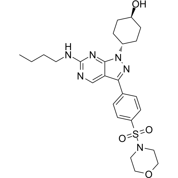
UNC1062 is a highly selective tyrosine kinase (MERTK) inhibitor with an IC50 of 1.1 nM (Morrison Ki = 0.33 nM). UNC1062 exhibits good selectivity for the TAM family (TYRO3 IC50 = 60 nM, AXL IC50 = 85 nM). UNC1062 exhibits significant anti-proliferative effects and induces apoptosis in various cancer models (such as melanoma, gastric cancer, and acute myeloid leukemia). UNC1062 inhibits multiple pathways, including MAPK/ERK, PI3K/AKT and JAK/STAT and affects the motility of head and neck squamous cell carcinoma (HNSCC) cells through the RhoA signaling pathway. UNC1062 inhibits macrophage efferocytosis, and it suitable for research on atherosclerosis.
For research use only. We do not sell to patients.
- Purity : 98.92%
- CAS No.: 1350549-36-8
- Formula: C25H34N6O4S
- Molecular Weight:514.64
-
Storage:Powder -20°C, 3 years , 4°C, 2 years ; In solvent -80°C, 6 months , -20°C, 1 month

To place orders, for customer services and technical support, please contact: MedChemExpress USA
Tel: 609-228-6898 E-mail: [email protected] [email protected]


-
 Biological Activity
-
Chemical Information
-
Solvent & Solubility
- Protocols
- Purity & Documentation
- References
-
 Help & FAQs


Publications Citing Use of MedChemExpress (MCE) UNC1062
More

Biological Activity
Description
IC50 & Target
[1]|
Axl 85 nM () |
Tyro3 60 nM () |
STAT6 |
Akt |
ERK1 |
ERK2 |
In Vitro
UNC1062 (0.5-10 μM, 72 h-21 d) inhibits acute myeloid leukemia (AML) cell lines (OCI/AML5, TMD7, THP-1 and HEL cells) and gastric cancer cell lines (HSC-60 and SNU-5) proliferation and inhibits melanoma cell lines (HMCB and G361 cells) and gastric cancer cells colony formation[1][2][3].
UNC1062 (0.5-2 μM, 48 h) induces apoptosis of melanoma cell lines (HMCB and G361 cells), AML cell lines (OCI/AML5, TMD7, THP-1 and HEL cells) and gastric cancer cell lines (HSC-60 and SNU-5)[1][2][3].
UNC1062 (1-5 μM, 24-72 h) causes G2 arrest in gastric cancer cell lines and HNSCC cell lines (G2 arrest cells and NC cells)[3][4].
UNC1062 (0.25-1 μM, 16-96 h) significantly weakens the migration and invasion ability of SKMEL119 cells and Detroit 562 cells[1][4].
UNC1062 (0.1-2 μM, 90-100 min) inhibits MERTK phosphorylation and significantly attenuates GAS6-induced activation of STAT6, AKT, and ERK1/2[1].
UNC1062 (20 μg/L, 18.5 h) attenuates the effects of Ru360 (a mitochondrial calcium uniporter (MCU) inhibitor) in reducing inflammatory cytokines and intracytoplasmic lipid content in primary mouse macrophages, it was confirmed that MCU affects inflammatory response by regulating efferocytosis[5].
MedChemExpress (MCE) has not independently confirmed the accuracy of these methods. They are for reference only.
-
Cell Line:Melanoma cell lines (HMCB and G361 cells) and AML cell lines (OCI/AML5, TMD7, THP-1 and HEL cells)
-
Concentration:0.5 and 1 μM for melanoma cell lines and 2 μM for AML cell lines
-
Incubation Time:48 h
-
Result:Increased cell death rate and PARP cleavage (caspase-3).
-
Cell Line:SKMEL119 cells and Detroit 562 cells
-
Concentration:1 μM for SKMEL119 cells and 0.25 μM for Detroit 562 cells
-
Incubation Time:16 h for SKMEL119 cells and 24 h Detroit 562 cells
-
Result:Exhibited significant 30% decrease in the velocity of individual SKMEL119 cell migration.
Exhibited an 40% reduction in invasion in Detroit 562 cells.
-
Cell Line:SKMEL119 cells and Detroit 562 cells
-
Concentration:1 μM for SKMEL119 cells and 0.25 μM for Detroit 562 cells
-
Incubation Time:96 h for SKMEL119 cells and 24 h Detroit 562 cells
-
Result:Exhibited an 89% reduction in invasion in SKMEL119 cells.
Exhibited an 45% reduction in invasion in Detroit 562 cells.
-
Cell Line:HMCB and G361 cells
-
Concentration:0.1, 0.2, 0.5, 1 and 2 μM
-
Incubation Time:After 90 min of pre-treatment, add GAS6 (HY-P75174) for 10 min
-
Result:Dose-dependently reduced MERTK phosphorylation.
Significantly attenuated GAS6-induced activation of STAT6, AKT, and ERK1/2.


Chemical Information
-
CAS No. 1350549-36-8
-
Appearance Solid
-
Molecular Weight 514.64
-
Formula C25H34N6O4S
-
Color White to off-white
-
SMILES
O[C@H]1CC[C@H](N2N=C(C3=CC=C(S(=O)(N4CCOCC4)=O)C=C3)C5=CN=C(NCCCC)N=C52)CC1
-
Shipping
Room temperature in continental US; may vary elsewhere.
-
Storage
Powder -20°C 3 years 4°C 2 years In solvent -80°C 6 months -20°C 1 month


Publications (2)
-
Journal Impact Factor
-
Most Recent
-
Immunol Lett
Modulation of Oxidized Low-density Lipoprotein-Affected Macrophage Efferocytosis by Mitochondrial Calcium Uniporter in a Murine Model. [Abstract]2023 Nov:263:14-24. PMID: 37689315 -


Solvent & Solubility
In Vitro:
DMSO : 10 mg/mL (19.43 mM; ultrasonic and warming and heat to 60°C; Hygroscopic DMSO has a significant impact on the solubility of product, please use newly opened DMSO)
Please refer to the solubility information to select the appropriate solvent. Once prepared, please aliquot and store the solution to prevent product inactivation from repeated freeze-thaw cycles.
Storage method and period of stock solution: -80°C, 6 months; -20°C, 1 month. When stored at -80°C, please use it within 6 months. When stored at -20°C, please use it within 1 month.
Please refer to the solubility information to select the appropriate solvent. Once prepared, please aliquot and store the solution to prevent product inactivation from repeated freeze-thaw cycles.
Storage method and period of stock solution: -80°C, 6 months; -20°C, 1 month. When stored at -80°C, please use it within 6 months. When stored at -20°C, please use it within 1 month.
Concentration (start) × Volume (start) = Concentration (final) × Volume (final)


Purity & Documentation
-
Data Sheet (282 KB)
-
SDS (251 KB)
- English - EN (251 KB)
- Français - FR (251 KB)
- Deutsch - DE (251 KB)
- Norwegian - NO (251 KB)
- Español - ES (251 KB)
- Swedish - SV (251 KB)
- Italian - IT (251 KB)
- Korean - KR (251 KB)
- Portuguese - PT (251 KB)
-
Handling Instructions (2659 KB)
References
[1]. Schlegel J, et al. MERTK receptor tyrosine kinase is a therapeutic target in melanoma. J Clin Invest. 2013;123(5):2257-2267. [Content Brief]
[2]. Koda Y, Itoh M, Tohda S. Effects of MERTK Inhibitors UNC569 and UNC1062 on the Growth of Acute Myeloid Leukaemia Cells. Anticancer Res. 2018 Jan;38(1):199-204. [Content Brief]
[3]. Yi JH, et al. MerTK is a novel therapeutic target in gastric cancer. Oncotarget. 2015 Apr 20;8(57):96656-96667. [Content Brief]
[4]. von Mässenhausen A, et al. MERTK as a novel therapeutic target in head and neck cancer. Oncotarget. 2016 May 31;7(22):32678-94. [Content Brief]
[5]. Lu N, et al. Modulation of oxidized low-density lipoprotein-affected macrophage efferocytosis by mitochondrial calcium uniporter in a murine model. Immunol Lett. 2023 Nov;263:14-24. [Content Brief]


Complete Stock Solution Preparation Table
Please refer to the solubility information to select the appropriate solvent. Once prepared, please aliquot and store the solution to prevent product inactivation from repeated freeze-thaw cycles.
Storage method and period of stock solution: -80°C, 6 months; -20°C, 1 month. When stored at -80°C, please use it within 6 months. When stored at -20°C, please use it within 1 month.
| Optional Solvent | Concentration Solvent Mass | 1 mg | 5 mg | 10 mg | 25 mg |
|---|---|---|---|---|---|
| DMSO | 1 mM | 1.9431 mL | 9.7155 mL | 19.4311 mL | 48.5776 mL |
| 5 mM | 0.3886 mL | 1.9431 mL | 3.8862 mL | 9.7155 mL | |
| 10 mM | 0.1943 mL | 0.9716 mL | 1.9431 mL | 4.8578 mL | |
| 15 mM | 0.1295 mL | 0.6477 mL | 1.2954 mL | 3.2385 mL |


UNC1062 Related Classifications
HY-117548 Related Classifications
 Powered by Bioz