



1-Monopalmitin

Based on 1 Customer Validation
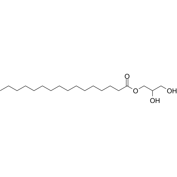
1-Monopalmitin (Glyceryl palmitate) is an activator of the PI3K/Akt pathway and an inhibitor of P-glycoprotein (P-gp). 1-Monopalmitin can induce G2/M arrest and caspase-dependent apoptosis in cancer cells, while inhibiting IAPs protein expression. 1-Monopalmitin can increase drug accumulation by inhibiting P-gp activity in intestinal Caco-2 cells. 1-Monopalmitin has the ability to induce protective autophagy and apoptosis of lung cancer cells (IC50=50-58 μg/mL), with low toxicity to normal cells.
For research use only. We do not sell to patients.
- Purity : 99.89%
- CAS No.: 542-44-9
- Formula: C19H38O4
- Molecular Weight:330.51
-
Storage:Powder -20°C, 3 years , 4°C, 2 years ; In solvent -80°C, 6 months , -20°C, 1 month

To place orders, for customer services and technical support, please contact: MedChemExpress USA
Tel: 609-228-6898 E-mail: [email protected] [email protected]


-
 Biological Activity
-
Chemical Information
-
Solvent & Solubility
- Protocols
- Purity & Documentation
- References
-
 Help & FAQs


-
HY-N1428
Citric acidOrganic Acid -
HY-N2512
1-MonomyristinFAAH/MAGL Inhibitor -

HY-W009141R
1-Monopalmitin (Standard)P-glycoprotein Inhibitor -
HY-128754
MonooleinBiological Lipid -

HY-Y1039
1-Stearoyl-rac-glycerolBiochemical Assay Reagent -
HY-W015309
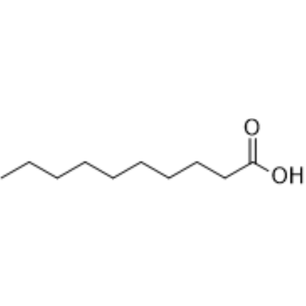
Decanoic acidTyrosinase Inhibitor; Melanosome Inhibitor; mTORC1 Signaling Inhibitor -
HY-50671
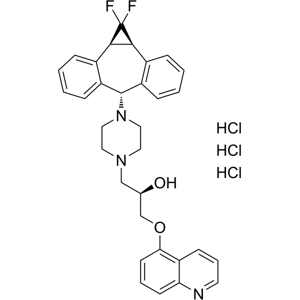
Zosuquidar trihydrochlorideP-gp Inhibitor -
HY-W127329
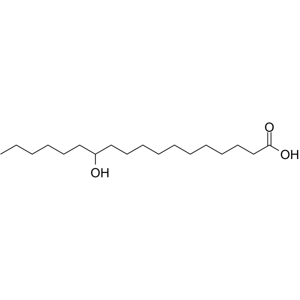
12-Hydroxystearic acidOrganogelator -
HY-N1916
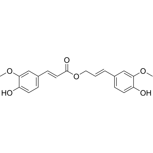
Coniferyl ferulateGST Inhibitor -
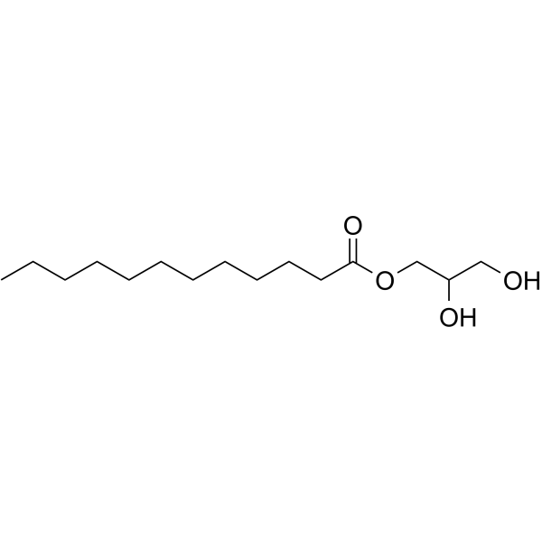
HY-121620
MonolaurinAntimicrobial Agent -
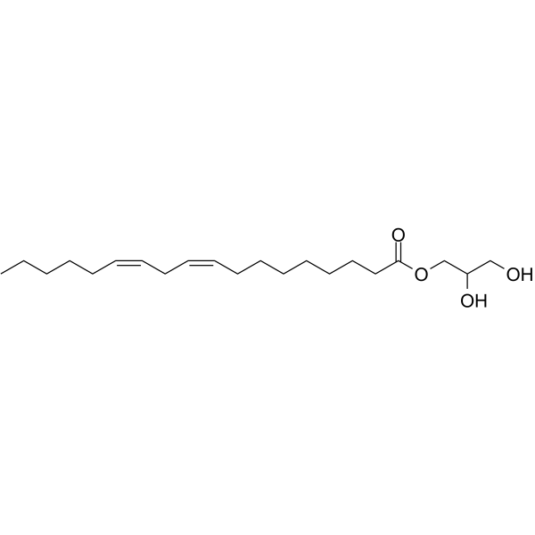
HY-111346
1-Linoleoyl GlycerolLpPLA2 Inhibitor -

HY-128754R
Monoolein (Standard)Biological Lipid
All Caspase Isoforms
More

Biological Activity
Description
In Vitro
1-Monopalmitin (0-100 μg/mL; 48 h) inhibits cell proliferation in lung cancer cells (A549, SPC-A1, H460, H1299) in a dose-dependent manner with low toxicity to normal bronchial epithelial cells HBE[1].
1-Monopalmitin (50 μg/mL; 20 h) induces G2/M arrest in A549 and SPC-A1 cells, upregulates p21 protein and downregulates Cyclin D1[1].
1-Monopalmitin (12.5-50 μg/mL; 48 h) promotes apoptosis of A549 and SPC-A1 cells, activates Caspase-3 and PARP cleavage, and inhibits the expression of inhibitor of apoptosis proteins (IAPs)[1].
1-Monopalmitin (50 μg/mL; 8 h) induces protective autophagy in A549 and SPC-A1 cells, as evidenced by LC3-II accumulation and p62 degradation[1].
1-Monopalmitin (0.1-300 μM; 2 h) increases Rhodamine-123 (HY-D0816) and Daunorubicin (HY-13062A) accumulation in a dose-dependent manner in intestinal Caco-2 cells, inhibits P-glycoprotein (P-gp) activity with non-cytotoxictiy[2].
MedChemExpress (MCE) has not independently confirmed the accuracy of these methods. They are for reference only.
-
Cell Line:A549, SPC-A1 (lung cancer cells); H460, H1299 (lung cancer cells); HBE (normal human bronchial epithelial cells)
-
Concentration:0, 12.5, 25, 50, 75, 100, 150, 200 μg/mL
-
Incubation Time:48 h
-
Result:Significantly reduced viability of A549 (IC50=50.12 μg/mL) and SPC-A1 (IC50=58.30 μg/mL) cells in a dose-dependent manner.
Showed higher IC50 values against H460 and H1299 cells of 176.75 and 165.53 μg/mL, respectively; while exhibited IC50 of 161.34 μg/mL against normal HBE cells, indicating minimal toxicity to normal cells.
- RNA extraction experimental
- Western Blot
- Detection of Apoptosis
- TUNEL staining for apoptotic DNA fragmentation
- Flow cytometric DNA-content cell-cycle staining
- Annexin V plus membrane-impermeant dye apoptosis staining
- Autophagy
- Apoptosis Solutions
- Protocol for Cell Cycle
- Lysosome and acidic-vesicle live-cell staining
- Macroautophagy Solutions
- Protocol For Protein Expression And Purification


Chemical Information
-
CAS No. 542-44-9
-
Appearance Solid
-
Molecular Weight 330.51
-
Formula C19H38O4
-
Color White to off-white
-
SMILES
OC(CO)COC(CCCCCCCCCCCCCCC)=O
-
Synonyms
Glyceryl palmitate
-
Initial Source
-
Shipping
Room temperature in continental US; may vary elsewhere.
-
Storage
Powder -20°C 3 years 4°C 2 years In solvent -80°C 6 months -20°C 1 month


Solvent & Solubility
In Vitro:
DMSO : 50 mg/mL (151.28 mM; ultrasonic and warming and heat to 60°C; Hygroscopic DMSO has a significant impact on the solubility of product, please use newly opened DMSO)
Ethanol : 50 mg/mL (151.28 mM; warming and heat to 60°C)
Please refer to the solubility information to select the appropriate solvent. Once prepared, please aliquot and store the solution to prevent product inactivation from repeated freeze-thaw cycles.
Storage method and period of stock solution: -80°C, 6 months; -20°C, 1 month. When stored at -80°C, please use it within 6 months. When stored at -20°C, please use it within 1 month.
Please refer to the solubility information to select the appropriate solvent. Once prepared, please aliquot and store the solution to prevent product inactivation from repeated freeze-thaw cycles.
Storage method and period of stock solution: -80°C, 6 months; -20°C, 1 month. When stored at -80°C, please use it within 6 months. When stored at -20°C, please use it within 1 month.
Concentration (start) × Volume (start) = Concentration (final) × Volume (final)
In Vivo:
Select the appropriate dissolution method based on your experimental animal and administration route.
- For the following dissolution methods, please ensure to first prepare a clear stock solution using an In Vitro approach and then sequentially add co-solvents:
- To ensure reliable experimental results, the clarified stock solution can be appropriately stored based on storage conditions. As for the working solution for In Vivo experiments, it is recommended to prepare freshly and use it on the same day.
- The percentages shown for the solvents indicate their volumetric ratio in the final prepared solution. If precipitation or phase separation occurs during preparation, heat and/or sonication can be used to aid dissolution.
In Vivo Dissolution Calculator
Please enter the basic information of animal experiments:
-
-
-
-
Recommended: Prepare an additional quantity of animals to account for potential losses during experiments.
Please enter your animal formula composition:
-
%DMSO +
Recommended: Keep the proportion of DMSO in working solution below 2% if your animal is weak.
-
%+
-
+%Tween-80 + +
-
%Saline +
The co-solvents required include: DMSO, . All of co-solvents are available by MedChemExpress (MCE). , Tween 80. All of co-solvents are available by MedChemExpress (MCE).
Working solution concentration: 0.22 mg/mL
Method for preparing stock solution: mg drug dissolved in μL DMSO. Stock solution concentration: mg/mL.
1. Take μL DMSO stock solution;
2. Add μL .
μL , mix evenly;
3. Then add μL Tween 80, mix evenly;
4. Then add μL
Please ensure that the stock solution in the first step is dissolved to a clear state, and add co-solvents in sequence. You can use ultrasonic heating (ultrasonic cleaner, recommended frequency 20-40 kHz), vortexing, etc. to assist dissolution.


Purity & Documentation
-
Data Sheet (268 KB)
-
SDS (392 KB)
- English - EN (392 KB)
- Français - FR (392 KB)
- Deutsch - DE (392 KB)
- Norwegian - NO (392 KB)
- Español - ES (392 KB)
- Swedish - SV (392 KB)
- Italian - IT (392 KB)
- Korean - KR (392 KB)
- Portuguese - PT (392 KB)
-
Handling Instructions (2659 KB)
References


Complete Stock Solution Preparation Table
Please refer to the solubility information to select the appropriate solvent. Once prepared, please aliquot and store the solution to prevent product inactivation from repeated freeze-thaw cycles.
Storage method and period of stock solution: -80°C, 6 months; -20°C, 1 month. When stored at -80°C, please use it within 6 months. When stored at -20°C, please use it within 1 month.
| Optional Solvent | Concentration Solvent Mass | 1 mg | 5 mg | 10 mg | 25 mg |
|---|---|---|---|---|---|
| DMSO / Ethanol | 1 mM | 3.0256 mL | 15.1281 mL | 30.2563 mL | 75.6407 mL |
| 5 mM | 0.6051 mL | 3.0256 mL | 6.0513 mL | 15.1281 mL | |
| 10 mM | 0.3026 mL | 1.5128 mL | 3.0256 mL | 7.5641 mL | |
| 15 mM | 0.2017 mL | 1.0085 mL | 2.0171 mL | 5.0427 mL | |
| 20 mM | 0.1513 mL | 0.7564 mL | 1.5128 mL | 3.7820 mL | |
| 25 mM | 0.1210 mL | 0.6051 mL | 1.2103 mL | 3.0256 mL | |
| 30 mM | 0.1009 mL | 0.5043 mL | 1.0085 mL | 2.5214 mL | |
| 40 mM | 0.0756 mL | 0.3782 mL | 0.7564 mL | 1.8910 mL | |
| 50 mM | 0.0605 mL | 0.3026 mL | 0.6051 mL | 1.5128 mL | |
| 60 mM | 0.0504 mL | 0.2521 mL | 0.5043 mL | 1.2607 mL | |
| 80 mM | 0.0378 mL | 0.1891 mL | 0.3782 mL | 0.9455 mL | |
| 100 mM | 0.0303 mL | 0.1513 mL | 0.3026 mL | 0.7564 mL |


1-Monopalmitin Related Classifications
HY-W009141 Related Classifications
 Powered by Bioz