



Umckalin
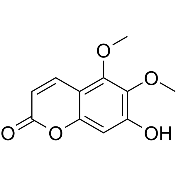
Umckalin is Coumarin (HY-N0709) derivative that exhibits anti-inflammatory properties. Umckalin reduces phosphorylation of p38 MAPK, JNK, and ERK, lowers TNF-α, IL-6, IL-1β, NO, and PGE2 production. Umckalin can be used for the research of chronic inflammatory diseases.
Nos produits utilisent uniquement pour la recherche. Nous ne vendons pas aux patients.
- CAS No.: 43053-62-9
- Formule: C11H10O5
- Masse moléculaire:222.19
-
Stockage:
Please store the product under the recommended conditions in the Certificate of Analysis.

To place orders, for customer services and technical support, please contact: MedChemExpress USA
Tel: 609-228-6898 E-mail: [email protected] [email protected]


-
 Activité biologique
-
Chemical Information
- Protocole
- Pureté et documentation
- Références
-
 Help & FAQs


Voir tous les produits spécifiques à Isoform TNF Receptor
More

Activité biologique
Description
In Vitro
Umckalin (25-400 µM; 72 h) does not reduce viability of B16F10 murine melanoma cells at concentrations up to 200 µM[2].
Umckalin (50-200 µM; 72 h) dose-dependently increases intracellular melanin content in B16F10 murine melanoma cells[2].
Umckalin (50-200 µM; 72 h) dose-dependently enhances tyrosinase activity in B16F10 murine melanoma cells[2].
Umckalin (50-200 µM; 24 h) upregulates TRP-1 and MITF protein expression in B16F10 murine melanoma cells, while having minimal to no effect on tyrosinase and TRP-2 expression[2].
Umckalin (50-200 µM; 24 h) activates the GSK-3β/β-catenin pathway in B16F10 murine melanoma cells by increasing Ser9 phosphorylation of GSK-3β, reducing β-catenin phosphorylation, and increasing total β-catenin levels[2].
Umckalin (50-200 µM; 4 h) suppresses MAPK pathway activity in B16F10 murine melanoma cells by reducing ERK and JNK phosphorylation, while not affecting p38 phosphorylation[2].
Umckalin (50-200 µM; 4 h) activates the PI3K/AKT pathway in B16F10 murine melanoma cells by increasing PI3K and AKT phosphorylationn[2].
MedChemExpress (MCE) has not independently confirmed the accuracy of these methods. They are for reference only.
-
Cell Line:B16F10 murine melanoma cells
-
Concentration:25; 50; 100; 200; 400 µM
-
Incubation Time:72 h
-
Result:Showed no cytotoxic effects at concentrations up to 200 µM.
Caused significant cytotoxicity at 400 µM.
-
Cell Line:B16F10 murine melanoma cells
-
Concentration:50; 100; 200 µM
-
Incubation Time:24 h
-
Result:Increased TRP-1 protein expression significantly in a dose-dependent manner (p < 0.001 at all concentrations).
Caused a small but significant increase in tyrosinase expression only at 200 µM.
Had no notable effect on TRP-2 expression.
Increased MITF protein expression significantly in a dose-dependent manner.
Increased Ser9 phosphorylation of GSK-3β significantly in a dose-dependent manner.
Increased total β-catenin levels significantly only at 200 µM.
Reduced β-catenin phosphorylation significantly at 200 µM.
-
Cell Line:B16F10 murine melanoma cells
-
Concentration:50-200 µM
-
Incubation Time:4 h
-
Result:Reduced ERK phosphorylation significantly in a dose-dependent manner.
Reduced JNK phosphorylation significantly in a dose-dependent manner.
Had no significant effect on p38 phosphorylation.\nIncreased PI3K phosphorylation significantly in a dose-dependent manner.
Increased AKT phosphorylation significantly in a dose-dependent manner.


Chemical Information
-
CAS No. 43053-62-9
-
Masse moléculaire 222.19
-
Formule C11H10O5
-
SMILES
O=C1C=CC2=C(OC)C(OC)=C(O)C=C2O1
-
Structure Classification
-
Initial Source
-
Livraison
Room temperature in continental US; may vary elsewhere.
-
Stockage
Please store the product under the recommended conditions in the Certificate of Analysis.


Pureté et documentation
Références

Calculators
Concentration (start) × Volume (start) = Concentration (final) × Volume (final)


Umckalin Related Classifications
HY-N8712 Related Classifications
 Powered by Bioz