



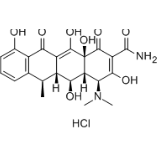
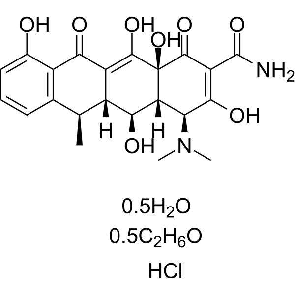
Doxycycline hydrochloride GMP is Doxycycline (hydrochloride) (HY-N0565A) produced by using GMP guidelines. GMP small molecules works appropriately as an auxiliary reagent for cell therapy manufacture. Doxycycline hydrochloride is an orally active highly lipophilic, tissue-permeable MMP inhibitor with broad-spectrum antibacterial activity. Doxycycline hydrochloride is also a semi-synthetic antibiotic with chelating properties, which blocks bacterial protein synthesis and inhibits extracellular matrix degradation through interactions with zinc and calcium atoms. Doxycycline hydrochloride also inhibits mitochondrial biogenesis, translation, and the expression of respiratory chain proteins. Doxycycline hydrochloride induces apoptosis, inhibits autophagy and EMT, downregulates stem cell markers, and activates the PI3K-AKT pathway, thereby effectively inhibiting the viability and proliferation of cancer cells such as breast cancer cells. Doxycycline hydrochloride also promotes the survival and self-renewal of embryonic stem cells and neural stem cells, and reduces the frequency of medium changes in culture. Doxycycline hydrochloride has been applied in studies related to breast cancer, prostate cancer, bladder cancer, and other cancers.
For research use only. We do not sell to patients.
- CAS No.: 10592-13-9
- Formula: C22H25ClN2O8
- Molecular Weight:480.90
-
Storage:
Please store the product under the recommended conditions in the Certificate of Analysis.


To place orders, for customer services and technical support, please contact: MedChemExpress USA
Tel: 609-228-6898 E-mail: [email protected] [email protected]


-
 Biological Activity
-
Chemical Information
- Protocols
- Purity & Documentation
- References
-
 Help & FAQs


-
HY-N0565A
Doxycycline hydrochlorideMMP Inhibitor -

HY-N0565AR
Doxycycline hydrochloride (Standard)Reference Standard -
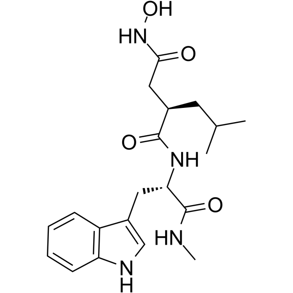
HY-15768
IlomastatMMP Inhibitor -
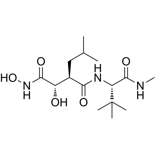
HY-12169
MarimastatMMP Inhibitor -
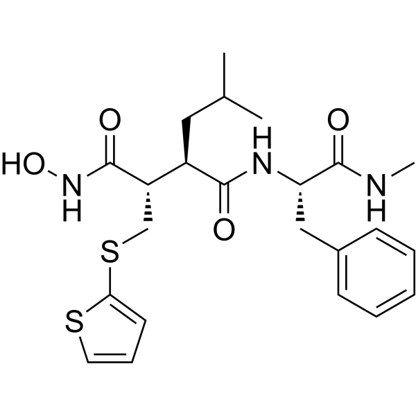
HY-13564
BatimastatMMP Inhibitor -
HY-N0565B
Doxycycline hyclateMMP Inhibitor -
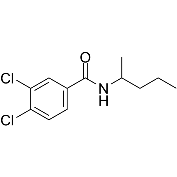
HY-15827
NSC 405020MMP14 Inhibitor -
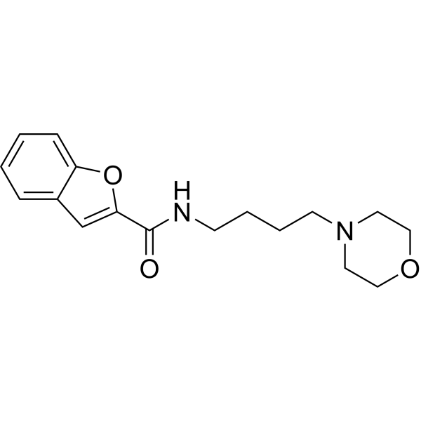
HY-100359
CL-82198MMP-13 Inhibitor -
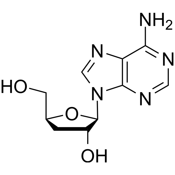
HY-N0262
CordycepinBacterial Inhibitor -
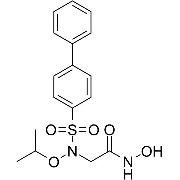
HY-103444
ARP-100MMP-2 Inhibitor -
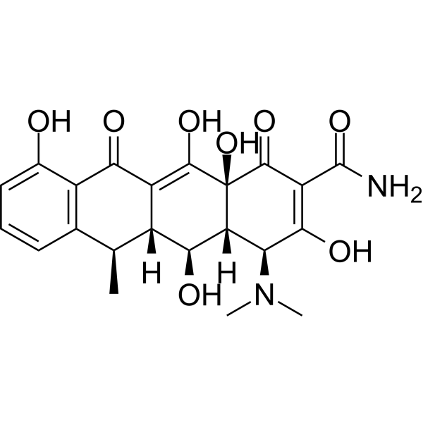
HY-N0565
DoxycyclineMMP Inhibitor -

HY-N0565BR
Doxycycline hyclate (Standard)Reference Standard


Biological Activity
Description
In Vitro
Doxycycline (11.39 μM + MCF-7; 7.13 μM + MDA-MB-468; 72 h) hydrochloride GMP significantly reduces the population of CD44+/CD24-/low breast cancer stem cells in MCF-7 and MDA-MB-468 cells[2].
Doxycycline (11.39 μM+MCF-7; 7.13 μM+MDA-MB-468; 72 h) hydrochloride GMP significantly downregulates the mRNA and protein expression of stem cell markers (Nanog, Oct4, Sox2, c-Myc, CD44) in MCF-7 and MDA-MB-468 breast cancer cells[2].
Doxycycline (1 μg/mL; 5 d) hydrochloride GMP increases the colony formation efficiency of undifferentiated human embryonic stem cells (H9, HSF6, H1, H7, HUES6) and human induced pluripotent stem cells (retrovirus-derived, lentivirus-derived, protein-induced lines), and elevates the number of colonies expressing undifferentiated markers under various culture conditions[3].
Doxycycline (1 μg/mL; 5 d) hydrochloride GMP promotes the survival and proliferation of H9 human embryonic stem cells (hESC) more effectively than Y-27632 (HY-10071), resulting in larger cell colonies, a higher proportion of undifferentiated AP+ cells, as well as downregulated expression of pro-apoptotic genes and upregulated expression of pluripotency-related genes[3].
Doxycycline (1 μg/mL; 6 d; passage 12) hydrochloride GMP reduces cell apoptosis, increases S-phase cell accumulation, supports the long-term expansion of H9 human embryonic stem cells (H9 hESCs) and Lenti-1 induced pluripotent stem cells (Lenti-1 hiPSCs) cultured in clusters on MEF feeder layers, and maintains the pluripotency and normal karyotype of the cells during repeated passaging[3].
Doxycycline (1 μg/mL; 7 d) hydrochloride GMP maintains the viability, proliferation and expression of undifferentiated markers of H9 human embryonic stem cells for up to 7 days without medium change, and reduces apoptotic cell death of newly seeded dissociated cells and preformed cell clusters within 3 days without medium change[5].
Doxycycline (1 μg/mL; 30-72 d) hydrochloride GMP supports long-term subculture of H9, HSF6 human embryonic stem cells as well as Retro-1, Lenti-1, Pro-1 human induced pluripotent stem cells. The culture medium is replaced every 3 days, and the growth rate remains consistent with that under the standard daily medium change condition during passages 5 to 12[5].
Doxycycline (1 μg/mL; 72 d) hydrochloride GMP maintains normal karyotype, expression of undifferentiated markers and pluripotency (the ability to differentiate into all cells of the three germ layers in vitro) of H9 human embryonic stem cells that are subcultured 12 times with medium replacement every 3 days[5].
MedChemExpress (MCE) has not independently confirmed the accuracy of these methods. They are for reference only. Further protocols information, click here.
-
Cell Line:MCF-7 human breast cancer monolayer cells, MDA-MB-468 human breast cancer monolayer cells, BCSC-enriched mammosphere cultures derived from MCF-7 and MDA-MB-468 cell lines
-
Concentration:1-250 μM; 37.5 μM (MCF-7 mammospheres); 29.1 μM (MDA-MB-468 mammospheres)
-
Incubation Time:72 h
-
Result:Inhibited breast cancer cell viability in a dose-dependent manner, with IC50 values of 11.39 μM for MCF-7 monolayer cells and 7.13 μM for MDA-MB-468 monolayer cells.
Reduced viability of BCSC-enriched mammosphere cultures with IC50 values 3-fold higher than their corresponding monolayer cultures (37.5 μM for MCF-7 mammospheres, 29.1 μM for MDA-MB-468 mammospheres).
-
Cell Line:human embryonic stem cell lines H9, HSF6; human induced pluripotent stem cell lines Retro-1, Lenti-1, Pro-1
-
Concentration:1 μg/mL
-
Incubation Time:30 days (5 passages, media changes every 3 days); 72 days (12 passages, media changes every 3 days)
-
Result:Supported H9 hESCs expansion 3684-fold over 5 passages, with an average population doubling level (PDL) of 3.18 per passage, comparable to cultures with daily media changes and doxycycline-free medium.
Enabled HSF6, Retro-1, Lenti-1, and Pro-1 lines to show identical growth (PDL values) between doxycycline-supplemented cultures with 3-day media changes and doxycycline-free cultures with daily media changes.
Maintained consistent PDL values (3.17 at passage 2 vs. 2.96 at passage 12) for H9 hESCs cultured for 12 passages in doxycycline-supplemented mTESR™-1 or PluriSTEM™ medium.
Supported steady accumulation of cell numbers over passages for H9 hESCs cultured for 12 passages.
In Vivo
Doxycycline (60 mg/kg; intraperitoneal injection; once daily for 15 consecutive days) hydrochloride GMP significantly inhibits the growth of CD44v9-expressing prostate cancer xenografts by reducing the proliferation of CD44v9-positive cells[4].
Supplementary culture of H9 human embryonic stem cells (H9 hESCs) with Doxycycline (1 μg/mL; medium changed every 3 days; up to passage 12) hydrochloride GMP maintains their pluripotency and enables in vivo teratoma formation, which contains all three embryonic germ layers[5].
Note:
Please do not refer to only one article to determine the experimental conditions. It is recommended to determine the optimal experimental conditions (animal strain, age, dosage, frequency and cycle, detection time and indicators, etc.) through preliminary experiments before the formal experiment.
Doxycycline and Tetracycline (HY-A0107), act systemically after absorption from the upper gastrointestinal tract. The main advantage of Doxycycline over Tetracycline is its longer activity, and it can be taken twice or once a day. The peak concentration of both agents is similar, but in the case of Doxycycline the time to peak concentration is shorter, and half life is significantly longer[6].
Doxycycline (Dox) is often used as an inducer in molecular biology studies to induce gene expression. In cells or model animals that have constructed tetracycline induced expression systems (Tet-On/Tet-Off systems), the expression of target genes can be precisely controlled by adding or removing Dox[7][8].
Dose reference for Dox induction[7][8]:
(1) Model animal: male Sprague-Dawley rats
Tet regulatory system: 20-3000 ppm of Dox is supplied in diet.
(2) Model animal: Cags mice
Tet regulatory system: 625 ppm of Dox is supplied in diet.
Administration: (for GDNF as targeted gene) 3g/kg (dietary with regular food) • po • once daily for 6 days
(2) 3 g/kg dietary DOX produced DOX serum levels equivalent to 1mg/ml DOX in drinking water.
MedChemExpress (MCE) has not independently confirmed the accuracy of these methods. They are for reference only.
-
Animal Model:Wistar rats with Cardiomyopathy (8-week-old male, 220 g)[1]
-
Dosage:25 mg/kg; 50 mg/kg
-
Administration:i.g.; twice daily; 10 consecutive days
-
Result:Caused minimal skeletal muscle injury (individual myocyte necrosis with mononuclear cell infiltration), slight but significant increases in creatine kinase (CK) and aspartate aminotransferase (AST) compared to controls, and maintained histologically normal cardiac muscle with normal troponin I (cTnI) levels at 25 mg/kg.
Resulted in 30% mortality, dullness, loss of appetite, dyspnoea, congested cava and pulmonary vein with scanty oedematous fluid in abdominal and pleural cavities, multifocal skeletal muscle degeneration/necrosis, mild to moderate epicardial lymphocytic infiltration with focal atrial myocarditis and myocardial necrosis, ventricular muscle degeneration/necrosis with myositis, pulmonary lesions including thickened interalveolar septa, alveolar histiocytosis, and haemosiderin-laden macrophages (heart failure cells), significant increases in alanine aminotransferase (ALT), AST, CK, and cTnI compared to control and 25 mg/kg groups, and a significant decrease in blood calcium levels at 50 mg/kg.


Chemical Information
-
CAS No. 10592-13-9
-
Molecular Weight 480.90
-
Formula C22H25ClN2O8
-
SMILES
O=C(C(C1=O)=C(O)[C@@H](N(C)C)[C@]2([H])[C@@H](O)[C@]3([H])[C@@H](C)C4=C(C(C3=C(O)[C@@]21O)=O)C(O)=CC=C4)N.Cl
-
Shipping
Room temperature in continental US; may vary elsewhere.
-
Storage
Please store the product under the recommended conditions in the Certificate of Analysis.

Protocols
-
Research Protocol for Infectious Diseases
Infectious-disease experiments test how pathogens interact with host barriers, innate immune receptors, inflammatory signaling, pathogen replication, and tissue injury; pattern-recognition receptors such as TLRs, RIG-I-like receptors, NOD-like receptors, and inflammasomes detect microbial molecules and activate NF-κB, interferon, and cytokine responses. The central hypothesis is that infection severity reflects the balance between pathogen burden and host response: protective inflammation restricts pathogen growth, whereas excessive or mislocalized inflammation contributes to tissue damage and disease phenotype. Unresolved questions include which host pathways are protective versus pathogenic, why some infection models fail to translate to human disease, and which combined readouts best predict clinically relevant infection outcomes.
-
Collagen: Sirius Red Staining
Sirius Red or picrosirius red staining is a histochemical method for visualizing collagen-rich extracellular matrix in tissue sections, and collagen fibers are detected as red-stained structures under bright-field microscopy with enhanced birefringence under polarized light. Picrosirius red is useful for assessing total collagen organization, distribution, and fibrosis burden, but polarized color should not be interpreted as a definitive collagen type I versus type III readout because color is affected by fiber orientation, thickness, and packing.
-
RNA extraction experimental
By lysing cells, releasing RNA, and removing impurities such as proteins and DNA, high-purity RNA products are finally obtained. The commonly used traditional method is the guanidine isothiocyanate/phenol/chloroform method (Trizol), which is suitable for a variety of animal materials including animal tissues, microorganisms, cultured cells, etc., and most plant materials.
-
Mitochondrial membrane-potential fluorescent assay
Mitochondrial membrane potential fluorescent assays estimate ΔΨm in living cells using lipophilic cationic dyes such as TMRM, TMRE, rhodamine 123, and JC-1, which accumulate in mitochondria according to membrane polarization; loss of signal after FCCP or CCCP treatment is interpreted as mitochondrial depolarization. TMRM/TMRE and rhodamine 123 are commonly used for semi-quantitative live-cell microscopy or flow cytometry, while JC-1 can report a shift from red aggregate fluorescence to green monomer fluorescence during depolarization; interpretation requires controls because dye concentration, quenching mode, cell type, dye efflux, and mitochondrial mass can affect fluorescence independently of ΔΨm.
-
Annexin V plus membrane-impermeant dye apoptosis staining
Annexin V-based apoptosis assays rely on the detection of phosphatidylserine (PS) externalization from the inner leaflet of the plasma membrane to the outer leaflet, an early biochemical hallmark of apoptosis. Fluorescently labeled Annexin V binds PS in a calcium-dependent manner, enabling identification of early apoptotic cells by flow cytometry or fluorescence microscopy. When combined with a membrane-impermeant DNA-binding dye (e. g. , propidium iodide), this approach allows discrimination between viable (Annexin V−/dye−), early apoptotic (Annexin V+/dye−), and late apoptotic or necrotic (Annexin V+/dye+) cell populations by assessing membrane integrity and PS exposure.
-
ECM-Embedded Organoid (Matrigel/Dome) Culture
ECM-embedded organoid dome culture embeds epithelial stem cells, crypts, organoid fragments, or tumor-derived epithelial cells in a basement-membrane-like hydrogel such as Matrigel, allowing 3D growth, self-organization, lumen formation, budding or cystic morphogenesis, and lineage maintenance under defined niche-factor-containing medium. The primary readouts are organoid establishment efficiency, growth, morphology, passaging capacity, lineage-marker expression, and, when fluorescently labeled lines are used, microscopy- or flow-cytometry-based quantification of population behavior in 3D culture.
-
Bacterial live/dead nucleic-acid viability staining
The LIVE/DEAD bacterial viability staining method is based on differential permeability of nucleic-acid-binding fluorescent dyes, most commonly SYTO 9 and propidium iodide (PI), which enables discrimination of bacterial populations with intact versus compromised cytoplasmic membranes. SYTO 9 penetrates both intact and damaged bacterial membranes and binds nucleic acids to produce green fluorescence, whereas propidium iodide penetrates only cells with compromised membranes and fluoresces red while also reducing SYTO 9 signal through competitive binding and fluorescence interactions. The resulting fluorescence pattern is interpreted as a proxy for membrane integrity, which is widely used as an indicator of bacterial viability in microscopy, flow cytometry, and spectroscopic platforms. However, mechanistic studies show that SYTO 9 and PI interactions involve displacement and fluorescence resonance energy transfer effects, which can influence signal interpretation depending on dye ratios a
-
Breast Cancer Modeling
Breast cancer is a heterogeneous cancer, and it has been distinguished into four subtypes: luminal A, luminal B, HER2-positive and basal-like. Molecular mutations, epigenetic alterations, hormone exposure and immune microenvironment are related to the progression of breast cancer.
-
Matrigel Transwell/Boyden Chamber Invasion Assay
Matrigel Transwell/Boyden chamber invasion assay measures the ability of cells to degrade or traverse an extracellular matrix-coated porous membrane and move from an upper chamber toward a chemoattractant in a lower chamber. Invasion is distinguished from migration by coating the membrane with Matrigel or basement membrane matrix; uncoated inserts measure migration, while coated inserts require cells to cross an ECM barrier before reaching the underside of the membrane.
-
Cell Viability Determination by MTT Colorimetric Assay
The following protocol uses the MTT colorimetric assay as a classic literature-established method for assessing cell viability/metabolic activity in cultured mammalian cells. MTT[3-(4,5-dimethylthiazol-2-yl)-2,5-diphenyltetrazolium bromide] is reduced by metabolically active cells to a colored formazan product; the amount of formazan is quantified spectrophotometrically and provides an indirect measure of metabolically active viable cells. Importantly, MTT reduction reflects cellular oxidoreductase/metabolic activity rather than an absolute direct count of living cells, so changes in cellular metabolism can alter the signal independently of cell number.
-
Fibrosis/Collagen Morphometry
Fibrosis and collagen morphometry is based on the quantitative visualization of fibrillar collagen deposition in tissue sections using histochemical stains such as Sirius Red (Picrosirius Red) or Masson's trichrome, followed by image-based or polarization-enhanced analysis to estimate collagen proportional area as a surrogate of extracellular matrix accumulation during fibrotic remodeling. Sirius Red combined with polarized light microscopy enhances detection of collagen fibers due to birefringence properties, enabling more specific visualization of collagen type I and III fibrils compared to conventional bright-field histology, while whole-section or region-restricted digital morphometry reduces field-selection bias in fibrosis assessment. Alternative quantitative approaches include second harmonic generation (SHG) and two-photon excited fluorescence microscopy, which enable label-free detection of fibrillar collagen and have been validated against histological staining and biochemica
-
Invadopodia/Fluorescent Gelatin Degradation Assay
Invadopodia/fluorescent gelatin degradation assay detects proteolytic extracellular matrix degradation by cancer-cell invadopodia, which are actin-rich protrusive structures associated with matrix remodeling, invasion, and metastasis. The readout is generated by culturing cells on fluorescent gelatin and measuring dark degraded areas where fluorescent substrate has been locally removed, often together with immunofluorescent detection of invadopodia markers such as F-actin, cortactin, and TKS5.
-
Apoptosis
Apoptosis, also called programmed cell death, is generally characterized by distinct morphological characteristics.
-
TUNEL staining for apoptotic DNA fragmentation
TUNEL staining detects DNA strand breaks by using terminal deoxynucleotidyl transferase to add labeled nucleotides to exposed 3′-OH DNA termini, generating either microscopic staining in fixed cells or tissue sections, or fluorescence/cytometric signal in cell suspensions. TUNEL positivity reflects DNA fragmentation but should not be interpreted alone as definitive apoptosis, because TUNEL can also label necrotic, autolytic, mechanically damaged, or DNA-repair-associated DNA breaks.
-
Mitochondrial membrane-potential and mitochondrial mass staining
Mitochondrial membrane potential staining measures the electrochemical polarization across the mitochondrial inner membrane in live cells using lipophilic cationic fluorescent probes; early rhodamine-based work showed that selective mitochondrial dye accumulation is lost when the mitochondrial transmembrane potential is dissipated. JC-1 reports mitochondrial polarization by shifting from green monomer fluorescence to red J-aggregate fluorescence as dye concentration increases within energized mitochondria; therefore, the red/green fluorescence ratio is used as a relative readout of mitochondrial membrane potential. TMRE or TMRM staining provides a single-channel relative readout because these cationic rhodamine esters accumulate in polarized mitochondria, and lower fluorescence indicates reduced mitochondrial polarization when acquisition and dye-loading conditions are controlled. Mitochondrial mass staining is commonly performed with MitoTracker Green FM or related MitoTracker dyes as
-
Autophagy
Autophagy is a process in which eukaryotic cells use lysosomes to degrade their own cytoplasmic proteins and damaged organelles under the regulation of autophagy related gene (Atg). Microtubule-associated proteins light chain 3 (LC3) is recognized as autophagy marker, which transfers from cytoplasmic LC3 (LC3-I) to membrane type (LC3-II). LC3-II/I ratio could be detected by Western Blot and fluorescence microscopy.
-
Apoptosis Solutions
Apoptosis is a regulated, generally non-lytic cell-death pathway that removes unwanted, damaged, infected, or abnormal cells through coordinated morphological changes, caspase activation, DNA fragmentation, and membrane remodeling. The intrinsic apoptosis pathway is controlled mainly by mitochondrial outer membrane permeabilization, BCL-2 family proteins, cytochrome c release, apoptosome formation, caspase-9 activation, and downstream executioner caspase-3/7 activation. The extrinsic apoptosis pathway is initiated by death receptors such as Fas, TNFR, and TRAIL receptors, which recruit adaptor proteins and activate caspase-8 before engaging executioner caspases or mitochondrial amplification through BID cleavage. Apoptosis is linked to many phenotypes, including cancer cell killing, tissue homeostasis, immune regulation, neurodegeneration, infection response, and treatment-induced cytotoxicity; unresolved questions include how apoptosis interacts with necroptosis, pyroptosis, ferroptos
-
Matrigel/ECM Transwell Invasion Assay
The Matrigel/ECM Transwell invasion assay measures the ability of cells to move toward a chemoattractant while crossing an extracellular-matrix barrier placed on a porous membrane; therefore, the readout reflects both chemotactic motility and matrix invasion rather than migration alone. Matrigel is a basement-membrane-rich matrix derived from Engelbreth-Holm-Swarm mouse sarcoma and has been used as a reconstituted basement membrane barrier in chemoinvasion assays. The assay readout is generated by quantifying cells that reach the underside of the insert membrane or lower compartment after incubation, commonly by staining and counting invaded cells or by fluorescence-based quantification.
-
Lysosome and acidic-vesicle live-cell staining
Lysosome and acidic-vesicle live-cell staining detects acidic intracellular compartments by using membrane-permeant acidotropic probes that accumulate in low-pH vesicles, including lysosomes, late endosomes, autolysosomes, and acidic phagosomes. LysoTracker staining is commonly used as an intensity-based readout of acidic lysosomal compartment abundance or enlargement, while acridine orange produces green fluorescence in less concentrated compartments and red fluorescence after concentration-dependent accumulation in acidic vesicular organelles. Loss or reduction of acridine-orange red signal can be used as a readout of lysosomal membrane permeabilization or reduced acidic-vesicle integrity. This protocol is designed for live cultured cells and can be adapted for fluorescence microscopy, high-content imaging, plate-reader readout, or flow cytometry when the selected literature supports the readout. Because these dyes report acidotropic accumulation rather than lysosome identity alone,
-
Macroautophagy Solutions
Macroautophagy is a conserved lysosome-dependent degradation pathway in which cytoplasmic material is sequestered into double-membrane autophagosomes and delivered to lysosomes for degradation and recycling. The pathway supports cellular homeostasis during nutrient limitation, organelle stress, protein-aggregate accumulation, infection, differentiation, and tissue remodeling by coupling cargo sequestration, autophagosome maturation, lysosomal fusion, and degradation of cargo-derived macromolecules. The core molecular sequence includes initiation by nutrient- and stress-regulated autophagy machinery, autophagosome nucleation, LC3/ATG8-family conjugation to autophagosomal membranes, cargo selection through receptors such as SQSTM1/p62, autophagosome-lysosome fusion, and lysosomal degradation. LC3 was identified as a mammalian homolog of yeast Atg8 that localizes to autophagosomal membranes after processing, and p62/SQSTM1 was shown to connect ubiquitinated cargo with autophagic degradati
-
hPSC maintenance and expansion
This protocol maintains and expands human pluripotent stem cells under feeder-free, chemically defined conditions using E8 medium and vitronectin-coated culture surfaces; the readout is sustained adherent colony growth with undifferentiated morphology and retained pluripotency-marker expression during serial passaging. E8-based hPSC culture relies on defined soluble factors and matrix-dependent adhesion rather than feeder cells; vitronectin supports hPSC attachment through integrin-mediated interactions, and EDTA passaging dissociates colonies as small aggregates without enzymatic digestion, centrifugation, or routine ROCK-inhibitor treatment.


Purity & Documentation
References
[1]. El-Neweshy MS, et al. Experimental doxycycline overdose in rats causes cardiomyopathy. Int J Exp Pathol. 2013;94(2):109-114. [Content Brief]
[2]. Zhang L, et al. Doxycycline inhibits the cancer stem cell phenotype and epithelial-to-mesenchymal transition in breast cancer. Cell Cycle. 2017;16(8):737-745. [Content Brief]
[5]. Chang MY, et al. Doxycycline supplementation allows for the culture of human ESCs/iPSCs with media changes at 3-day intervals. Stem Cell Res. 2015;15(3):608-613. [Content Brief]
[6]. Manfredsson FP, et al. Tight Long-term dynamic doxycycline responsive nigrostriatal GDNF using a single rAAV vector. Mol Ther. 2009 Nov;17(11):1857-67. [Content Brief] [Content Brief]
[7]. Kistner A, et al. Doxycycline-mediated quantitative and tissue-specific control of gene expression in transgenic mice. Proc Natl Acad Sci U S A. 1996 Oct 1;93(20):10933-8. [Content Brief]
[8]. Niv Y. Doxycycline in Eradication Therapy of Helicobacter pylori--a Systematic Review and Meta-Analysis. Digestion. 2016;93(2):167-73. [Content Brief]

Calculators
Concentration (start) × Volume (start) = Concentration (final) × Volume (final)


Doxycycline (hydrochloride) (GMP) Related Classifications
HY-N0565AG Related Classifications
 Powered by Bioz
Keywords