



(+)-Erinacin A

Based on 1 Customer Validation
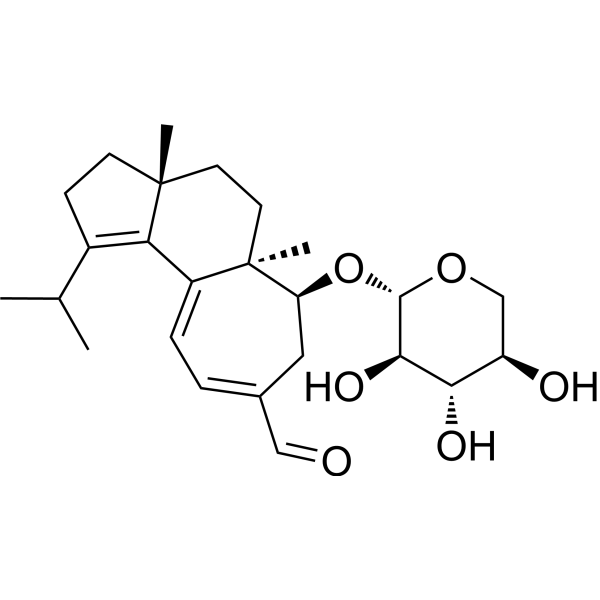
(+)-Erinacin A (Erinacine A) is a cyanoditerpenoid isolated from Hericium erinaceus with anticancer, anti-inflammatory and neuroprotective activities. (+)-Erinacin A can induce cancer cell death by activating extrinsic and intrinsic apoptosis pathways. (+)-Erinacin A can also inhibit the expression of NO synthase (iNOS) and the production of nitrotyrosine to exert inflammatory and neuroprotective effects, thereby reducing ischemic brain damage.
For research use only. We do not sell to patients.
- Purity : 99.81%
- CAS No.: 156101-08-5
- Formula: C25H36O6
- Molecular Weight:432.55
-
Storage:
-20°C, protect from light
* In solvent : -80°C, 6 months; -20°C, 1 month (protect from light)

To place orders, for customer services and technical support, please contact: MedChemExpress USA
Tel: 609-228-6898 E-mail: [email protected] [email protected]


-
 Biological Activity
-
Chemical Information
-
Solvent & Solubility
- Protocols
- Purity & Documentation
- References
-
 Help & FAQs


-
HY-N0721
NeoandrographolideMAPK Inhibitor -
HY-N1500
PulegoneMonoterpene Ketone Compound -
HY-133971
Cholesterol 5α,6α-epoxideBiochemical Assay Reagent -
HY-100347
SRI-011381TGF-β/Smad Agonist -
HY-119486
TavapadonDopamine D1/D5 Receptor Agonist -
HY-124729
BL-918ULK1 Activator -
HY-156958
S-72Tubulin Inhibitor -

HY-137936
Terrecyclic AcidSesquiterpene -

HY-N0721R
Neoandrographolide (Standard)Natural Product -
HY-139931S
1-Stearoyl-2-Arachidonoyl-d8-sn-glycero-3-PCStable Isotope -
HY-105003
ST 1535A2A Adenosine Antagonist -

HY-N1500R
Pulegone (Standard)Monoterpene Ketone Compound
All Caspase Isoforms
More

Biological Activity
Description
In Vitro
(+)-Erinacin A (30 μM; 3-24 h) activates the extrinsic and intrinsic apoptosis pathways in DLD-1 cells and reduces the expression of anti-apoptotic proteins[4].
MedChemExpress (MCE) has not independently confirmed the accuracy of these methods. They are for reference only. Further protocols information, click here.
-
Cell Line:DLD-1 Cells
-
Concentration:30 µM
-
Incubation Time:3-24 h
-
Result:Increased active forms of caspase-3, caspase-9 and caspase-8. Decreased the expression of the antiapoptotic proteins Bcl-2 and Bcl-XL. Upregulated the levels of phosphorylated JNK Thr183 and Tyr185, NFκB, and p300 proteins.
In Vivo
MedChemExpress (MCE) has not independently confirmed the accuracy of these methods. They are for reference only.
-
Animal Model:DLD-1 xenograft mouse model[5].
-
Dosage:1, 2 and 5 mg/kg
-
Administration:Intraperitoneal injection (i.p.) once a day for 5 days
-
Result:Inhibited tumor volume by 60%, 32% and 26%.
-
Animal Model:SAM rats with transient stroke[4].
-
Dosage:1, 5 and 10 mg/kg
-
Administration:Intraperitoneal injection (i.p.) single dose
-
Result:Induced NeuN immune response in the entire brain region of SAM rats, and increased the number of NeuN cells. Inhibited the activation of iNOS and p38/MAPK pathways and the expression of CHOP and nitrotyrosine proteins in rat brain tissue.


Chemical Information
-
CAS No. 156101-08-5
-
Appearance Solid
-
Molecular Weight 432.55
-
Formula C25H36O6
-
Color Light yellow to yellow
-
SMILES
O[C@H]1[C@H](O)[C@@H](O)[C@H](O[C@@H]2[C@@](CC[C@]3(C)C4=C(C(C)C)CC3)(C)C4=CC=C(C=O)C2)OC1
-
Synonyms
Erinacine A
-
Structure Classification
-
Initial Source
Hericium erinaceus
-
Shipping
Room temperature in continental US; may vary elsewhere.
-
Storage
-20°C, protect from light
* In solvent : -80°C, 6 months; -20°C, 1 month (protect from light)


Solvent & Solubility
In Vitro:
Ethanol : 10 mg/mL (23.12 mM; Need ultrasonic)
Please refer to the solubility information to select the appropriate solvent. Once prepared, please aliquot and store the solution to prevent product inactivation from repeated freeze-thaw cycles.
Storage method and period of stock solution: -80°C, 6 months; -20°C, 1 month (protect from light). When stored at -80°C, please use it within 6 months. When stored at -20°C, please use it within 1 month.
Please refer to the solubility information to select the appropriate solvent. Once prepared, please aliquot and store the solution to prevent product inactivation from repeated freeze-thaw cycles.
Storage method and period of stock solution: -80°C, 6 months; -20°C, 1 month (protect from light). When stored at -80°C, please use it within 6 months. When stored at -20°C, please use it within 1 month.
Concentration (start) × Volume (start) = Concentration (final) × Volume (final)

Protocols
-
RNA extraction experimental
By lysing cells, releasing RNA, and removing impurities such as proteins and DNA, high-purity RNA products are finally obtained. The commonly used traditional method is the guanidine isothiocyanate/phenol/chloroform method (Trizol), which is suitable for a variety of animal materials including animal tissues, microorganisms, cultured cells, etc., and most plant materials.
-
Apoptosis
Apoptosis, also called programmed cell death, is generally characterized by distinct morphological characteristics.
-
TUNEL staining for apoptotic DNA fragmentation
TUNEL staining detects DNA strand breaks by using terminal deoxynucleotidyl transferase to add labeled nucleotides to exposed 3′-OH DNA termini, generating either microscopic staining in fixed cells or tissue sections, or fluorescence/cytometric signal in cell suspensions. TUNEL positivity reflects DNA fragmentation but should not be interpreted alone as definitive apoptosis, because TUNEL can also label necrotic, autolytic, mechanically damaged, or DNA-repair-associated DNA breaks.
-
Cell Cytotoxicity Assay
Cytotoxicity assays are usually based on the assessment of cell membrane damage, which can also be indirectly detected by measuring cell viability. Detection methods include MTT assay, CKK-8 assay, LDH assay and ATP assay, etc.
-
Mammalian live/dead viability and cytotoxicity staining
Live/dead viability and cytotoxicity staining assays are based on the simultaneous detection of intracellular esterase activity in metabolically active (viable) cells and membrane integrity loss in non-viable cells. In commonly used dual-staining approaches, membrane-permeant fluorogenic substrates are converted by intracellular esterases into fluorescent products in live cells, while impermeant DNA-binding dyes selectively enter cells with compromised plasma membranes and label nucleic acids in dead or dying cells, enabling discrimination between viable and non-viable populations by fluorescence microscopy or flow cytometry.
-
Annexin V plus membrane-impermeant dye apoptosis staining
Annexin V-based apoptosis assays rely on the detection of phosphatidylserine (PS) externalization from the inner leaflet of the plasma membrane to the outer leaflet, an early biochemical hallmark of apoptosis. Fluorescently labeled Annexin V binds PS in a calcium-dependent manner, enabling identification of early apoptotic cells by flow cytometry or fluorescence microscopy. When combined with a membrane-impermeant DNA-binding dye (e. g. , propidium iodide), this approach allows discrimination between viable (Annexin V−/dye−), early apoptotic (Annexin V+/dye−), and late apoptotic or necrotic (Annexin V+/dye+) cell populations by assessing membrane integrity and PS exposure.
-
Apoptosis Solutions
Apoptosis is a regulated, generally non-lytic cell-death pathway that removes unwanted, damaged, infected, or abnormal cells through coordinated morphological changes, caspase activation, DNA fragmentation, and membrane remodeling. The intrinsic apoptosis pathway is controlled mainly by mitochondrial outer membrane permeabilization, BCL-2 family proteins, cytochrome c release, apoptosome formation, caspase-9 activation, and downstream executioner caspase-3/7 activation. The extrinsic apoptosis pathway is initiated by death receptors such as Fas, TNFR, and TRAIL receptors, which recruit adaptor proteins and activate caspase-8 before engaging executioner caspases or mitochondrial amplification through BID cleavage. Apoptosis is linked to many phenotypes, including cancer cell killing, tissue homeostasis, immune regulation, neurodegeneration, infection response, and treatment-induced cytotoxicity; unresolved questions include how apoptosis interacts with necroptosis, pyroptosis, ferroptos
-
Research Protocol for Inflammation-related Diseases
The NLRP3 inflammasome is a cytosolic innate immune signaling platform that integrates priming signals and danger-signal activation to promote caspase-1 activation, maturation of IL-1β and IL-18, and gasdermin D-mediated pyroptotic cell death. The core experimental logic is to determine whether inflammatory disease phenotypes are driven by increased NLRP3 expression, ASC-containing inflammasome assembly, caspase-1 cleavage, GSDMD cleavage, and extracellular release of IL-1β/IL-18 rather than by nonspecific cell injury alone. The pathway is strongly linked to inflammation-related disease phenotypes because monosodium urate crystals activate NALP3/NLRP3 inflammasome signaling in gout-like crystal inflammation, cholesterol crystals activate NLRP3 inflammasomes in atherogenesis models, and DSS-induced intestinal inflammation has been reported to involve NLRP3 inflammasome activity. However, experimental colitis studies also show context-dependent protective effects of NLRP3 inflammasome co


Purity & Documentation
-
Data Sheet (279 KB)
-
SDS (252 KB)
- English - EN (252 KB)
- Français - FR (252 KB)
- Deutsch - DE (252 KB)
- Norwegian - NO (252 KB)
- Español - ES (252 KB)
- Swedish - SV (252 KB)
- Italian - IT (252 KB)
- Korean - KR (252 KB)
- Portuguese - PT (252 KB)
-
Handling Instructions (2659 KB)
References
[1]. Bailly C, et.al. Erinacine A and related cyathane diterpenoids: Molecular diversity and mechanisms underlying their neuroprotection and anticancer activities. Pharmacol Res. 2020 Sep;159:104953. [Content Brief]
[2]. Tsai-Teng T, et al. Erinacine A-enriched Hericium erinaceus mycelium ameliorates Alzheimer’s disease-related pathologies in APPswe/PS1dE9 transgenic mice[J]. Journal of biomedical science, 2016, 23: 1-12. [Content Brief]
[3]. Prasher P, et al. Key oncologic pathways inhibited by Erinacine A: a perspective for its development as an anticancer molecule[J]. Biomedicine & Pharmacotherapy, 2023, 160: 114332. [Content Brief]
[4]. Lee K F, et al. Protective effects of Hericium erinaceus mycelium and its isolated erinacine A against ischemia-injury-induced neuronal cell death via the inhibition of iNOS/p38 MAPK and nitrotyrosine[J]. International journal of molecular sciences, 2014, 15(9): 15073-15089. [Content Brief]
[5]. Lee K C, et al. Induction apoptosis of erinacine A in human colorectal cancer cells involving the expression of TNFR, Fas, and Fas ligand via the JNK/p300/p50 signaling pathway with histone acetylation[J]. Frontiers in Pharmacology, 2019, 10: 1174. [Content Brief]


Complete Stock Solution Preparation Table
Please refer to the solubility information to select the appropriate solvent. Once prepared, please aliquot and store the solution to prevent product inactivation from repeated freeze-thaw cycles.
Storage method and period of stock solution: -80°C, 6 months; -20°C, 1 month (protect from light). When stored at -80°C, please use it within 6 months. When stored at -20°C, please use it within 1 month.
| Optional Solvent | Concentration Solvent Mass | 1 mg | 5 mg | 10 mg | 25 mg |
|---|---|---|---|---|---|
| Ethanol | 1 mM | 2.3119 mL | 11.5594 mL | 23.1187 mL | 57.7968 mL |
| 5 mM | 0.4624 mL | 2.3119 mL | 4.6237 mL | 11.5594 mL | |
| 10 mM | 0.2312 mL | 1.1559 mL | 2.3119 mL | 5.7797 mL | |
| 15 mM | 0.1541 mL | 0.7706 mL | 1.5412 mL | 3.8531 mL | |
| 20 mM | 0.1156 mL | 0.5780 mL | 1.1559 mL | 2.8898 mL |


(+)-Erinacin A Related Classifications
HY-111954 Related Classifications
 Powered by Bioz