



Q-Peptide

Based on 1 Customer Validation
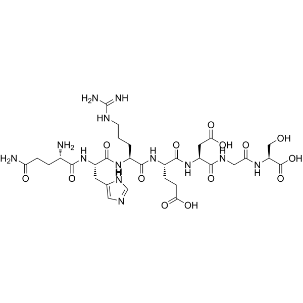
Q-Peptide is an angiopoietin-1 derived peptide (QHREDGS). Q-Peptide interacts with β1-integrin, binds to integrins on the surface of osteoblasts, and serves as an acyl donor substrate for Streptomyces mobaraensis transglutaminase. Q-Peptide activates Akt, MAPKp42/44, ILK, ERK1/2, and downregulates caspase-3/7. Q-Peptide inhibits cell apoptosis, enhances cell adhesion and migration, and promotes osteoblast differentiation, bone matrix deposition and mineralization. Q-Peptide can be used in studies related to myocardial infarction, bone regeneration, diabetic wound repair and human induced pluripotent stem cells.
For research use only. We do not sell to patients.
- CAS No.: 1361235-89-3
- Formula: C31H49N13O14
- Molecular Weight:827.80
-
Storage:
Sealed storage, away from moisture.
Powder -80°C, 2 years , -20°C, 1 year* In solvent : -80°C, 6 months; -20°C, 1 month (sealed storage, away from moisture)

To place orders, for customer services and technical support, please contact: MedChemExpress USA
Tel: 609-228-6898 E-mail: [email protected] [email protected]


-
 Biological Activity
-
Chemical Information
-
Solvent & Solubility
- Protocols
- Purity & Documentation
- References
-
 Help & FAQs


All Caspase Isoforms
More

Biological Activity
Description
|
Akt |
p38 MAPK |
ERK1 |
ERK2 |
Caspase 3 |
Caspase-7 |
In Vitro
Q-Peptide (QHREDGS) (5-500 μM; 5 continuous passages, 7 days) improves human induced pluripotent stem cell (BJ1D, 0901B, IMR90) expansion and viability in feeder-dependent culture by reducing caspase-dependent apoptosis, without altering proliferation, while having no significant effect on human embryonic stem cells (H9)[1].
Q-Peptide (50 μM; 5 continuous passages) preserves the in vitro pluripotency of human induced pluripotent stem cells (BJ1D, 0901B)[1].
Q-Peptide (5-500 μM; 48h) significantly increases colony number and size in feeder-free human induced pluripotent stem cells cultured on Matrigel[1].
Q-Peptide (50 μM; 0min-24h) increases integrin-linked kinase (ILK) expression, and increases expression and activation of extracellular signal-regulated kinases 1/2 (ERK1/2) across multiple time points, without affecting AKT activation[1].
Q-Peptide (7.2-29.0 mM; 6h) dose-dependently enhances human induced pluripotent stem cell adhesion to non-adhesive PEG hydrogels, and this interaction is mediated by β1-integrins[1].
Q-Peptide (200 μM; 40 min) potently acts as a substrate for Streptomyces mobaraensis transglutaminase (STG), with a cross-linking conversion yield of 93.0%[4].
MedChemExpress (MCE) has not independently confirmed the accuracy of these methods. They are for reference only.
-
Cell Line:human induced pluripotent stem cells
-
Concentration:50 μM
-
Incubation Time:0min, 30min, 1.5h, 2h, 4h, 24h
-
Result:Increased ILK protein expression.
Significantly upregulated ERK1/2 expression.
Showed no increase in AKT phosphorylation.
In Vivo
QP-derived functional peptide hydrogel (1% w/v, for local single injection) mediated mesenchymal stem cell transplantation can improve cardiac function (EF and FS), reduce infarct size, collagen content and cardiomyocyte apoptosis in rat myocardial infarction models. This treatment also elevates the survival rate of transplanted cells and promotes angiogenesis. Ctf1 pretreatment can further enhance the therapeutic effects of QP-based hydrogel mediated mesenchymal stem cell transplantation[5].
MedChemExpress (MCE) has not independently confirmed the accuracy of these methods. They are for reference only.
-
Animal Model:BKS.Cg-Dock7m+/+Leprdb/J (db/db) (8-week-old male, leptin-receptor deficient type 2 diabetic, full-thickness 8-mm dorsal excisional wounds)[3]
-
Dosage:2.2 nmol
5.9 nmol -
Administration:topical; single application
-
Result:Showed faster wound closure starting on day 8.
Reduced normalized wound area significantly by day 14 compared to blank or peptide-free hydrogel controls.
Reduced epithelial gap significantly compared to controls.
Increased re-epithelialization percentage significantly compared to controls.
Enlarged granulation tissue area significantly compared to controls.
Thinned advancing epidermal tongue significantly compared to controls.
Increased total number of blood vessels in the wound significantly compared to blank and peptide-free hydrogel controls.
Was significantly superior to clinically approved collagen dressings in accelerating diabetic wound healing in db/db mice, achieving near-complete wound closure by day 21
- Detection of Apoptosis
- TUNEL staining for apoptotic DNA fragmentation
- Annexin V plus membrane-impermeant dye apoptosis staining
- Human iPSC generation/reprogramming culture
- Cell migration
- Mesenchymal stromal/stem cell osteogenic differentiation
- Apoptosis Solutions
- Research Protocol for Cardiovascular Diseases
- CRISPR-Cas9 editing of human pluripotent stem cells


Chemical Information
-
CAS No. 1361235-89-3
-
Appearance Solid
-
Molecular Weight 827.80
-
Formula C31H49N13O14
-
Color White to off-white
-
Sequence
Gln-His-Arg-Glu-Asp-Gly-Ser
-
Sequence Shortening
QHREDGS
-
Shipping
Room temperature in continental US; may vary elsewhere.
-
Storage
Sealed storage, away from moisture
Powder -80°C 2 years -20°C 1 year * In solvent : -80°C, 6 months; -20°C, 1 month (sealed storage, away from moisture)


Solvent & Solubility
In Vitro:
DMSO : 100 mg/mL (120.80 mM; Need ultrasonic; Hygroscopic DMSO has a significant impact on the solubility of product, please use newly opened DMSO)
Please refer to the solubility information to select the appropriate solvent. Once prepared, please aliquot and store the solution to prevent product inactivation from repeated freeze-thaw cycles.
Storage method and period of stock solution: -80°C, 6 months; -20°C, 1 month (sealed storage, away from moisture). When stored at -80°C, please use it within 6 months. When stored at -20°C, please use it within 1 month.
Please refer to the solubility information to select the appropriate solvent. Once prepared, please aliquot and store the solution to prevent product inactivation from repeated freeze-thaw cycles.
Storage method and period of stock solution: -80°C, 6 months; -20°C, 1 month (sealed storage, away from moisture). When stored at -80°C, please use it within 6 months. When stored at -20°C, please use it within 1 month.
Concentration (start) × Volume (start) = Concentration (final) × Volume (final)
In Vivo:
Select the appropriate dissolution method based on your experimental animal and administration route.
- For the following dissolution methods, please ensure to first prepare a clear stock solution using an In Vitro approach and then sequentially add co-solvents:
- To ensure reliable experimental results, the clarified stock solution can be appropriately stored based on storage conditions. As for the working solution for In Vivo experiments, it is recommended to prepare freshly and use it on the same day.
- The percentages shown for the solvents indicate their volumetric ratio in the final prepared solution. If precipitation or phase separation occurs during preparation, heat and/or sonication can be used to aid dissolution.
Add each solvent one by one: 10% DMSO 40% PEG300 5% Tween-80 45% Saline
Solubility: ≥ 2.5 mg/mL (3.02 mM); Clear solution
This protocol yields a clear solution of ≥ 2.5 mg/mL (saturation unknown).
Taking 1 mL working solution as an example, add 100 μL DMSO stock solution (25.0 mg/mL) to 400 μL PEG300, and mix evenly; then add 50 μL Tween-80 and mix evenly; then add 450 μL Saline to adjust the volume to 1 mL.
Preparation of Saline: Dissolve 0.9 g sodium chloride in ddH₂O and dilute to 100 mL to obtain a clear Saline solution.
Add each solvent one by one: 10% DMSO 90% (20% SBE-β-CD in Saline)
Solubility: ≥ 2.5 mg/mL (3.02 mM); Clear solution
This protocol yields a clear solution of ≥ 2.5 mg/mL (saturation unknown).
Taking 1 mL working solution as an example, add 100 μL DMSO stock solution (25.0 mg/mL) to 900 μL 20% SBE-β-CD in Saline, and mix evenly.
Preparation of 20% SBE-β-CD in Saline (4°C, storage for one week): 2 g SBE-β-CD powder is dissolved in 10 mL Saline, completely dissolve until clear.
In Vivo Dissolution Calculator
Please enter the basic information of animal experiments:
-
-
-
-
Recommended: Prepare an additional quantity of animals to account for potential losses during experiments.
Please enter your animal formula composition:
-
%DMSO +
Recommended: Keep the proportion of DMSO in working solution below 2% if your animal is weak.
-
%+
-
+%Tween-80 + +
-
%Saline +
The co-solvents required include: DMSO, . All of co-solvents are available by MedChemExpress (MCE). , Tween 80. All of co-solvents are available by MedChemExpress (MCE).
Working solution concentration: 0.22 mg/mL
Method for preparing stock solution: mg drug dissolved in μL DMSO. Stock solution concentration: mg/mL. * In solvent : -80°C, 6 months; -20°C, 1 month (sealed storage, away from moisture)
1. Take μL DMSO stock solution;
2. Add μL .
μL , mix evenly;
3. Then add μL Tween 80, mix evenly;
4. Then add μL
Please ensure that the stock solution in the first step is dissolved to a clear state, and add co-solvents in sequence. You can use ultrasonic heating (ultrasonic cleaner, recommended frequency 20-40 kHz), vortexing, etc. to assist dissolution.


Purity & Documentation
-
Data Sheet (282 KB)
-
SDS (252 KB)
- English - EN (252 KB)
- Français - FR (252 KB)
- Deutsch - DE (252 KB)
- Norwegian - NO (252 KB)
- Español - ES (252 KB)
- Swedish - SV (252 KB)
- Italian - IT (252 KB)
- Korean - KR (252 KB)
- Portuguese - PT (252 KB)
-
Handling Instructions (2659 KB)
References
[1]. Dang LT, et al. Inhibition of apoptosis in human induced pluripotent stem cells during expansion in a defined culture using angiopoietin-1 derived peptide QHREDGS. Biomaterials. 2014;35(27):7786-7799. [Content Brief]
[2]. Feric N, et al. Angiopoietin-1 peptide QHREDGS promotes osteoblast differentiation, bone matrix deposition and mineralization on biomedical materials. Biomater Sci. 2014;2(10):1384-1398. [Content Brief]
[3]. Xiao Y, et al. Diabetic wound regeneration using peptide-modified hydrogels to target re-epithelialization. Proc Natl Acad Sci U S A. 2016;113(40):E5792-E5801. [Content Brief]
[4]. Lee JH, et al. Glutamine (Q)-peptide screening for transglutaminase reaction using mRNA display. Biotechnol Bioeng. 2013;110(2):353-362. [Content Brief]
[5]. Cai H, et al. Self-assembling peptide modified with QHREDGS as a novel delivery system for mesenchymal stem cell transplantation after myocardial infarction. FASEB J. 2019;33(7):8306-8320. [Content Brief]


Complete Stock Solution Preparation Table
Please refer to the solubility information to select the appropriate solvent. Once prepared, please aliquot and store the solution to prevent product inactivation from repeated freeze-thaw cycles.
Storage method and period of stock solution: -80°C, 6 months; -20°C, 1 month (sealed storage, away from moisture). When stored at -80°C, please use it within 6 months. When stored at -20°C, please use it within 1 month.
| Optional Solvent | Concentration Solvent Mass | 1 mg | 5 mg | 10 mg | 25 mg |
|---|---|---|---|---|---|
| DMSO | 1 mM | 1.2080 mL | 6.0401 mL | 12.0802 mL | 30.2005 mL |
| 5 mM | 0.2416 mL | 1.2080 mL | 2.4160 mL | 6.0401 mL | |
| 10 mM | 0.1208 mL | 0.6040 mL | 1.2080 mL | 3.0201 mL | |
| 15 mM | 0.0805 mL | 0.4027 mL | 0.8053 mL | 2.0134 mL | |
| 20 mM | 0.0604 mL | 0.3020 mL | 0.6040 mL | 1.5100 mL | |
| 25 mM | 0.0483 mL | 0.2416 mL | 0.4832 mL | 1.2080 mL | |
| 30 mM | 0.0403 mL | 0.2013 mL | 0.4027 mL | 1.0067 mL | |
| 40 mM | 0.0302 mL | 0.1510 mL | 0.3020 mL | 0.7550 mL | |
| 50 mM | 0.0242 mL | 0.1208 mL | 0.2416 mL | 0.6040 mL | |
| 60 mM | 0.0201 mL | 0.1007 mL | 0.2013 mL | 0.5033 mL | |
| 80 mM | 0.0151 mL | 0.0755 mL | 0.1510 mL | 0.3775 mL | |
| 100 mM | 0.0121 mL | 0.0604 mL | 0.1208 mL | 0.3020 mL |


Q-Peptide Related Classifications
HY-P4866 Related Classifications
 Powered by Bioz