Delivery of temperature sensitive items including proteins and kits will be paused on 6/19 for the Juneteenth holiday. For urgent orders please contact customer service.
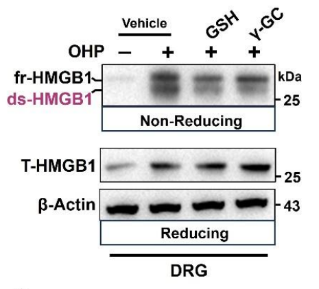
Administration of glutathione (GSH) or Gamma-glutamylcysteine (γ-GC, a cell-permeable GSH analog) (600 mg/kg; p.o.; single dose) targeted to the DRG during OIPN. Western blots showing that oral γ-GC (which elevates intracellular GSH) significantly decreases DRG ds-HMGB1 compared to untreated OIPN controls (and more effectively than equimolar GSH).
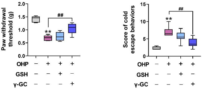
Behavioral assessments demonstrating that Gamma-glutamylcysteine (γ-GC, 600 mg/kg; p.o.; single dose) treatment mitigates pain: mechanical allodynia thresholds and cold-plate latencies/scores in OIPN mice at week 1. γ-GC reverses oxaliplatin-induced hypersensitivity, whereas GSH has a more modest effect.
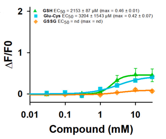
Dose-response curves of hTAS1R1/rTAS1R3 stimulated with GSH, Gamma-glutamylcysteine (Glu-Cys), and GSSG. The results showed that the application of Glu-Cys produced weak agonist activity, with an EC50 value of 3204 ± 1543 μM and a max ∆F/F0 of 0.42 ± 0.07.
Gamma-glutamylcysteine TFA (γ-Glu-Cys TFA) is an orally active, blood-brain barrier permeable dipeptide. Gamma-glutamylcysteine TFA activates AMPK, SIRT1, IL-4/STAT6, AC/cAMP/PI3K, IGF-1R/IRS1/PI3K, and Nrf2 signaling pathways; it inhibits NF-κB, JAK1/STAT1/3, MAPKs, cadmium-induced p38 MAPK, JNK, and PI3K/Akt signaling pathways. Gamma-glutamylcysteine TFA regulates macrophage polarization, modulates the trafficking of CD36 and GLUT4, induces glutathione synthesis, improves metabolic dysfunction, reduces lipid deposition, ameliorates glucose homeostasis, inhibits apoptosis (Apoptosis), stabilizes mitochondria, suppresses lipid peroxidation, iron accumulation and ferroptosis (Ferroptosis), reduces ds-HMGB1 levels, reverses mechanical hyperalgesia, and alleviates hepatic lipid droplet formation. Gamma-glutamylcysteine TFA is applicable to research related to inflammatory bowel disease, type 2 diabetes, cadmium-induced neurotoxicity, Alzheimer's disease, cerebral ischemia/reperfusion injury, neuropathy, and alcoholic liver disease[1][2][3][4][5][6][8][9].
IC50 & Target
IL-10
IL-6
IL-1β
Human Endogenous Metabolite
In Vitro
Gamma-glutamylcysteine TFA (0-80 μM; 24 h) inhibits M1 polarization of Raw264.7 macrophages by suppressing the JAK1/STAT1/3, AKT, MAPKs and NF-κB signaling pathways, while activating the AMPK/SIRT1 axis[1]. Gamma-glutamylcysteine TFA (0-80 μM; 24 h) promotes M2 polarization of Raw264.7 macrophages, and drives their repolarization from M1 to M2 via activation of the IL-4/STAT6 and AMPK/SIRT1 signaling pathways[1]. Gamma-glutamylcysteine TFA (20-80 μM; 24 h) induces the polarization of human Jurcat CD4+ T lymphocytes toward Th2 by promoting IL-4 secretion[1]. Gamma-glutamylcysteine TFA (2 mM; 6 h) activates the Nrf2 signaling pathway via nuclear translocation and upregulates the expression of antioxidant target genes and proteins in primary intestinal epithelial cells (IECs) of chicken embryos[2]. Gamma-glutamylcysteine TFA (2 mM; 6 h) inhibits oxidative stress induced by Salmonella typhimurium in primary wild-type chicken embryonic intestinal epithelial cells (IECs) by maintaining Nrf2 activation[2]. Gamma-glutamylcysteine TFA (2 mM; 6 h) protects primary intestinal epithelial cells (IECs) from wild-type chicken embryos against intestinal barrier disruption and inflammatory injury induced by Salmonella typhimurium, and this protective effect depends on the Nrf2 signaling pathway[2]. Gamma-glutamylcysteine TFA (20-80 μM; administered for 24 h after 24 h of pre-treatment with insulin + PA) dose-dependently activates the IGF-1R/IRS1/PI3K/Akt signaling pathway in insulin-resistant HepG2 cells, primary mouse hepatocytes and C2C12 myotubes induced by Insulin (HY-P701240) + Palmitic acid (HY-N0830) (PA), by increasing the phosphorylation levels of key pathway proteins[3]. Gamma-glutamylcysteine TFA (2-4 mM; 2 h pretreatment followed by 12 h cadmium exposure) inhibits the activation of JNK/p38 MAPK and PI3K/Akt signaling pathways in cadmium-treated cells by downregulating the expression of pro-apoptotic markers and normalizing the Bax/Bcl-2 ratio. This consequently suppresses cadmium-induced changes in apoptosis-related proteins, inhibits cadmium-induced cell apoptosis, and prevents cadmium-induced mitochondrial transmembrane potential depolarization in PC12 cells[4]. Gamma-glutamylcysteine TFA (2-4 mM; 2 h pretreatment followed by 12 h cadmium exposure) dose-dependently inhibits cadmium-induced oxidative stress in PC12 cells by reducing ROS and lipid peroxidation levels, restoring antioxidant enzyme activity, and maintaining intracellular GSH homeostasis[4]. Gamma-glutamylcysteine TFA (0.25-16 mM; 24 h) shows no toxicity to BV-2 cells at concentrations up to 4 mM after 24 h of incubation, while higher concentrations (8, 16 mM) reduce cell viability in a dose-dependent manner[5]. Gamma-glutamylcysteine TFA (2-4 mM; 30 min pretreatment, 24 h AβO exposure) dose-dependently inhibits the release of proinflammatory mediators (TNF-α, IL-1β, NO) and the expression of proinflammatory proteins (iNOS, COX-2) induced by AβO in BV-2 cells, suppresses AβO-induced oxidative stress, and restores the antioxidant capacity of cells[5]. Gamma-glutamylcysteine TFA (4 mM; 30 min pretreatment, followed by 6-24 h of AβO exposure) inhibits AβO-induced activation of the NF-κB signaling pathway in BV-2 cells, upregulates and maintains the expression of Nurr1 mRNA and protein, and thereby exerts anti-inflammatory effects by suppressing the binding of NF-κB p65 to the iNOS promoter induced by AβO in BV-2 cells[5]. Gamma-glutamylcysteine TFA (2-4 mM; 30 min pretreatment, followed by 24 h AβO exposure) dose-dependently inhibits the release of proinflammatory mediators (TNF-α, IL-1β, NO) induced by AβO in primary mouse microglia, suppresses oxidative stress, and restores the antioxidant capacity of cells[5]. Gamma-glutamylcysteine TFA (0.25-2 mM; 12 h) increases GSH levels, GSH/GSSG ratio, GPX activity, and cell viability in primary cortical neurons treated with OGD/R, with the strongest effect observed at 2 mM for 12 h[6]. Gamma-glutamylcysteine TFA (0.85-7 mM; 12 h) increases GSH level, GSH/GSSG ratio, GPX activity and cell viability in PC12 cells treated with oxygen-glucose deprivation/reoxygenation (OGD/R)[6]. Gamma-glutamylcysteine TFA (1.7-7 mM; 0-12 h, time-course 0-12 h) regulates the mRNA and protein levels of GSS in OGD/R-treated PC12 cells, promotes nuclear translocation of Nrf2 in cells, reduces the interaction between Nrf2 and Keap1, inhibits the upregulation of Keap1, decreases MDA and Fe2+ levels, improves cell viability, and thereby inhibits ferroptosis in cells[6]. Gamma-glutamylcysteine TFA (3.5 mM; 12 h, time-course 0-12 h) activates Nrf2 in OGD/R-treated PC12 cells by increasing the level of phosphorylated PKC-ε[6]. Gamma-glutamylcysteine TFA (200 μM; 15 min pre-incubation, 24 h co-incubation with oligomeric Aβ40) protects primary human astrocytes against oligomeric Aβ40-induced cytotoxicity, apoptosis, oxidative stress, neuroinflammation, and dysregulated metalloproteinase activity, while restoring antioxidant status and GSH levels[7]. Gamma-glutamylcysteine TFA (20-80 μM; 2 h pretreatment followed by 24 h ethanol exposure; 400 μM; 24 h single treatment) protects human L02 hepatocytes against ethanol-induced injury by dose-dependently increasing cell viability, reducing hepatic enzyme release, inhibiting cell apoptosis, suppressing oxidative stress and mitochondrial damage, and attenuating the activation of pro-inflammatory signaling pathways[9].
MedChemExpress (MCE) has not independently confirmed the accuracy of these methods. They are for reference only.
Significantly increased phosphorylation of p-IGF-1R (Tyr1135), p-IRS1 (Tyr612), p-PI3K, and p-Akt (Ser473) in a dose-dependent manner relative to insulin + PA-only treated cells across all three cell types, with significant increases observed at 20, 40, and 80 μM.
Down-regulated the ratio of Bax/Bcl-2 in cadmium-treated PC12 cells. Reduced the protein levels of cytosolic cytopigment c, cleaved-caspase-9, cleaved-caspase-3, and cleaved-PARP in cadmium-treated PC12 cells.
Dose-dependently decreased AβO-induced upregulation of iNOS and COX-2 protein expression. Caused a greater reduction in both proteins at 4 mM than at 2 mM.
Inhibited AβO-induced phosphorylation of IKKα and IκBα. Inhibited AβO-induced degradation of IκBα.
In Vivo
Gamma-glutamylcysteine TFA (600-1200 mg/kg; p.o.; daily; ~3 days) significantly increases the survival rate of mice with TNBS-induced inflammatory bowel disease, alleviates colon damage, and shifts macrophage polarization from the pro-inflammatory M1 phenotype to the anti-inflammatory M2 phenotype, with both doses exhibiting therapeutic efficacy[1]. Gamma-glutamylcysteine TFA (250-500 mg/kg; p.o.; daily; 8 weeks) dose-dependently improves glycemic control, insulin sensitivity, β-cell function and hepatic steatosis, while alleviating diabetes-related organ damage in db/db mice[3]. Gamma-glutamylcysteine TFA (100-400 mg/kg/d; p.o.; daily; 20 days) dose-dependently inhibits AβO-induced neuroinflammation in male ICR mice[5]. Gamma-glutamylcysteine TFA (688 mg/kg; p.o.; single dose) upregulates GSH by activating the PKC-ε/Nrf2/GSS pathway, and significantly reduces cerebral infarction volume, neurological dysfunction, and neuronal ferroptosis induced by cerebral ischemia/reperfusion injury in male Sprague-Dawley rats[6]. Gamma-glutamylcysteine TFA (600 mg/kg; p.o.; once) significantly reduces the level of ds-HMGB1 in DRG of OIPN mice and reverses oxaliplatin-induced mechanical hyperalgesia[8]. Gamma-glutamylcysteine TFA (700-1200 mg/kg; p.o.; daily; 7 days) alleviates acute ethanol-induced hepatotoxicity in male C57BL/6JNifdc mice in a dose-dependent manner by reducing hepatic enzyme release, restoring hepatic antioxidant levels, alleviating histopathological damage, and inhibiting inflammatory signaling pathways[9].
MedChemExpress (MCE) has not independently confirmed the accuracy of these methods. They are for reference only.
Conferred significant protection against lethality. Reversed TNBS-induced body weight loss. Reduced the increase in Disease Activity Index (DAI). Mitigated colon shortening, bleeding, and ulcerations. Lowered histological injury scores. Significantly reduced colon tissue mRNA levels of M1 markers (Inos, Il-1β). Significantly increased colon tissue mRNA levels of M2 markers (Cd206, Arg1). Significantly reduced serum TNF-α levels relative to the TNBS model group. Confirmed reduced M1 marker (iNOS, IL-1β) and increased M2 marker (CD206, ARG1) protein levels in colon tissue via Western blot and immunohistochemistry. Significantly reduced TNBS-induced phosphorylation of JAK1, STAT1, STAT3, AKT, JNK, ERK, p38, IKKα/β, and IkBα in colon lamina propria mononuclear cells. Increased phosphorylation of STAT6 and serum IL-4 levels.
Significantly decreased food intake, food efficiency, and water intake in db/db mice at 500 mg/kg dose. Decreased water intake in db/db mice at 250 mg/kg dose. Dose-dependently decreased fasting blood glucose, serum HbA1c, serum insulin levels, and HOMA-IR index, while increased HOMA-β index in db/db mice. Dose-dependently improved glucose tolerance (reduced oral glucose tolerance test AUC) and insulin sensitivity (reduced insulin tolerance test AUC) in db/db mice. Dose-dependently improved organ coefficients (reduced liver coefficient, increased cardiac and kidney coefficients) and reduced tissue damage (cardiomyocyte degeneration, kidney glomerular/tubular damage, hepatocyte ballooning, pancreatic islet loss) in db/db mice, with 500 mg/kg treatment more effective than metformin. Dose-dependently reduced urinary MAU/ALB levels and increased hepatic glycogen content in db/db mice. Dose-dependently reduced subcutaneous adipose tissue (SAT) and visceral adipose tissue (VAT) weight, and decreased adipocyte size in db/db mice. Dose-dependently reduced hepatic triglyceride (TG), total cholesterol (TC), and serum TG, TC, LDL-C levels, while increased serum HDL-C levels in db/db mice; 500 mg/kg treatment reduced hepatic TG more effectively than metformin. Dose-dependently reduced hepatic lipid droplet accumulation (Oil Red O staining) and decreased serum ALT and AST levels in db/db mice. Dose-dependently reduced hepatic CD36, PPARα, and CPT1A protein expression, and increased hepatic p-IGF-1R, p-IRS1, p-PI3K, and p-Akt protein expression in db/db mice. Showed no significant effect on body weight, fasting blood glucose, organ coefficients, or tissue histology in C57BL/6J mice at both doses.
Animal Model:
ICR mice (male, 22-25 g, Alzheimer's disease model induced by intracerebroventricular administration of 2 μg AβO)[5]
Dosage:
100 mg/kg/d; 400 mg/kg/d
Administration:
p.o.; daily; 20 days
Result:
Suppressed AβO-induced microglial activation (reduced Iba1 fluorescence intensity in the hippocampus), and inhibited upregulation of hippocampal COX-2, iNOS, and Iba1 protein expression (100 mg/kg/d dose). Suppressed AβO-induced microglial activation with greater reduction in Iba1 fluorescence intensity than the 100 mg/kg/d dose, and exhibited stronger inhibition of AβO-induced upregulation of hippocampal COX-2, iNOS, and Iba1 protein expression compared to the 100 mg/kg/d dose (400 mg/kg/d dose).
Animal Model:
Sprague-Dawley (SD) (male, 260-300 g, transient focal cerebral ischemia induced by 90-minute middle cerebral artery occlusion followed by reperfusion)[6]
Dosage:
688 mg/kg
Administration:
p.o.; single dose (administered 1.5 hours after MCAO)
Result:
Significantly reduced cerebral infarction volume. Significantly lowered neurological deficit scores. Significantly increased the number of Nissl-positive neurons in the ipsilateral cerebral cortex. Significantly reduced the number of FJB+/NeuN+ (dying) neurons in the cerebral cortex. Alleviated MCAO/R-induced neuronal mitochondrial damage, including reduced loss of mitochondrial cristae and outer membrane rupture. Significantly decreased cortical H2O2, malondialdehyde (MDA), and Fe2+ levels, and reduced 4-HNE-positive neuronal counts. Restored MCAO/R-induced alterations in ferroptosis-related protein and mRNA levels: increased FTH1 and GPX4, and decreased ACSL4 and TF (no effect on SLC7A11). Significantly elevated cortical GSH levels, GSH/GSSG ratio, and glutathione peroxidase (GPX) activity. Inhibited MCAO/R-induced reduction of cortical GSS mRNA and protein levels, and increased GSS/NeuN-positive neuron counts. Increased total Nrf2 and phosphorylated Nrf2 protein levels in cortical tissue, and elevated Nrf2/NeuN and p-Nrf2/NeuN-positive neuron counts. Increased phosphorylated PKC protein levels in cerebral cortical tissue.
p.o.; 2 hours prior to each weekly oxaliplatin injection
Result:
Reduced ds-HMGB1 levels in the DRG to ~2.0 relative units. Produced a smaller reduction in serum ds-HMGB1 to ~1.6 relative units. Increased paw withdrawal thresholds to ~1.2 g, reversing oxaliplatin-induced mechanical allodynia. Reduced cold escape behavior scores to ~4.0, modestly improving cold sensitivity. Exhibited greater efficacy at reducing DRG ds-HMGB1 levels and improving pain responses than equimolar glutathione.
Reduced ethanol-induced elevations in serum ALT, AST, and TG levels in a dose-dependent manner. Reversed ethanol-induced depletion of hepatic GSH levels. Reduced hepatic lipid droplet formation and inflammatory cell infiltration. Decreased ethanol-induced increases in hepatic protein levels of iNOS, p-p65, p-IKKα/β, and p-IκBα in a dose-dependent manner.
DMSO : 100 mg/mL (274.50 mM; Need ultrasonic; Hygroscopic DMSO has a significant impact on the solubility of product, please use newly opened DMSO)
H2O : 100 mg/mL (274.50 mM; Need ultrasonic)
Preparing Stock Solutions
ConcentrationSolventMass
1 mg
5 mg
10 mg
1 mM
2.7450 mL
13.7250 mL
27.4499 mL
5 mM
0.5490 mL
2.7450 mL
5.4900 mL
10 mM
0.2745 mL
1.3725 mL
2.7450 mL
View the Complete Stock Solution Preparation Table
*Please refer to the solubility information to select the appropriate solvent. Once prepared, please aliquot and store the solution to prevent product inactivation from repeated freeze-thaw cycles. Storage method and period of stock solution: -80°C, 6 months; -20°C, 1 month (sealed storage, away from moisture). When stored at -80°C, please use it within 6 months. When stored at -20°C, please use it within 1 month.
*
Note: If you choose water as the stock solution, please dilute it to the working solution,
then filter and sterilize it with a 0.22 μm filter before use.
For the following dissolution methods, please ensure to first prepare a clear stock solution using an In Vitro approach and then sequentially add co-solvents:
To ensure reliable experimental results, the clarified stock solution can be appropriately stored based on storage conditions. As for the working solution for in vivo experiments, it is recommended to prepare freshly and use it on the same day. The percentages shown for the solvents indicate their volumetric ratio in the final prepared solution. If precipitation or phase separation occurs during preparation, heat and/or sonication can be used to aid dissolution.
This protocol yields a clear solution of ≥ 2.5 mg/mL (saturation unknown).
Taking 1 mL working solution as an example, add 100 μLDMSO stock solution (25.0 mg/mL) to 400 μL PEG300, and mix evenly; then add 50 μL Tween-80 and mix evenly; then add 450 μL Saline to adjust the volume to 1 mL.
Preparation of Saline: Dissolve 0.9 g sodium chloride in ddH₂O and dilute to 100 mL to obtain a clear Saline solution.
Protocol 2
Add each solvent one by one: 10% DMSO 90% (20% SBE-β-CD in Saline)
Solubility: ≥ 2.5 mg/mL (6.86 mM); Clear solution
This protocol yields a clear solution of ≥ 2.5 mg/mL (saturation unknown).
Taking 1 mL working solution as an example, add 100 μLDMSO stock solution (25.0 mg/mL) to 900 μL 20% SBE-β-CD in Saline, and mix evenly.
Preparation of 20% SBE-β-CD in Saline (4°C, storage for one week): 2 g SBE-β-CD powder is dissolved in 10 mL Saline, completely dissolve until clear.
Protocol 3
Add each solvent one by one: 10% DMSO 90% Corn Oil
Solubility: ≥ 2.5 mg/mL (6.86 mM); Clear solution
This protocol yields a clear solution of ≥ 2.5 mg/mL (saturation unknown). If the continuous dosing period exceeds half a month, please choose this protocol carefully.
Taking 1 mL working solution as an example, add 100 μLDMSO stock solution (25.0 mg/mL) to 900 μLCorn oil, and mix evenly.
In Vivo Dissolution Calculator
Please enter the basic information of animal experiments:
Dosage
mg/kg
Animal weight (per animal)
g
Dosing volume (per animal)
μL
Number of animals
Recommended: Prepare an additional quantity of animals to account for potential losses during experiments.
Calculation results:
Working solution concentration:
mg/mL
This product has good water solubility, please refer to the measured solubility data in water/PBS/Saline for details.
The concentration of the stock solution you require exceeds the measured solubility. The following solution is for reference only.If necessary, please contact MedChemExpress (MCE).
*Please refer to the solubility information to select the appropriate solvent. Once prepared, please aliquot and store the solution to prevent product inactivation from repeated freeze-thaw cycles. Storage method and period of stock solution: -80°C, 6 months; -20°C, 1 month (sealed storage, away from moisture). When stored at -80°C, please use it within 6 months. When stored at -20°C, please use it within 1 month.
Optional Solvent
ConcentrationSolventMass
1 mg
5 mg
10 mg
25 mg
DMSO / H2O
1 mM
2.7450 mL
13.7250 mL
27.4499 mL
68.6248 mL
5 mM
0.5490 mL
2.7450 mL
5.4900 mL
13.7250 mL
10 mM
0.2745 mL
1.3725 mL
2.7450 mL
6.8625 mL
15 mM
0.1830 mL
0.9150 mL
1.8300 mL
4.5750 mL
20 mM
0.1372 mL
0.6862 mL
1.3725 mL
3.4312 mL
25 mM
0.1098 mL
0.5490 mL
1.0980 mL
2.7450 mL
30 mM
0.0915 mL
0.4575 mL
0.9150 mL
2.2875 mL
40 mM
0.0686 mL
0.3431 mL
0.6862 mL
1.7156 mL
50 mM
0.0549 mL
0.2745 mL
0.5490 mL
1.3725 mL
60 mM
0.0457 mL
0.2287 mL
0.4575 mL
1.1437 mL
80 mM
0.0343 mL
0.1716 mL
0.3431 mL
0.8578 mL
100 mM
0.0274 mL
0.1372 mL
0.2745 mL
0.6862 mL
*
Note: If you choose water as the stock solution, please dilute it to the working solution,
then filter and sterilize it with a 0.22 μm filter before use.
Gamma-glutamylcysteine TFA Related Classifications
Species cross-reactivity must be investigated individually for each product. Many human cytokines will produce a nice response in mouse cell lines, and many mouse proteins will show activity on human cells. Other proteins may have a lower specific activity when used in the opposite species.
Customer Validation
Administration of glutathione (GSH) or Gamma-glutamylcysteine (γ-GC, a cell-permeable GSH analog) (600 mg/kg; p.o.; single dose) targeted to the DRG during OIPN. Western blots showing that oral γ-GC (which elevates intracellular GSH) significantly decreases DRG ds-HMGB1 compared to untreated OIPN controls (and more effectively than equimolar GSH).
Customer Validation
Behavioral assessments demonstrating that Gamma-glutamylcysteine (γ-GC, 600 mg/kg; p.o.; single dose) treatment mitigates pain: mechanical allodynia thresholds and cold-plate latencies/scores in OIPN mice at week 1. γ-GC reverses oxaliplatin-induced hypersensitivity, whereas GSH has a more modest effect.
Customer Validation
Dose-response curves of hTAS1R1/rTAS1R3 stimulated with GSH, Gamma-glutamylcysteine (Glu-Cys), and GSSG. The results showed that the application of Glu-Cys produced weak agonist activity, with an EC50 value of 3204 ± 1543 μM and a max ∆F/F0 of 0.42 ± 0.07.
MedChemExpress values your privacy and your trust is important to us. We use cookies to enhance your website experience. Some cookies are necessary to run the website.
Privacy and Cookie Policy


 Powered by Bioz


