



Mesaconic acid

Based on 1 Customer Validation
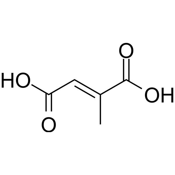
Mesaconic acid (Citronic acid; Methylfumaric acid) is an orally active anti-inflammatory and antioxidant agent. Mesaconic acid reduces the level of NF-κB in colon tissues, downregulates the expression of Keap1 and Bax, upregulates the expression of Nrf2 and Bcl2, and decreases the expression of Caspase-1. Mesaconic acid reduces the levels of NLRP3, ASC and Casp-1 in colon, liver and kidney tissues. Mesaconic acid reduces pro-inflammatory cytokine levels, increases the level of the anti-inflammatory cytokine IL-10, elevates NAD+ levels, regulates oxidative stress markers and antioxidant enzyme levels, and upregulates intestinal barrier proteins in colon, liver and kidney tissues. Mesaconic acid increases the abundance of beneficial gut bacteria and reduces the abundance of harmful gut bacteria in rapidly aging mice, and exhibits anti-aging properties. Mesaconic acid acts as a flame retardant and serves as a competitive inhibitor of fumarate reduction. Mesaconic acid can be used in the research of aging-related inflammation.
For research use only. We do not sell to patients.
- Purity : 99.95%
- CAS No.: 498-24-8
- Formula: C5H6O4
- Molecular Weight:130.10
-
Storage:Powder -20°C, 3 years , 4°C, 2 years ; In solvent -80°C, 6 months , -20°C, 1 month

To place orders, for customer services and technical support, please contact: MedChemExpress USA
Tel: 609-228-6898 E-mail: [email protected] [email protected]


-
 Biological Activity
-
Chemical Information
-
Solvent & Solubility
- Protocols
- Purity & Documentation
- References
-
 Help & FAQs


-
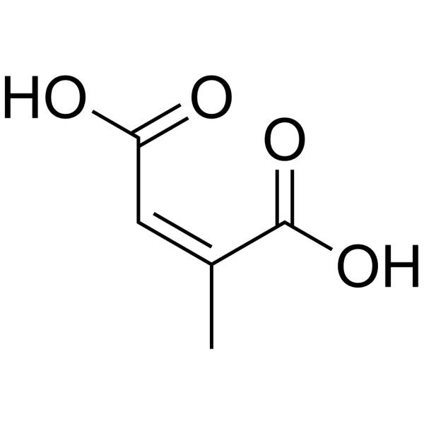
HY-113298
Citraconic acidNLRP3/Keap1-Nrf2 Inhibitor -
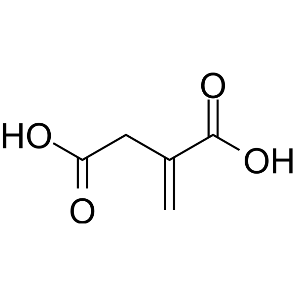
HY-Y0520
Itaconic acidFungi Metabolite -

HY-78036R
Mesaconic acid (Standard)Active Product -
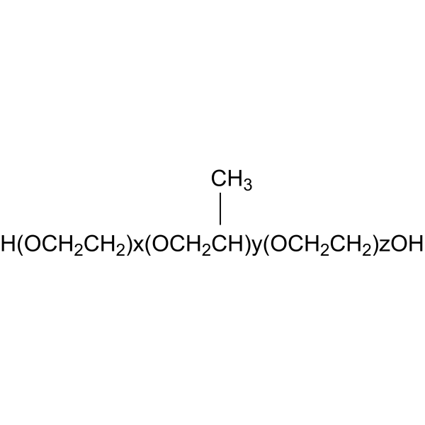
HY-D1005
Poloxamer 407 (F127)LPL Receptor Inhibitor -
HY-D1005A
Poloxamer 188Linear Copolymer -
HY-D1005H
Poloxamer 188 (F68)Nonionic Linear Copolymer -

HY-D1005A6
Poloxamer 124 (L44)Biochemical Assay Agent -
HY-W110551
2-Methacryloyloxyethyl phosphorylcholinePolymer Monomer -
HY-12444
Y15FAK Inhibitor -
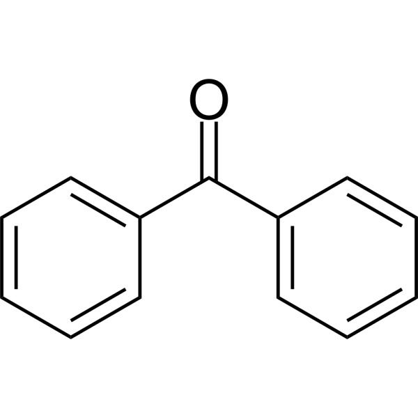
HY-Y0546
BenzophenonePhotosensitizer -

HY-D1005A22
Poloxamer 338 (F108)Biochemical Assay Agent -
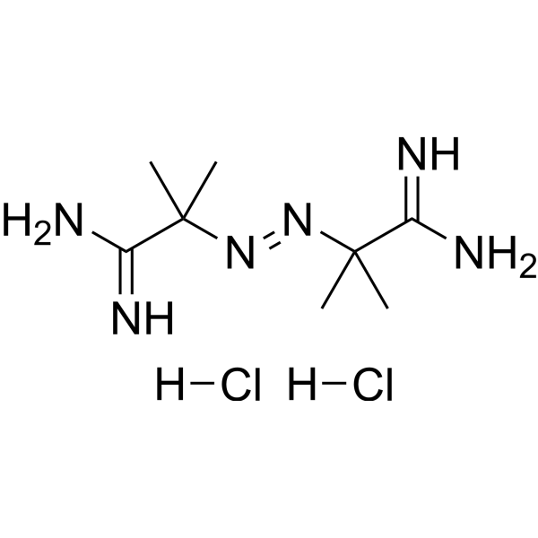
HY-Y0525
AAPHRadical Generator


Biological Activity
Description
In Vitro
Mesaconic acid is an unsaturated dibasic acid naturally present in fresh cabbage leaves[2].
MedChemExpress (MCE) has not independently confirmed the accuracy of these methods. They are for reference only. Further protocols information, click here.
In Vivo
MedChemExpress (MCE) has not independently confirmed the accuracy of these methods. They are for reference only.
-
Animal Model:SAMP8 (SPF grade; 6 mice per group)[1]
-
Dosage:200 mg/kg
-
Administration:p.o.; daily; 9 weeks
-
Result:Significantly reduced levels of pro-inflammatory factors IL-1β, IL-6, IL-5, and TNF-α in colon, liver, and kidney tissues.
Significantly increased levels of anti-inflammatory factor IL-10 in colon, liver, and kidney tissues.
Significantly decreased levels of NLRP3, ASC, and Casp-1 in colon, liver, and kidney tissues.
Significantly increased levels of GSH-Px, SOD, T-AOC, and CAT in colon, liver, and kidney tissues.
Significantly reduced levels of ROS, MDA, carbonyl protein, NF-κB, P38 protein, and P65 protein in colon, liver, and kidney tissues.
Significantly increased levels of NAD+ in colon, liver, and kidney tissues.
Significantly increased levels of ZO-1, occludin, and claudin-1 in colon tissue.
Significantly decreased serum levels of D-LA, DAO, and endotoxin.
Significantly increased relative mRNA expression levels of GSH-Px, SOD, TrxR, Nrf2, and Bcl2 in colon tissue.
Significantly reduced relative mRNA expression levels of Keap1, Bax, NF-κB, NLRP3, and Caspase-1 in colon tissue.
Significantly increased observed species, Chao1, ACE, and Shannon indices of gut microbiota.
Increased abundance of beneficial bacteria including Ligilactobacillus, Erysipelatoclostridium, Dubosiella, Parasutterella, Haemophilus, Parabacteroides, Rikenellaceae RC9 gut group, and Bacteroides.
Decreased abundance of harmful bacteria Helicobacter pylori, Escherichia-Shigella, and Staphylococcus.
Identified Dubosiella as a key differential beneficial genus in the mesaconic acid group via LEfSe analysis.
Detected 260 positive mode metabolites (155 upregulated, 105 downregulated) and 131 negative mode metabolites (61 upregulated, 70 downregulated) with significant differences compared to the model group.
Significantly upregulated metabolites including taurochenodeoxycholic acid (sodium salt), 3-hydroxybutyric acid, tauroursodeoxycholic acid, 7-ketolithocholic acid, and deoxycholic acid.
Showed intact mucosa, neatly arranged epithelial cells, no inflammatory cell infiltration, increased goblet cells, positive PAS staining (PAS++), and moderately positive MUC2 immunohistochemistry (MUC2++) in colon tissue histology.


Chemical Information
-
CAS No. 498-24-8
-
Appearance Solid
-
Molecular Weight 130.10
-
Formula C5H6O4
-
Color White to off-white
-
SMILES
C/C(C(O)=O)=C\C(O)=O
-
Synonyms
Citronic acid; Methylfumaric acid
-
Structure Classification
-
Initial Source
-
Shipping
Room temperature in continental US; may vary elsewhere.
-
Storage
Powder -20°C 3 years 4°C 2 years In solvent -80°C 6 months -20°C 1 month


Solvent & Solubility
In Vitro:
Methanol : ≥ 116.67 mg/mL (896.77 mM)
H2O : 50 mg/mL (384.32 mM; Need ultrasonic)
* "≥" means soluble, but saturation unknown.
Please refer to the solubility information to select the appropriate solvent. Once prepared, please aliquot and store the solution to prevent product inactivation from repeated freeze-thaw cycles.
Storage method and period of stock solution: -80°C, 6 months; -20°C, 1 month. When stored at -80°C, please use it within 6 months. When stored at -20°C, please use it within 1 month.
* Note: If you choose water as the stock solution, please dilute it to the working solution, then filter and sterilize it with a 0.22 μm filter before use.
Please refer to the solubility information to select the appropriate solvent. Once prepared, please aliquot and store the solution to prevent product inactivation from repeated freeze-thaw cycles.
Storage method and period of stock solution: -80°C, 6 months; -20°C, 1 month. When stored at -80°C, please use it within 6 months. When stored at -20°C, please use it within 1 month.
* Note: If you choose water as the stock solution, please dilute it to the working solution, then filter and sterilize it with a 0.22 μm filter before use.
Concentration (start) × Volume (start) = Concentration (final) × Volume (final)

Protocols
-
RNA extraction experimental
By lysing cells, releasing RNA, and removing impurities such as proteins and DNA, high-purity RNA products are finally obtained. The commonly used traditional method is the guanidine isothiocyanate/phenol/chloroform method (Trizol), which is suitable for a variety of animal materials including animal tissues, microorganisms, cultured cells, etc., and most plant materials.
-
ROS/oxidative-stress fluorescent staining
ROS/oxidative-stress fluorescent staining uses cell-permeant fluorogenic probes that become fluorescent after oxidation inside cells or tissues; commonly used examples include DCFH-DA/DCFDA for broad cellular oxidant detection, DHE for superoxide-related signal detection, MitoSOX for mitochondrial superoxide-related signal detection, and CellROX probes for oxidative-stress-associated fluorescence readouts. The assay detects probe oxidation rather than a single ROS species unless the probe and analysis method have been chemically validated for that species. DCFH-DA enters cells, is deacetylated by intracellular esterases to DCFH, and produces fluorescent DCF after oxidation, so the readout is used as an operational measure of total cellular oxidative stress rather than a species-specific ROS measurement. DHE and MitoSOX can report superoxide-related oxidation, but red fluorescence alone can include non-specific ethidium-like oxidation products; HPLC or optimized spectral approaches are
-
Apoptosis Solutions
Apoptosis is a regulated, generally non-lytic cell-death pathway that removes unwanted, damaged, infected, or abnormal cells through coordinated morphological changes, caspase activation, DNA fragmentation, and membrane remodeling. The intrinsic apoptosis pathway is controlled mainly by mitochondrial outer membrane permeabilization, BCL-2 family proteins, cytochrome c release, apoptosome formation, caspase-9 activation, and downstream executioner caspase-3/7 activation. The extrinsic apoptosis pathway is initiated by death receptors such as Fas, TNFR, and TRAIL receptors, which recruit adaptor proteins and activate caspase-8 before engaging executioner caspases or mitochondrial amplification through BID cleavage. Apoptosis is linked to many phenotypes, including cancer cell killing, tissue homeostasis, immune regulation, neurodegeneration, infection response, and treatment-induced cytotoxicity; unresolved questions include how apoptosis interacts with necroptosis, pyroptosis, ferroptos
-
Research Protocol for Inflammation-related Diseases
The NLRP3 inflammasome is a cytosolic innate immune signaling platform that integrates priming signals and danger-signal activation to promote caspase-1 activation, maturation of IL-1β and IL-18, and gasdermin D-mediated pyroptotic cell death. The core experimental logic is to determine whether inflammatory disease phenotypes are driven by increased NLRP3 expression, ASC-containing inflammasome assembly, caspase-1 cleavage, GSDMD cleavage, and extracellular release of IL-1β/IL-18 rather than by nonspecific cell injury alone. The pathway is strongly linked to inflammation-related disease phenotypes because monosodium urate crystals activate NALP3/NLRP3 inflammasome signaling in gout-like crystal inflammation, cholesterol crystals activate NLRP3 inflammasomes in atherogenesis models, and DSS-induced intestinal inflammation has been reported to involve NLRP3 inflammasome activity. However, experimental colitis studies also show context-dependent protective effects of NLRP3 inflammasome co
-
Pyroptosis Solutions
Pyroptosis is a lytic inflammatory cell-death pathway executed by gasdermin pores, most classically through inflammasome-mediated activation of caspase-1, cleavage of gasdermin D, membrane pore formation, LDH release, and secretion of IL-1β and IL-18. The canonical pathway is commonly modeled by priming cells with an inflammatory signal such as LPS to induce pro-IL-1β and inflammasome components, followed by an activation signal such as ATP or nigericin to activate NLRP3, ASC speck formation, caspase-1 cleavage, GSDMD cleavage, cytokine release, and pyroptotic membrane rupture. The non-canonical pathway is triggered when cytosolic LPS activates mouse caspase-11 or human caspase-4/5, leading to GSDMD cleavage and pyroptosis, and this can secondarily activate NLRP3-dependent IL-1β release. Pyroptosis is linked to inflammatory injury, infection, cancer, liver disease, ocular disease, placental inflammation, and other disease phenotypes, but unresolved questions include which gasdermin fam


Purity & Documentation
-
Data Sheet (275 KB)
-
SDS (394 KB)
- English - EN (394 KB)
- Français - FR (394 KB)
- Deutsch - DE (394 KB)
- Norwegian - NO (394 KB)
- Español - ES (394 KB)
- Swedish - SV (394 KB)
- Italian - IT (394 KB)
- Korean - KR (394 KB)
- Portuguese - PT (394 KB)
-
Handling Instructions (2659 KB)
References
[1]. Wu L, et al. Mesaconic acid as a key metabolite with anti-inflammatory and anti-aging properties produced by Lactobacillus plantarum 124 from centenarian gut microbiota. NPJ biofilms and microbiomes. 2025 Aug 19;11(1):165. [Content Brief]
[2]. Buston HW. Note on the isolation of mesaconic acid from cabbage leaves. Biochem J. 1928;22(6):1523-5. [Content Brief]


Complete Stock Solution Preparation Table
Please refer to the solubility information to select the appropriate solvent. Once prepared, please aliquot and store the solution to prevent product inactivation from repeated freeze-thaw cycles.
Storage method and period of stock solution: -80°C, 6 months; -20°C, 1 month. When stored at -80°C, please use it within 6 months. When stored at -20°C, please use it within 1 month.
| Optional Solvent | Concentration Solvent Mass | 1 mg | 5 mg | 10 mg | 25 mg |
|---|---|---|---|---|---|
| H2O / Methanol | 1 mM | 7.6864 mL | 38.4320 mL | 76.8639 mL | 192.1599 mL |
| 5 mM | 1.5373 mL | 7.6864 mL | 15.3728 mL | 38.4320 mL | |
| 10 mM | 0.7686 mL | 3.8432 mL | 7.6864 mL | 19.2160 mL | |
| 15 mM | 0.5124 mL | 2.5621 mL | 5.1243 mL | 12.8107 mL | |
| 20 mM | 0.3843 mL | 1.9216 mL | 3.8432 mL | 9.6080 mL | |
| 25 mM | 0.3075 mL | 1.5373 mL | 3.0746 mL | 7.6864 mL | |
| 30 mM | 0.2562 mL | 1.2811 mL | 2.5621 mL | 6.4053 mL | |
| 40 mM | 0.1922 mL | 0.9608 mL | 1.9216 mL | 4.8040 mL | |
| 50 mM | 0.1537 mL | 0.7686 mL | 1.5373 mL | 3.8432 mL | |
| 60 mM | 0.1281 mL | 0.6405 mL | 1.2811 mL | 3.2027 mL | |
| 80 mM | 0.0961 mL | 0.4804 mL | 0.9608 mL | 2.4020 mL | |
| 100 mM | 0.0769 mL | 0.3843 mL | 0.7686 mL | 1.9216 mL |
* Note: If you choose water as the stock solution, please dilute it to the working solution, then filter and sterilize it with a 0.22 μm filter before use.


Mesaconic acid Related Classifications
HY-78036 Related Classifications
 Powered by Bioz