



RGB-286638
Based on 3 publication(s) in Google Scholar
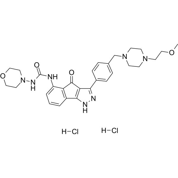
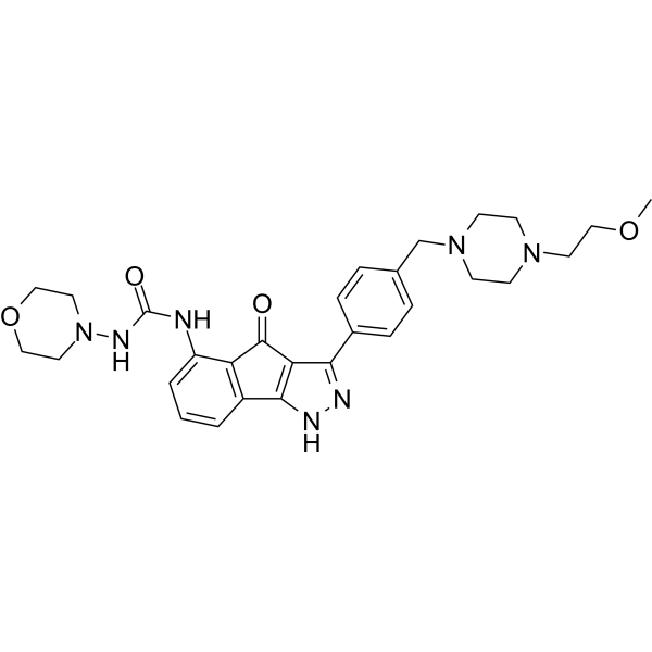
RGB-286638 is a potent inhibitor of CDK and MAPK9. RGB-286638 inhibits the activities of cyclin T1-CDK9, cyclin B1-CDK1, cyclin E-CDK2, cyclin D1-CDK4, cyclin E-CDK3 and p35-CDK5, with IC50 values of 1, 2, 3, 4, 5 and 5 nM, respectively. RGB-286638 inhibits GSK-3β and TAK1, with IC50 values of 3 nM and 5 nM, respectively. RGB-286638 induces caspase-dependent Apoptosis in cells. RGB-286638 delays tumor growth in an orthotopic glioblastoma mouse model. RGB-286638 extends survival in multiple myeloma models. RGB-286638 can be used in research related to glioblastoma and multiple myeloma.
Nur für Forschungszwecke. Wir verkaufen nicht an Patienten.
- Reinheit : 99.67%
- CAS. Nr.: 784210-87-3
- Formel: C29H37Cl2N7O4
- Molecular Weight:618.55
-
Speicherung:
4°C, sealed storage, away from moisture
* In solvent : -80°C, 6 months; -20°C, 1 month (sealed storage, away from moisture)

To place orders, for customer services and technical support, please contact: MedChemExpress USA
Tel: 609-228-6898 E-mail: [email protected] [email protected]


-
 Biologische Aktivität
-
Chemical Information
-
Lösungsmittel & Löslichkeit
- Protokoll
- Reinheit & Dokumentation
- Verweise
-
 Help & FAQs


-
HY-15504A
RGB-286638 free baseCDK/MAPK9 Inhibitor -
HY-10492
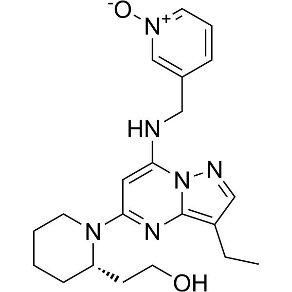
DinaciclibCDK2/5/9 Inhibitor -
HY-10008
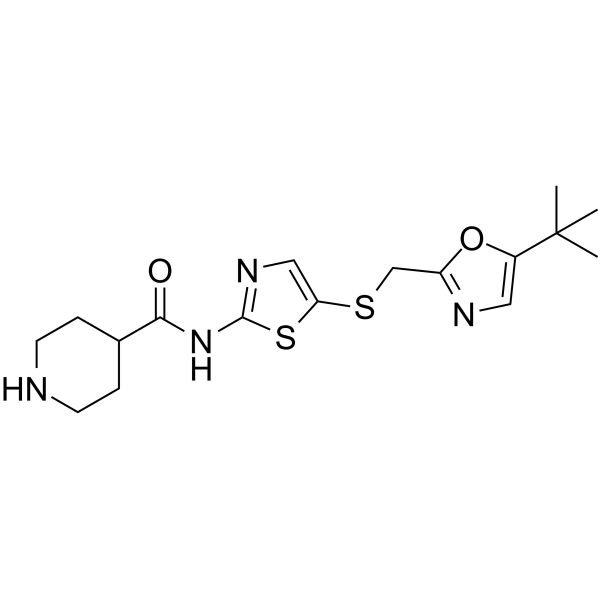
SNS-032CDK Inhibitor -

HY-10227
BortezomibProteasome Inhibitor -
HY-129492
GNF4877DYRK1A/GSK3β Inhibitor -
HY-B1357
DigitoxinAnti-cancer Agent -
HY-19980
Eprenetapoptp53 Activator -
HY-10452
Ixazomib citrateProteasome Inhibitor -
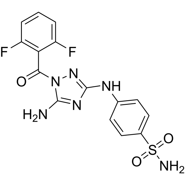
HY-10329
JNJ-7706621Aurora Kinase/CDK Inhibitor -
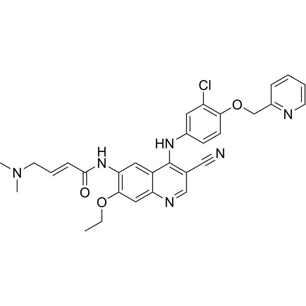
HY-32721
NeratinibHER2/EGFR Inhibitor -
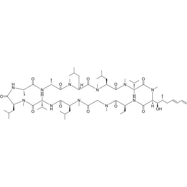
HY-106638
VoclosporinPP2B Inhibitor -
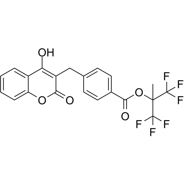
HY-14854
TecarfarinVKOR Antagonist
Publications Citing Use of MedChemExpress (MCE) RGB-286638
MoreAlle MEK Isoform-spezifische Produkte anzeigen
More

Biologische Aktivität
Beschreibung
|
T1-CDK9 1 nM (IC50) |
cyclin B1-CDK1 2 nM (IC50) |
cyclin E-CDK2 3 nM (IC50) |
cyclin D1-CDK4 4 nM (IC50) |
cyclin E-CDK3 5 nM (IC50) |
p35-CDK5 5 nM (IC50) |
cyclin H-CDK7 44 nM (IC50) |
cyclin D3-CDK6 55 nM (IC50) |
GSK-3β 3 nM (IC50) |
JAK2 50 nM (IC50) |
MEK1 54 nM (IC50) |
JNK2 |
Cellular Effect
|
Cell Line

|
Type | Value | Description | References |
|---|---|---|---|---|
| U-251 | IC50 |
0.01 μM
|
Antiproliferative activity against human U251 glioblastoma cells assessed as reduction in cell viability incubated for 72 hrs by MTT assay.
Antiproliferative activity against human U251 glioblastoma cells assessed as reduction in cell viability incubated for 72 hrs by MTT assay.
|
33392504 |
| U-251 | IC50 |
0.03 μM
|
Antiproliferative activity against human U251 glioblastoma cells assessed as reduction in cell viability incubated for 72 hrs by MTT assay.
Antiproliferative activity against human U251 glioblastoma cells assessed as reduction in cell viability incubated for 72 hrs by MTT assay.
|
33392504 |
| T98G | IC50 |
0.01 μM
|
Antiproliferative activity against human T98G glioblastoma cells assessed as reduction in cell viability incubated for 72 hrs by MTT assay.
Antiproliferative activity against human T98G glioblastoma cells assessed as reduction in cell viability incubated for 72 hrs by MTT assay.
|
33392504 |
| T98G | IC50 |
0.03 μM
|
Antiproliferative activity against human T98G glioblastoma cells assessed as reduction in cell viability incubated for 72 hrs by MTT assay.
Antiproliferative activity against human T98G glioblastoma cells assessed as reduction in cell viability incubated for 72 hrs by MTT assay.
|
33392504 |
| U138-MG | IC50 |
0.01 μM
|
Antiproliferative activity against human U138 glioblastoma cells assessed as reduction in cell viability incubated for 72 hrs by MTT assay.
Antiproliferative activity against human U138 glioblastoma cells assessed as reduction in cell viability incubated for 72 hrs by MTT assay.
|
33392504 |
| U138-MG | IC50 |
0.03 μM
|
Antiproliferative activity against human U138 glioblastoma cells assessed as reduction in cell viability incubated for 72 hrs by MTT assay.
Antiproliferative activity against human U138 glioblastoma cells assessed as reduction in cell viability incubated for 72 hrs by MTT assay.
|
33392504 |
| HT-29 | IC50 |
0.01 μM
|
Antiproliferative activity against human HT29 colorectal cells assessed as reduction in cell viability incubated for 72 hrs by MTT assay.
Antiproliferative activity against human HT29 colorectal cells assessed as reduction in cell viability incubated for 72 hrs by MTT assay.
|
33392504 |
| HT-29 | IC50 |
0.03 μM
|
Antiproliferative activity against human HT29 colorectal cells assessed as reduction in cell viability incubated for 72 hrs by MTT assay.
Antiproliferative activity against human HT29 colorectal cells assessed as reduction in cell viability incubated for 72 hrs by MTT assay.
|
33392504 |
In Vitro
MedChemExpress (MCE) has not independently confirmed the accuracy of these methods. They are for reference only.
-
Cell Line:U87, U251, T98G, U138, HT29, GBM8, patient-derived glioblastoma sphere cultures (GSCs)
-
Concentration:0-20 μM
-
Incubation Time:72 h
-
Result:Induced near-complete viability inhibition in U87, U251, T98G, U138, and HT29 cell lines, with IC50 values ranging from 0.01 to 0.03 μM.
Caused approximately one-third of GSC models to show high sensitivity matching established cell lines, one-third to show incomplete response with 5-30% viability after exposure to 1 μM, and one-third to show resistance.
In Vivo
RGB-286638 (30-40 mg/kg; i.v.; daily; 5 consecutive days) achieves a log10 cell kill of 1.6, maximum tumor growth inhibition of 85.06-86.34%, and prolonged survival in a mouse multiple myeloma xenograft model[2].
MedChemExpress (MCE) has not independently confirmed the accuracy of these methods. They are for reference only.
-
Animal Model:athymic nude-Fox1nu (female, 6-8 weeks old, orthotopic glioblastoma model via intracranial injection of luciferase- and mCherry-expressing GBM8 primary GBM cells)[1]
-
Dosage:40 mg/kg
-
Administration:i.v.; daily; 5 consecutive days
-
Result:Reduced in vivo luminescence signals (tumor growth measure) significantly compared to vehicle and control groups.
Showed no significant difference in median overall survival (53 days) versus vehicle-treated mice (47 days).
-
Animal Model:CB-17 severe combined immunodeficient (SCID) (male, 5-6 weeks old, irradiated with 2 Gy 24 hours prior to subcutaneous inoculation of MM.1S cells)[2]
-
Dosage:30 mg/kg; 40 mg/kg
-
Administration:i.v.; daily; 5 consecutive days
-
Result:Achieved a maximum tumor growth inhibition (TGI) of 85.06% at day 14 post-treatment.
Achieved a maximum tumor growth inhibition (TGI) of 86.34% at day 14 post-treatment.
Achieved a log10 cell kill of 1.6.
Prolonged survival, with the first death occurring at day 43 compared to day 24 in controls.
Caused maximum body weight loss of 8.4% at day 5, with subsequent weight recovery within two weeks.
Caused maximum body weight loss of 9.9% at day 15, with subsequent weight recovery within two weeks.
Resulted in no toxic deaths during the study.
- Detection of Apoptosis
- TUNEL staining for apoptotic DNA fragmentation
- Annexin V plus membrane-impermeant dye apoptosis staining
- Patient-Derived Xenograft (PDX)
- Apoptosis Solutions
- Patient-Derived Orthotopic Xenograft (PDOX)
- Subcutaneous Cell-Line-Derived Xenograft
- Orthotopic Cell-Line Xenograft
- How to Choose the Right Model Animal
Clinical Trial
| NCT Number | Sponsor | Condition | Start Date |
Phase
|
|---|---|---|---|---|
| NCT01329991 | Plexxikon| | 2011-05 | PHASE1 |


Chemical Information
-
CAS. Nr. 784210-87-3
-
Appearance Solid
-
Molecular Weight 618.55
-
Formel C29H37Cl2N7O4
-
Color Light yellow to yellow
-
SMILES
O=C(NN1CCOCC1)NC2=CC=CC(C3=C4C(C5=CC=C(CN6CCN(CCOC)CC6)C=C5)=NN3)=C2C4=O.Cl.Cl
-
Versand
Room temperature in continental US; may vary elsewhere.
-
Speicherung
4°C, sealed storage, away from moisture
* In solvent : -80°C, 6 months; -20°C, 1 month (sealed storage, away from moisture)


Publications (3)
-
Journal Impact Factor
-
Most Recent
-
Science
2017 Dec 1;358(6367):eaan4368. PMID: 29191878 -
Cancers (Basel)
Identification of New Vulnerabilities in Conjunctival Melanoma Using Image-Based High Content Drug Screening. [Abstract]2022 Mar 19;14(6):1575. PMID: 35326726 -


Lösungsmittel & Löslichkeit
In Vitro:
DMSO : ≥ 150 mg/mL (242.50 mM; Hygroscopic DMSO has a significant impact on the solubility of product, please use newly opened DMSO)
H2O : 25 mg/mL (40.42 mM; Need ultrasonic)
* "≥" means soluble, but saturation unknown.
Please refer to the solubility information to select the appropriate solvent. Once prepared, please aliquot and store the solution to prevent product inactivation from repeated freeze-thaw cycles.
Storage method and period of stock solution: -80°C, 6 months; -20°C, 1 month (sealed storage, away from moisture). When stored at -80°C, please use it within 6 months. When stored at -20°C, please use it within 1 month.
* Note: If you choose water as the stock solution, please dilute it to the working solution, then filter and sterilize it with a 0.22 μm filter before use.
Please refer to the solubility information to select the appropriate solvent. Once prepared, please aliquot and store the solution to prevent product inactivation from repeated freeze-thaw cycles.
Storage method and period of stock solution: -80°C, 6 months; -20°C, 1 month (sealed storage, away from moisture). When stored at -80°C, please use it within 6 months. When stored at -20°C, please use it within 1 month.
* Note: If you choose water as the stock solution, please dilute it to the working solution, then filter and sterilize it with a 0.22 μm filter before use.
Konzentration (Stammlösung) × Volumen (Stammlösung) = Konzentration (Ziellösung) × Volumen (Ziellösung)
In Vivo:
Select the appropriate dissolution method based on your experimental animal and administration route.
- For the following dissolution methods, please ensure to first prepare a clear stock solution using an In Vitro approach and then sequentially add co-solvents:
- To ensure reliable experimental results, the clarified stock solution can be appropriately stored based on storage conditions. As for the working solution for In Vivo experiments, it is recommended to prepare freshly and use it on the same day.
- The percentages shown for the solvents indicate their volumetric ratio in the final prepared solution. If precipitation or phase separation occurs during preparation, heat and/or sonication can be used to aid dissolution.
Add each solvent one by one: 10% DMSO 40% PEG300 5% Tween-80 45% Saline
Solubility: ≥ 7.5 mg/mL (12.13 mM); Clear solution
This protocol yields a clear solution of ≥ 7.5 mg/mL (saturation unknown).
Taking 1 mL working solution as an example, add 100 μL DMSO stock solution (75.0 mg/mL) to 400 μL PEG300, and mix evenly; then add 50 μL Tween-80 and mix evenly; then add 450 μL Saline to adjust the volume to 1 mL.
Preparation of Saline: Dissolve 0.9 g sodium chloride in ddH₂O and dilute to 100 mL to obtain a clear Saline solution.
Add each solvent one by one: 10% DMSO 90% (20% SBE-β-CD in Saline)
Solubility: ≥ 7.5 mg/mL (12.13 mM); Clear solution
This protocol yields a clear solution of ≥ 7.5 mg/mL (saturation unknown).
Taking 1 mL working solution as an example, add 100 μL DMSO stock solution (75.0 mg/mL) to 900 μL 20% SBE-β-CD in Saline, and mix evenly.
Preparation of 20% SBE-β-CD in Saline (4°C, storage for one week): 2 g SBE-β-CD powder is dissolved in 10 mL Saline, completely dissolve until clear.
In Vivo Dissolution Calculator
Please enter the basic information of animal experiments:
-
-
-
-
Recommended: Prepare an additional quantity of animals to account for potential losses during experiments.
Working solution concentration: 0.22 mg/mL
This product has good water solubility, please refer to the measured solubility data in water/PBS/Saline for details.


Reinheit & Dokumentation
-
Data Sheet (290 KB)
-
SDS (252 KB)
- English - EN (252 KB)
- Français - FR (252 KB)
- Deutsch - DE (252 KB)
- Norwegian - NO (252 KB)
- Español - ES (252 KB)
- Swedish - SV (252 KB)
- Italian - IT (252 KB)
- Korean - KR (252 KB)
- Portuguese - PT (252 KB)
-
Handling Instructions (2659 KB)
Verweise


Complete Stock Solution Preparation Table
Please refer to the solubility information to select the appropriate solvent. Once prepared, please aliquot and store the solution to prevent product inactivation from repeated freeze-thaw cycles.
Storage method and period of stock solution: -80°C, 6 months; -20°C, 1 month (sealed storage, away from moisture). When stored at -80°C, please use it within 6 months. When stored at -20°C, please use it within 1 month.
| Optional Solvent | Concentration Solvent Mass | 1 mg | 5 mg | 10 mg | 25 mg |
|---|---|---|---|---|---|
| H2O / DMSO | 1 mM | 1.6167 mL | 8.0834 mL | 16.1668 mL | 40.4171 mL |
| 5 mM | 0.3233 mL | 1.6167 mL | 3.2334 mL | 8.0834 mL | |
| 10 mM | 0.1617 mL | 0.8083 mL | 1.6167 mL | 4.0417 mL | |
| 15 mM | 0.1078 mL | 0.5389 mL | 1.0778 mL | 2.6945 mL | |
| 20 mM | 0.0808 mL | 0.4042 mL | 0.8083 mL | 2.0209 mL | |
| 25 mM | 0.0647 mL | 0.3233 mL | 0.6467 mL | 1.6167 mL | |
| 30 mM | 0.0539 mL | 0.2694 mL | 0.5389 mL | 1.3472 mL | |
| 40 mM | 0.0404 mL | 0.2021 mL | 0.4042 mL | 1.0104 mL | |
| DMSO | 50 mM | 0.0323 mL | 0.1617 mL | 0.3233 mL | 0.8083 mL |
| 60 mM | 0.0269 mL | 0.1347 mL | 0.2694 mL | 0.6736 mL | |
| 80 mM | 0.0202 mL | 0.1010 mL | 0.2021 mL | 0.5052 mL | |
| 100 mM | 0.0162 mL | 0.0808 mL | 0.1617 mL | 0.4042 mL |
* Note: If you choose water as the stock solution, please dilute it to the working solution, then filter and sterilize it with a 0.22 μm filter before use.


RGB-286638 Related Classifications
HY-15504 Related Classifications
 Powered by Bioz