



Rafutrombopag (tautomerism)

Based on 1 Customer Validation
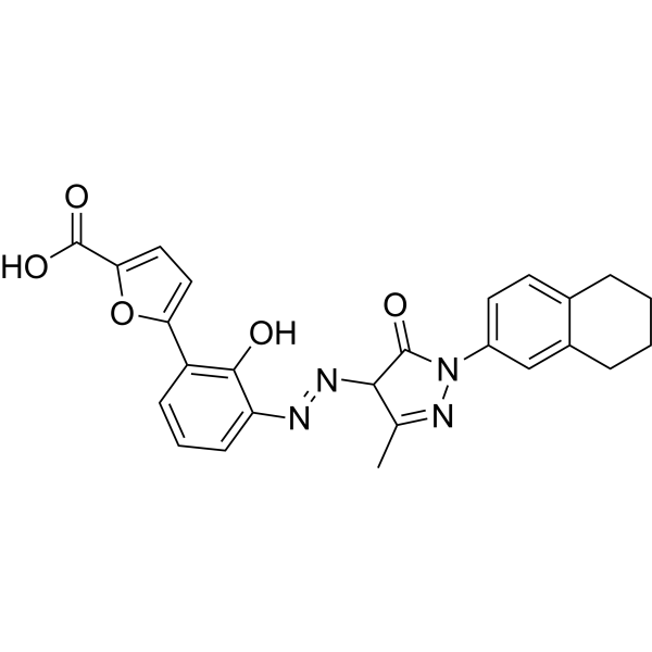
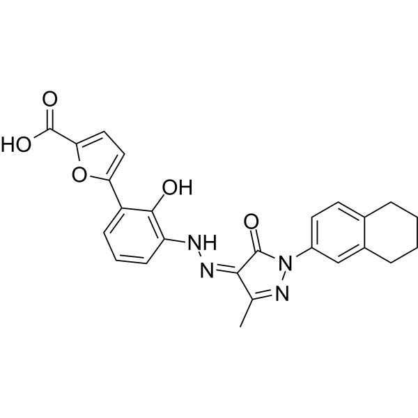
Rafutrombopag (tautomerism) (Hetrombopag) is an orally active nonpeptide thrombopoietin receptor (TPOR/MPL) agonist. Rafutrombopag can chelate iron and alleviate iron overload while promoting haematopoiesis. Rafutrombopag specifically stimulates proliferation and differentiation of human TPOR‐expressing cells, including 32D‐ MPL and human hematopoietic stem cells through stimulation of STAT, PI3K and ERK signalling pathways. Rafutrombopag effectively up-regulates G1-phase-related proteins, including p-RB, Cyclin D1 and CDK4/6, normalizes progression of the cell cycle, and prevents apoptosis by modulating BCL-XL/BAK expression in 32D-MPL cells. Rafutrombopag protects cardiomyocyte survival from oxidative stress damage as an enhancer of stem cells. Rafutrombopag can be used for the study of immune thrombocytopenia and oxidative stress-related cardiovascular disease.
For research use only. We do not sell to patients.
- Purity : 98.00%
- CAS No.: 2114365-78-3
- Formula: C25H22N4O5
- Molecular Weight:458.47
-
Storage:Powder -20°C, 3 years , 4°C, 2 years ; In solvent -80°C, 6 months , -20°C, 1 month

To place orders, for customer services and technical support, please contact: MedChemExpress USA
Tel: 609-228-6898 E-mail: [email protected] [email protected]


-
 Biological Activity
-
Chemical Information
-
Solvent & Solubility
- Protocols
- Purity & Documentation
- References
-
 Help & FAQs


-
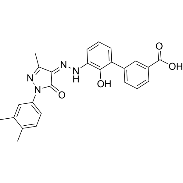
HY-15306
EltrombopagTPO Receptor Agonist -
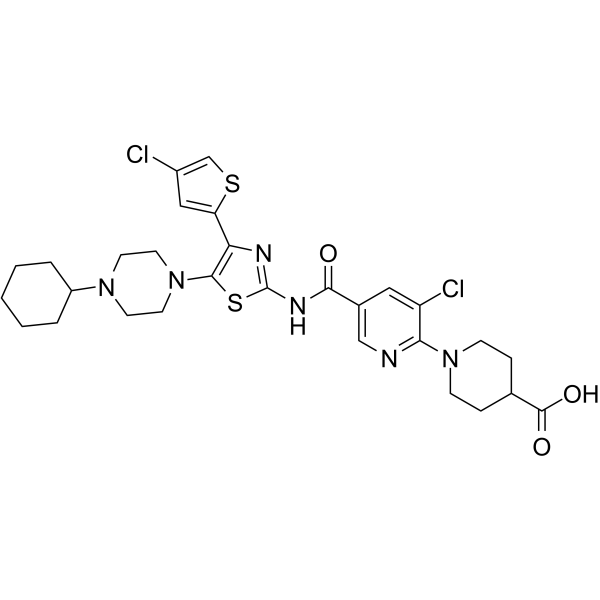
HY-13463
AvatrombopagTPO Receptor Agonist -
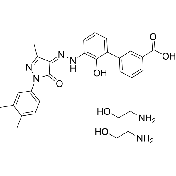
HY-15306A
Eltrombopag OlamineTPO Receptor Agonist -
HY-100380
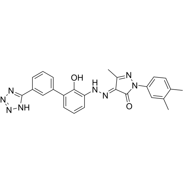
TPO agonist 1Thrombopoietin Receptor Agonist -

HY-15306AR
Eltrombopag Olamine (Standard)TPO Receptor Agonist -

HY-13463R
Avatrombopag (Standard)TPO Receptor Agonist -
HY-19883
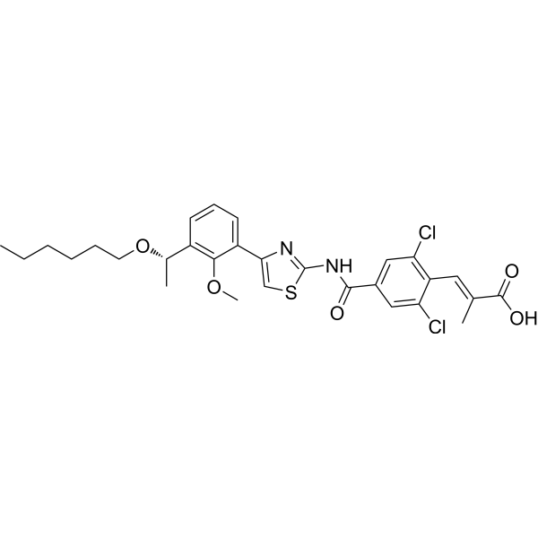
LusutrombopagThrombopoietin Receptor Agonist -
HY-13463A
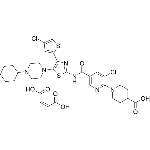
Avatrombopag maleateTPO Receptor Agonist -
HY-13463B
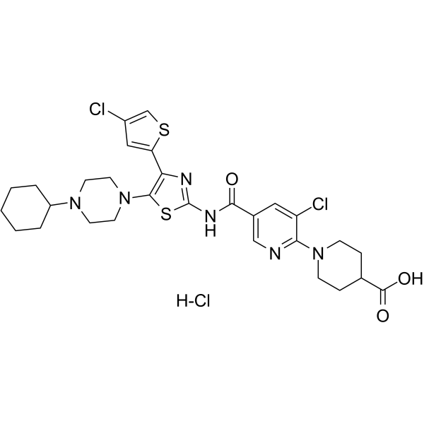
Avatrombopag hydrochlorideTPO Receptor Agonist -
HY-145589
RafutrombopagTPOR/MPL Agonist -

HY-15306R
Eltrombopag (Standard)TPO Receptor Agonist -

HY-19883R
Lusutrombopag (Standard)Thrombopoietin Receptor Agonist


Biological Activity
Description
IC50 & Target
[1]|
CDK4 |
CDK6 |
PI3K |
In Vitro
Rafutrombopag (0.01-1000 nM, 0-7 days) stimulates intracellular TPO signalling pathways and promotes 32D-MPL (EC50 = 0.4 nM) and BaF3/h TPOP ( EC50 = 1.2 nM) cell proliferation in a TPOR-dependent manner[1][2].
Rafutrombopag (0.01-10 μM, 0-10 days) promotes proliferation (EC50 = 2.3 nM) and differentiation of human cord blood-derived CD34+ cells[1][2].
Rafutrombopag (0-3 μM, 24-72 h) normalizes cell-cycle progression and prevents apoptosis in 32D-MPL cells and rat cardiac myocytes[1].
Rafutrombopag (0.01-1000 nM, 30 min-72 h) interacts specifically with TPOR and exerts an additive agonistic effect with rhTPO[1].
Rafutrombopag (0.3-3 μM, 12 h) enhances the beneficial effects of human UCB MNCs in increasing the survival of injured cardiomyocytes during free oxygen radical stress by enhancing human UCB MNCs viability and increasing the secretion of paracrine factors[2].
Rafutrombopag (3-30 μM) has anti-inflammatory activity and significantly reduces the production of NO and TNF-a in LPS (HY-D1056) stimulated macrophage RAW264.7 cells[2].
MedChemExpress (MCE) has not independently confirmed the accuracy of these methods. They are for reference only.
-
Cell Line:32D-MPL cells
-
Concentration:0.3.1 and 3 nM
-
Incubation Time:24 h
-
Result:Caused cells to re-enter the cell cycle, increasing the proportion of cells in G2 and S phases and decreasing the proportion of cells in G1 phase.
Reversed the decrease in G1-phase-related proteins, including p-RB, Cyclin D and CDK4/6, induced by cytokine withdrawal.
-
Cell Line:32D-MPL cells
-
Concentration:0.3.1 and 3 nM
-
Incubation Time:72 h
-
Result:Reduced the apoptosis effect.
Increased expression of the antiapoptotic family members BCL-XL and MCL-1, and decreased expression of proapoptotic BAK.
-
Cell Line:32D-MPL cells
-
Concentration:0.1, 1, 3, 10, 30, 100 nM
-
Incubation Time:0, 0.5, 1, 2, 4, 8, 12, 24h
-
Result:Induced phosphorylation of the major components of TPO‐mediated signalling, including STAT3, STAT5, ERK1/2, and AKT.
Stimulated the phosphorylation of these TPOR downstream effectors in a concentration-dependent manner.
-
Cell Line:human UCB MNCs
-
Concentration:0.3.1 and 3 μM
-
Incubation Time:12 h
-
Result:Increased myocyte viability by 20.3%, 43.7% and 46.8% at 0.3, 1 and 3 μM, respectively.
Showed weak antioxidant activity, and increased the myocyte viability by 15.7%.
In Vivo
Rafutrombopag (18 mg/kg, p.o., once daily for 12 days) significantly stimulates proliferation and prevents apoptosis of 32D-MPL cells in hollow fibres in a time-dependent manner in mice[1].
MedChemExpress (MCE) has not independently confirmed the accuracy of these methods. They are for reference only.
-
Animal Model:32D-MPL cell-containing hollow fibres assay established in mice[1]
-
Dosage:18 mg/kg
-
Administration:Oral administration (p.o.), once daily for 12 days
-
Result:Significantly stimulated proliferation and prevented apoptosis of 32D-MPL cells in hollow fibres in a time-dependent manner.
Reached the number of 32D-MPL cells a maximum after 3 days and then decreased within 12 days.
Had no effect on the counts of white blood cells, reticulocytes, or platelets in the peripheral blood.
- RNA extraction experimental
- Detection of Apoptosis
- TUNEL staining for apoptotic DNA fragmentation
- Flow cytometric DNA-content cell-cycle staining
- Annexin V plus membrane-impermeant dye apoptosis staining
- BrdU Incorporation Assay
- ROS/oxidative-stress fluorescent staining
- Apoptosis Solutions
- Protocol for Cell Cycle
- Research Protocol for Cardiovascular Diseases
- Perls' Prussian Blue Iron Staining
- Cell Viability Determination by MTT Colorimetric Assay


Chemical Information
-
CAS No. 2114365-78-3
-
Appearance Solid
-
Molecular Weight 458.47
-
Formula C25H22N4O5
-
Color Brown to reddish brown
-
SMILES
O=C(C1=CC=C(C2=C(O)C(/N=N/C3C(N(C4=CC=C5C(CCCC5)=C4)N=C3C)=O)=CC=C2)O1)O
-
Synonyms
Hetrombopag (tautomerism); SHR-8735 (tautomerism)
-
Shipping
Room temperature in continental US; may vary elsewhere.
-
Storage
Powder -20°C 3 years 4°C 2 years In solvent -80°C 6 months -20°C 1 month


Solvent & Solubility
In Vitro:
DMSO : 10 mg/mL (21.81 mM; ultrasonic and warming and heat to 60°C; Hygroscopic DMSO has a significant impact on the solubility of product, please use newly opened DMSO)
DMF : 6.25 mg/mL (13.63 mM; ultrasonic and warming and heat to 60°C)
Please refer to the solubility information to select the appropriate solvent. Once prepared, please aliquot and store the solution to prevent product inactivation from repeated freeze-thaw cycles.
Storage method and period of stock solution: -80°C, 6 months; -20°C, 1 month. When stored at -80°C, please use it within 6 months. When stored at -20°C, please use it within 1 month.
Please refer to the solubility information to select the appropriate solvent. Once prepared, please aliquot and store the solution to prevent product inactivation from repeated freeze-thaw cycles.
Storage method and period of stock solution: -80°C, 6 months; -20°C, 1 month. When stored at -80°C, please use it within 6 months. When stored at -20°C, please use it within 1 month.
Concentration (start) × Volume (start) = Concentration (final) × Volume (final)


Purity & Documentation
-
Data Sheet (282 KB)
-
SDS (252 KB)
- English - EN (252 KB)
- Français - FR (252 KB)
- Deutsch - DE (252 KB)
- Norwegian - NO (252 KB)
- Español - ES (252 KB)
- Swedish - SV (252 KB)
- Italian - IT (252 KB)
- Korean - KR (252 KB)
- Portuguese - PT (252 KB)
-
Handling Instructions (2659 KB)
References


Complete Stock Solution Preparation Table
Please refer to the solubility information to select the appropriate solvent. Once prepared, please aliquot and store the solution to prevent product inactivation from repeated freeze-thaw cycles.
Storage method and period of stock solution: -80°C, 6 months; -20°C, 1 month. When stored at -80°C, please use it within 6 months. When stored at -20°C, please use it within 1 month.
| Optional Solvent | Concentration Solvent Mass | 1 mg | 5 mg | 10 mg | 25 mg |
|---|---|---|---|---|---|
| DMF / DMSO | 1 mM | 2.1812 mL | 10.9058 mL | 21.8117 mL | 54.5292 mL |
| 5 mM | 0.4362 mL | 2.1812 mL | 4.3623 mL | 10.9058 mL | |
| 10 mM | 0.2181 mL | 1.0906 mL | 2.1812 mL | 5.4529 mL | |
| DMSO | 15 mM | 0.1454 mL | 0.7271 mL | 1.4541 mL | 3.6353 mL |
| 20 mM | 0.1091 mL | 0.5453 mL | 1.0906 mL | 2.7265 mL |


Rafutrombopag (tautomerism) Related Classifications
HY-122620 Related Classifications
 Powered by Bioz