



Licoflavone A
Based on 3 publication(s) in Google Scholar
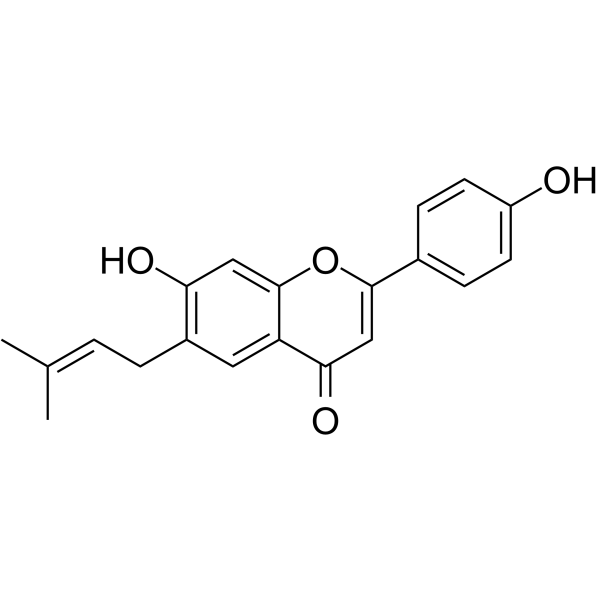
Licoflavone A is an orally active PTP1B/VEGFR-2 inhibitor, with an IC50 of 54.5 μM against PTP1B, an IC50 of 14.36 μM and a Kd of 142.38 nM against human VEGFR-2. Licoflavone A blocks the PI3K/AKT and MEK/ERK signaling pathways. Licoflavone A induces G1 phase cell cycle arrest, apoptosis via the intrinsic mitochondrial pathway (apoptosis), and inhibits migration, invasion and epithelial-mesenchymal transition (EMT) of gastric cancer cells. Licoflavone A inhibits the proliferation of gastric cancer cells in vitro and in xenograft models. Licoflavone A reduces the expression levels of HIF-1α, GLUT1, LDHA, PKM2 and HK2 in hypoxic gastric cancer cells, decreases glucose uptake and suppresses glycolysis. Licoflavone A can be used in research related to gastric cancer, type 2 diabetes and obesity.
For research use only. We do not sell to patients.
- Purity : 99.95%
- CAS No.: 61153-77-3
- Formula: C20H18O4
- Molecular Weight:322.35
-
Storage:
4°C, protect from light
* In solvent : -80°C, 6 months; -20°C, 1 month (protect from light)

To place orders, for customer services and technical support, please contact: MedChemExpress USA
Tel: 609-228-6898 E-mail: [email protected] [email protected]


-
 Biological Activity
-
Chemical Information
-
Solvent & Solubility
- Protocols
- Purity & Documentation
- References
-
 Help & FAQs


-
HY-N0377
LiquiritigeninEstrogen Receptor/ERR Agonist -
HY-14616
ShogaolAutophagy Inducer -
HY-N0783
EupatilinAutophagy Inducer -
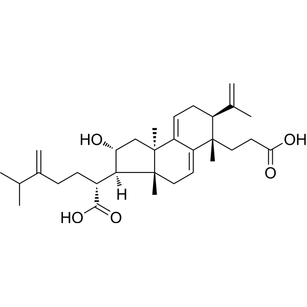
HY-N2995
Poricoic acid ANatural Product -

HY-N1366
HerniarinAnticancer Agent -
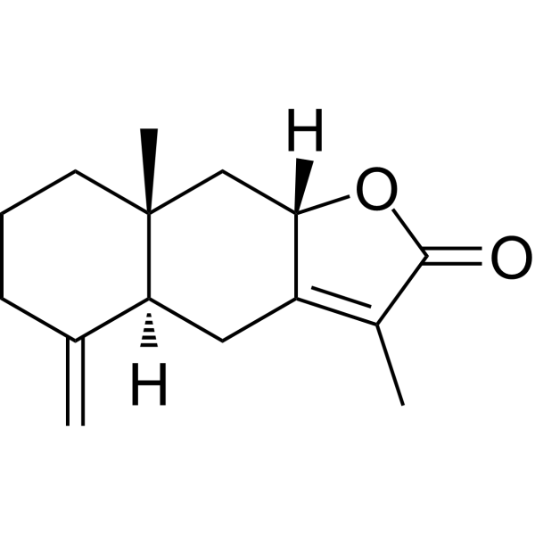
HY-N0202
Atractylenolide IIApoptosis Inducer -

HY-D0852
Sodium orthovanadateProtein Tyrosine/Alkaline Phosphatase Inhibitor -
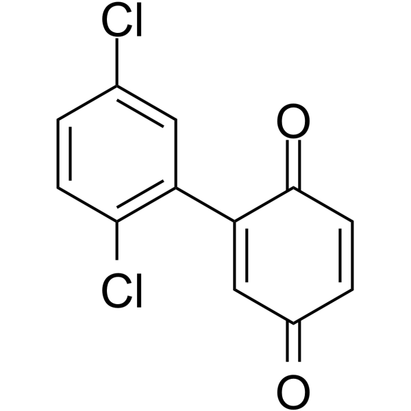
HY-100463
TPI-1SHP-1 Inhibitor -
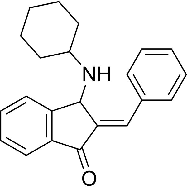
HY-115502
BCIDUSP6 Inhibitor -
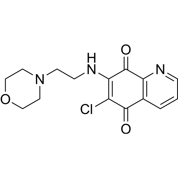
HY-100034
NSC 663284Cdc25 Inhibitor -
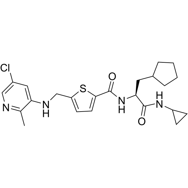
HY-15832
GSK 2830371Wip1 Phosphatase Inhibitor -
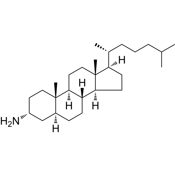
HY-19776
3α-AminocholestanePhosphatase Inhibitor
Publications Citing Use of MedChemExpress (MCE) Licoflavone A
MoreAll VEGFR Isoforms
MoreAll MEK Isoforms
More

Biological Activity
Description
|
VEGFR2 14.36 μM (IC50) |
VEGFR2 142.38 nM (Kd) |
PTP1B 54.5 μM (IC50) |
GLUT1 |
HIF-1α |
LDHA |
PKM2 |
In Vitro
Licoflavone A (6.25-100 μM; 24-72 h) inhibits the proliferation of human gastric cancer SGC-7901, MKN-45, MGC-803 cells and normal GES-1 cells in a dose- and time-dependent manner. Among these cell lines, it exhibits the strongest activity against MKN-45 cells (72 h IC50 = 12.19 μM) and the weakest activity against GES-1 cells[1].
Licoflavone A (25-100 μM; 14 days) dose-dependently inhibits colony formation of human gastric cancer MKN-45 cells stimulated by VEGF[1].
Licoflavone A (25-100 μM; 0-96 h) inhibits VEGF-stimulated 3D spheroid formation in human gastric cancer MKN-45 cells in a dose-dependent manner[1].
Licoflavone A (25-100 μM; 24-48 h) inhibits the proliferation of hypoxic human gastric cancer AGS cells in a time- and concentration-dependent manner[3].
Licoflavone A (25-100 μM; 10-14 d) reduces the colony-forming ability of human gastric cancer AGS cells under hypoxic conditions in a dose-dependent manner, and the 100 μM concentration decreases the colony formation rate to 38.78%[3].
Licoflavone A (25-100 μM; 24-48 h) potently and dose-dependently inhibits the migration of human gastric cancer AGS cells under hypoxic conditions, and the 100 μM concentration reduces the 48 h scratch wound healing rate to 14.31%[3].
Licoflavone A (25-100 μM; 72 h) arrests VEGF-stimulated human gastric cancer MKN-45 cells at the G0 phase of the cell cycle in a dose-dependent manner by downregulating cyclin D1 and c-Myc[1].
Licoflavone A (25-100 μM; 72 h) dose-dependently induces apoptosis in VEGF-stimulated human gastric cancer MKN-45 cells via the mitochondrial intrinsic pathway, which involves an increased Bax/Bcl-2 ratio, cytochrome c release, and caspase activation[1].
Licoflavone A (25-100 μM; 48 h) dose-dependently inhibits the migration and invasion abilities of VEGF-stimulated human gastric cancer MKN-45 cells[1].
Licoflavone A (25-100 μM; 72 h) inhibits epithelial-mesenchymal transition (EMT) in VEGF-stimulated human gastric cancer MKN-45 cells in a dose-dependent manner by regulating EMT-related proteins[1].
Licoflavone A (25-100 μM; 72 h) blocks the PI3K/AKT and MEK/ERK signaling pathways in VEGF-stimulated human gastric cancer MKN-45 cells in a dose-dependent manner by downregulating PI3K/AKT transcription and inhibiting MEK/ERK phosphorylation[1].
Licoflavone A (25-100 μM; 48 h) downregulates the mRNA expression of key glycolytic genes in human gastric cancer AGS cells under hypoxic conditions, and the 100 μM concentration significantly inhibits the mRNA expression of GLUT1, LDHA, PKM2 and HK2[3].
Licoflavone A (25-100 μM; 48 h) significantly downregulates the expression of HIF-1α and key glycolysis-related proteins in hypoxic human gastric cancer AGS cells at the concentration of 100 μM[3].
Licoflavone A (25-100 μM; 48 h) inhibits glucose uptake and HK activity in hypoxic human gastric cancer AGS cells at the concentration of 100 μM[3].
MedChemExpress (MCE) has not independently confirmed the accuracy of these methods. They are for reference only. Further protocols information, click here.
-
Cell Line:human gastric cancer SGC-7901, MKN-45, MGC-803 cells, normal human GES-1 cells
-
Concentration:6.25, 12.5, 25, 50, 100 μM
-
Incubation Time:24 h, 48 h, 72 h
-
Result:Decreased cell viability in a dose-dependent and time-dependent manner across all four cell lines.
Exhibited stronger inhibitory effect on gastric cancer cells than normal GES-1 cells, with effect increasing with longer incubation times.
Achieved IC50 values of 78.08 μM (SGC-7901, 24 h), 40.83 μM (SGC-7901, 48 h), 23.74 μM (SGC-7901, 72 h), 43.26 μM (MKN-45, 24 h), 23.67 μM (MKN-45, 48 h), 12.19 μM (MKN-45, 72 h), 124.50 μM (MGC-803, 24 h), 75.75 μM (MGC-803, 48 h), 47.19 μM (MGC-803, 72 h), 180.30 μM (GES-1, 24 h), 130.30 μM (GES-1, 48 h), 110.00 μM (GES-1, 72 h).
-
Cell Line:VEGF-stimulated human gastric cancer MKN-45 cells
-
Concentration:25, 50, 100 μM
-
Incubation Time:72 h
-
Result:Dose-dependently inhibited the proliferation of VEGF-stimulated MKN-45 cells, with statistically significant reductions compared to VEGF-only controls (p < 0.01).
-
Cell Line:VEGF-stimulated human gastric cancer MKN-45 cells
-
Concentration:25, 50, 100 μM
-
Incubation Time:72 h
-
Result:Dose-dependently blocked the G1 to S cell cycle transition, increasing the proportion of cells in G0 phase and decreasing the proportion in S phase.
Dose-dependently downregulated G1 phase proteins cyclin D1 and c-Myc via Western blot analysis.
-
Cell Line:VEGF-stimulated human gastric cancer MKN-45 cells
-
Concentration:25, 50, 100 μM
-
Incubation Time:72 h
-
Result:Induced dose-dependent increases in apoptotic features (apoptotic bodies, condensed/fragmented nuclei) detected via Hoechst 33342 staining.
Increased apoptotic cell populations in a dose-dependent manner, with apoptosis rates rising from ~2% in VEGF-only controls to ~50% at 100 μM, detected via flow cytometry.
Caused dose-dependent decreases in mitochondrial membrane potential, with increased green fluorescence (JC-10 monomer) indicating depolarization, detected via MMP assay.
Dose-dependently upregulated Bax, Cyt C, caspase 9, and cleaved-caspase 3, and downregulated Bcl-2, increasing the Bax/Bcl-2 ratio, detected via Western blot.
-
Cell Line:VEGF-stimulated human gastric cancer MKN-45 cells
-
Concentration:25, 50, 100 μM
-
Incubation Time:72 h
-
Result:Dose-dependently downregulated the expression of MMP2, MMP9, and N-cadherin.
Dose-dependently upregulated the expression of E-cadherin.
-
Cell Line:human gastric cancer AGS cells (hypoxic conditions)
-
Concentration:25, 50, 100 μM
-
Incubation Time:24 h, 48 h
-
Result:Inhibited cell proliferation by 7.98% at 25 μmol·L-1 for 24 h (no significant difference), 13.99% at 25 μM for 48 h, 21.94% at 50 μM for 24 h, 25.02% at 50 μM for 48 h, 23.00% at 100 μM for 24 h, and 41.74% at 100 μM for 48 h.
-
Cell Line:human gastric cancer AGS cells (hypoxic conditions)
-
Concentration:25, 50, 100 μM
-
Incubation Time:48 h
-
Result:Downregulated HK2 mRNA expression to 1.50 at 25 μM; downregulated GLUT1 mRNA expression to 1.00, LDHA mRNA expression to 2.23, and HK2 mRNA expression to 1.36 at 50 μM; downregulated GLUT1 mRNA expression to 0.84, LDHA mRNA expression to 1.98, PKM2 mRNA expression to 2.93, and HK2 mRNA expression to 1.04 at 100 μmol·L-1.
-
Cell Line:human gastric cancer AGS cells (hypoxic conditions)
-
Concentration:25, 50, 100 μM
-
Incubation Time:48 h
-
Result:Reduced HK2 protein expression to 1.00 at 25 μM; reduced HK2 protein expression to 1.14 at 50 μM; reduced HIF-1α protein expression to 0.82, GLUT1 protein expression to 1.11, LDHA protein expression to 0.87, PKM2 protein expression to 0.99, and HK2 protein expression to 0.53 at 100 μmol·L-1.
In Vivo
MedChemExpress (MCE) has not independently confirmed the accuracy of these methods. They are for reference only.
-
Animal Model:BALB/c-nude (male, 4-6 weeks old, 18 g, subcutaneous xenograft model)[1]
-
Dosage:50 mg/kg
-
Administration:p.o.; daily; 14 days
-
Result:Reduced tumor volume significantly.
Reduced tumor weight significantly.
Caused no significant changes in body weight.


Chemical Information
-
CAS No. 61153-77-3
-
Appearance Solid
-
Molecular Weight 322.35
-
Formula C20H18O4
-
Color Off-white to yellow
-
SMILES
O=C1C=C(C2=CC=C(O)C=C2)OC3=CC(O)=C(C/C=C(C)\C)C=C13
-
Structure Classification
-
Initial Source
-
Shipping
Room temperature in continental US; may vary elsewhere.
-
Storage
4°C, protect from light
* In solvent : -80°C, 6 months; -20°C, 1 month (protect from light)


Publications (3)
-
Journal Impact Factor
-
Most Recent
-
Food Chem
Effects of sun drying combined with baking processes on the flavor quality of Chongqing Tuocha raw tea. [Abstract]2025 Dec 30:497:146992. PMID: 41285060 -
Food Chem
Flavonoid-mediated metabolic underpinning quality variation in red bud-sport pear mutants. [Abstract]2025 Oct 15:489:144992. PMID: 40466530 -
Genomics
Transcriptomics and metabolomics reveal the induction of flavonoid biosynthesis pathway in the interaction of Stylosanthes-Colletotrichum gloeosporioides. [Abstract]2021 Jul;113(4):2702-2716. PMID: 34111523


Solvent & Solubility
In Vitro:
DMSO : 100 mg/mL (310.22 mM; Need ultrasonic; Hygroscopic DMSO has a significant impact on the solubility of product, please use newly opened DMSO)
H2O : < 0.1 mg/mL (insoluble)
* Licoflavone A is usually formulated as a suspension.
Please refer to the solubility information to select the appropriate solvent. Once prepared, please aliquot and store the solution to prevent product inactivation from repeated freeze-thaw cycles.
Storage method and period of stock solution: -80°C, 6 months; -20°C, 1 month (protect from light). When stored at -80°C, please use it within 6 months. When stored at -20°C, please use it within 1 month.
Please refer to the solubility information to select the appropriate solvent. Once prepared, please aliquot and store the solution to prevent product inactivation from repeated freeze-thaw cycles.
Storage method and period of stock solution: -80°C, 6 months; -20°C, 1 month (protect from light). When stored at -80°C, please use it within 6 months. When stored at -20°C, please use it within 1 month.
Concentration (start) × Volume (start) = Concentration (final) × Volume (final)
In Vivo:
Select the appropriate dissolution method based on your experimental animal and administration route.
- For the following dissolution methods, please ensure to first prepare a clear stock solution using an In Vitro approach and then sequentially add co-solvents:
- To ensure reliable experimental results, the clarified stock solution can be appropriately stored based on storage conditions. As for the working solution for In Vivo experiments, it is recommended to prepare freshly and use it on the same day.
- The percentages shown for the solvents indicate their volumetric ratio in the final prepared solution. If precipitation or phase separation occurs during preparation, heat and/or sonication can be used to aid dissolution.
Add each solvent one by one: 10% DMSO 40% PEG300 5% Tween-80 45% Saline
Solubility: ≥ 2.5 mg/mL (7.76 mM); Clear solution
This protocol yields a clear solution of ≥ 2.5 mg/mL (saturation unknown).
Taking 1 mL working solution as an example, add 100 μL DMSO stock solution (25.0 mg/mL) to 400 μL PEG300, and mix evenly; then add 50 μL Tween-80 and mix evenly; then add 450 μL Saline to adjust the volume to 1 mL.
Preparation of Saline: Dissolve 0.9 g sodium chloride in ddH₂O and dilute to 100 mL to obtain a clear Saline solution.
In Vivo Dissolution Calculator
Please enter the basic information of animal experiments:
-
-
-
-
Recommended: Prepare an additional quantity of animals to account for potential losses during experiments.
Please enter your animal formula composition:
-
%DMSO +
Recommended: Keep the proportion of DMSO in working solution below 2% if your animal is weak.
-
%+
-
+%Tween-80 + +
-
%Saline +
The co-solvents required include: DMSO, . All of co-solvents are available by MedChemExpress (MCE). , Tween 80. All of co-solvents are available by MedChemExpress (MCE).
Working solution concentration: 0.22 mg/mL
Method for preparing stock solution: mg drug dissolved in μL DMSO. Stock solution concentration: mg/mL. * In solvent : -80°C, 6 months; -20°C, 1 month (protect from light)
1. Take μL DMSO stock solution;
2. Add μL .
μL , mix evenly;
3. Then add μL Tween 80, mix evenly;
4. Then add μL
Please ensure that the stock solution in the first step is dissolved to a clear state, and add co-solvents in sequence. You can use ultrasonic heating (ultrasonic cleaner, recommended frequency 20-40 kHz), vortexing, etc. to assist dissolution.

Protocols
-
RNA extraction experimental
By lysing cells, releasing RNA, and removing impurities such as proteins and DNA, high-purity RNA products are finally obtained. The commonly used traditional method is the guanidine isothiocyanate/phenol/chloroform method (Trizol), which is suitable for a variety of animal materials including animal tissues, microorganisms, cultured cells, etc., and most plant materials.
-
Annexin V plus membrane-impermeant dye apoptosis staining
Annexin V-based apoptosis assays rely on the detection of phosphatidylserine (PS) externalization from the inner leaflet of the plasma membrane to the outer leaflet, an early biochemical hallmark of apoptosis. Fluorescently labeled Annexin V binds PS in a calcium-dependent manner, enabling identification of early apoptotic cells by flow cytometry or fluorescence microscopy. When combined with a membrane-impermeant DNA-binding dye (e. g. , propidium iodide), this approach allows discrimination between viable (Annexin V−/dye−), early apoptotic (Annexin V+/dye−), and late apoptotic or necrotic (Annexin V+/dye+) cell populations by assessing membrane integrity and PS exposure.
-
BrdU Incorporation Assay
Bromodeoxyuridine (BrdU) incorporation assay is based on the principle that BrdU, a thymidine analog, is incorporated into newly synthesized DNA during the S phase of the cell cycle, thereby serving as a marker of DNA replication and cellular proliferation. Incorporated BrdU can be detected using anti-BrdU antibodies following DNA denaturation, enabling visualization or quantification of proliferating cells through immunochemical detection methods such as immunofluorescence or immunohistochemistry.
-
Cell migration
Cell migration is a method that plays an important role in wound healing, cell differentiation, embryonic development, etc.
-
Protocol for Cell Cycle
Cell-cycle analysis by flow cytometry measures DNA content in single cells to estimate the fraction of cells in G0/G1, S, and G2/M phases. Propidium iodide intercalates into DNA, and after RNA removal with RNase, fluorescence intensity reflects cellular DNA content: 2N cells are assigned to G0/G1, cells between 2N and 4N to S phase, and 4N cells to G2/M. DNA-content analysis alone cannot reliably separate G0 from G1 or G2 from M. Ki-67 can distinguish quiescent G0 cells from cycling cells, EdU or BrdU incorporation marks active DNA synthesis in S phase, and phospho-histone H3 staining identifies mitotic cells within the 4N population.
-
Research Protocol for Metabolic Diseases
AMP-activated protein kinase, AMPK, is a conserved cellular energy sensor that responds to reduced cellular energy status and coordinates metabolism by increasing ATP-generating catabolic pathways while suppressing ATP-consuming anabolic processes. In metabolic disease research, the AMPK pathway is experimentally relevant because it regulates hepatic lipid synthesis, fatty acid oxidation, glucose production, skeletal-muscle glucose disposal, mTORC1-linked biosynthesis, autophagy, mitochondrial homeostasis, and whole-body energy balance. The central pathway logic is that energy stress, metformin, exercise-like stimulation, or direct AMPK activators increase AMPKα Thr172 phosphorylation and downstream substrate phosphorylation, including ACC and RAPTOR. Phosphorylation of ACC suppresses lipogenesis and supports fatty acid oxidation, whereas phosphorylation of RAPTOR suppresses mTORC1 signaling and links cellular energy status to growth and protein synthesis control. The pathway is linked
-
Cell Viability Determination by MTT Colorimetric Assay
The following protocol uses the MTT colorimetric assay as a classic literature-established method for assessing cell viability/metabolic activity in cultured mammalian cells. MTT[3-(4,5-dimethylthiazol-2-yl)-2,5-diphenyltetrazolium bromide] is reduced by metabolically active cells to a colored formazan product; the amount of formazan is quantified spectrophotometrically and provides an indirect measure of metabolically active viable cells. Importantly, MTT reduction reflects cellular oxidoreductase/metabolic activity rather than an absolute direct count of living cells, so changes in cellular metabolism can alter the signal independently of cell number.
-
Apoptosis
Apoptosis, also called programmed cell death, is generally characterized by distinct morphological characteristics.
-
TUNEL staining for apoptotic DNA fragmentation
TUNEL staining detects DNA strand breaks by using terminal deoxynucleotidyl transferase to add labeled nucleotides to exposed 3′-OH DNA termini, generating either microscopic staining in fixed cells or tissue sections, or fluorescence/cytometric signal in cell suspensions. TUNEL positivity reflects DNA fragmentation but should not be interpreted alone as definitive apoptosis, because TUNEL can also label necrotic, autolytic, mechanically damaged, or DNA-repair-associated DNA breaks.
-
Cell Cytotoxicity Assay
Cytotoxicity assays are usually based on the assessment of cell membrane damage, which can also be indirectly detected by measuring cell viability. Detection methods include MTT assay, CKK-8 assay, LDH assay and ATP assay, etc.
-
Flow cytometric DNA-content cell-cycle staining
Flow cytometric DNA-content cell-cycle staining measures the fluorescence intensity of DNA-bound fluorochromes in single cells or nuclei to estimate DNA content distributions, allowing assignment of populations to G0/G1, S, and G2/M phases by DNA histogram deconvolution. Propidium iodide (PI) intercalates into DNA, and PI fluorescence is proportional to cellular DNA content when staining is performed under conditions that make DNA accessible and minimize non-DNA signal. Cells with G2/M DNA content are expected to show approximately twice the fluorescence intensity of G0/G1 cells, while S-phase cells occupy intermediate fluorescence values. PI-based DNA-content analysis can also detect cells with fractional DNA content, often reported as sub-G1, when DNA fragmentation and extraction during staining reduce retained DNA signal in apoptotic cells. DAPI is an alternative DNA fluorochrome for univariate DNA-content analysis, while bivariate approaches combining DNA content with proliferation
-
Patient-Derived Xenograft (PDX)
Patient-derived xenograft (PDX) models are generated by engrafting primary human tumor tissue directly into immunodeficient mice, allowing in vivo propagation of patient tumor biology without initial in vitro adaptation. These models are used to preserve key histopathological and molecular characteristics of the original tumor and enable assessment of tumor growth dynamics and therapeutic response in a living organism. The biological readout is tumor engraftment and subsequent growth in the murine host, which reflects the ability of human tumor cells to survive, vascularize, and expand in an immunocompromised microenvironment.
-
Apoptosis Solutions
Apoptosis is a regulated, generally non-lytic cell-death pathway that removes unwanted, damaged, infected, or abnormal cells through coordinated morphological changes, caspase activation, DNA fragmentation, and membrane remodeling. The intrinsic apoptosis pathway is controlled mainly by mitochondrial outer membrane permeabilization, BCL-2 family proteins, cytochrome c release, apoptosome formation, caspase-9 activation, and downstream executioner caspase-3/7 activation. The extrinsic apoptosis pathway is initiated by death receptors such as Fas, TNFR, and TRAIL receptors, which recruit adaptor proteins and activate caspase-8 before engaging executioner caspases or mitochondrial amplification through BID cleavage. Apoptosis is linked to many phenotypes, including cancer cell killing, tissue homeostasis, immune regulation, neurodegeneration, infection response, and treatment-induced cytotoxicity; unresolved questions include how apoptosis interacts with necroptosis, pyroptosis, ferroptos
-
Cell invasion
Cell invasion is the ability of cells to migrate from one area to another via the extracellular matrix. Cell invasion is the response of normal and cancer cells to chemical and mechanical stimuli. Before migrating to a new region, the extracellular matrix is degraded by proteases within the cell. Cell invasion often occurs during wound repair, vascularization and inflammation, abnormal tissue invasion, and tumor cell metastasis.
-
Splenic/Portal-Vein Liver Metastasis Xenograft
Splenic and portal-vein liver metastasis xenograft models deliver tumor cells into the portal circulation so that cells reach the liver first and form hepatic metastatic lesions; splenic injection uses the spleen as an access route to the portal system, while direct portal-vein injection introduces cells into the portal vein without requiring splenectomy. The assay detects liver colonization, intrahepatic tumor growth, tumor distribution, treatment response, survival, and liver-metastasis microenvironment changes; readouts include bioluminescence or fluorescence imaging, gross liver nodule counts, liver weight or tumor burden, histology, and survival.
-
Subcutaneous Cell-Line-Derived Xenograft
Subcutaneous cell-line-derived xenograft (CDX) models are established by implanting cultured human cancer cell lines into immunodeficient mice, where the injected cells form localized tumors that can be monitored in vivo as a measure of tumorigenic potential, growth kinetics, and treatment response. These models are widely used in oncology research because they allow reproducible tumor formation and enable comparative assessment of tumor growth between different cell lines or genetic manipulations in a controlled in vivo microenvironment. Subcutaneous implantation of cancer cells in immunodeficient mice is a standard approach for evaluating tumor growth behavior and therapeutic response across multiple cancer types, including prostate, esophageal, pancreatic, and colon cancer models.
-
Orthotopic Cell-Line Xenograft
Orthotopic cell-line xenograft models involve implantation of human cancer cell lines into the anatomically corresponding organ of immunodeficient mice to reproduce tumor growth within a native microenvironment, enabling more clinically relevant tumor behavior compared with subcutaneous models. These models are widely used because orthotopic placement better recapitulates tumor progression, including invasion and metastatic spread, which are often underrepresented in heterotopic implantation systems. Compared with conventional xenografts, orthotopic implantation is described as more technically complex but provides improved simulation of tumor-microenvironment interactions and metastatic behavior, making it particularly valuable for translational oncology research. Surgical orthotopic implantation approaches have been emphasized as enabling faithful reproduction of clinical cancer features, including metastasis and disease progression patterns that align with the tumor’s organ of origi
-
Intraperitoneal/Peritoneal Dissemination Xenograft
Intraperitoneal (IP) or peritoneal dissemination xenograft models are based on the introduction of human cancer cells into the peritoneal cavity of immunodeficient mice, where they attach to peritoneal surfaces, form multicellular aggregates or spheroids, and progressively generate disseminated tumor nodules that mimic advanced peritoneal metastatic disease. These models are widely used to study ovarian cancer progression, tumor-microenvironment interactions, and intraperitoneal therapeutic responses, often incorporating bioluminescence or fluorescence imaging to longitudinally monitor tumor burden in vivo. The biological principle relies on the capacity of tumor cells such as SKOV3 or related ovarian carcinoma lines to survive in suspension, aggregate within ascites-like fluid, adhere to mesothelial surfaces, and invade peritoneal organs, thereby recapitulating human peritoneal carcinomatosis patterns observed in advanced disease.


Purity & Documentation
-
Data Sheet (295 KB)
-
SDS (252 KB)
- English - EN (252 KB)
- Français - FR (252 KB)
- Deutsch - DE (252 KB)
- Norwegian - NO (252 KB)
- Español - ES (252 KB)
- Swedish - SV (252 KB)
- Italian - IT (252 KB)
- Korean - KR (252 KB)
- Portuguese - PT (252 KB)
-
Handling Instructions (2659 KB)
References
[1]. Hongxia G, et al. Licoflavone A Suppresses Gastric Cancer Growth and Metastasis by Blocking the VEGFR-2 Signaling Pathway. J Oncol. 2022 Apr 25;2022:5497991. [Content Brief]
[2]. Li S, et al. Prenylflavonoids from Glycyrrhiza uralensis and their protein tyrosine phosphatase-1B inhibitory activities. Bioorg Med Chem Lett. 2010 Sep 15;20(18):5398-401. [Content Brief]


Complete Stock Solution Preparation Table
Please refer to the solubility information to select the appropriate solvent. Once prepared, please aliquot and store the solution to prevent product inactivation from repeated freeze-thaw cycles.
Storage method and period of stock solution: -80°C, 6 months; -20°C, 1 month (protect from light). When stored at -80°C, please use it within 6 months. When stored at -20°C, please use it within 1 month.
| Optional Solvent | Concentration Solvent Mass | 1 mg | 5 mg | 10 mg | 25 mg |
|---|---|---|---|---|---|
| DMSO | 1 mM | 3.1022 mL | 15.5111 mL | 31.0222 mL | 77.5555 mL |
| 5 mM | 0.6204 mL | 3.1022 mL | 6.2044 mL | 15.5111 mL | |
| 10 mM | 0.3102 mL | 1.5511 mL | 3.1022 mL | 7.7555 mL | |
| 15 mM | 0.2068 mL | 1.0341 mL | 2.0681 mL | 5.1704 mL | |
| 20 mM | 0.1551 mL | 0.7756 mL | 1.5511 mL | 3.8778 mL | |
| 25 mM | 0.1241 mL | 0.6204 mL | 1.2409 mL | 3.1022 mL | |
| 30 mM | 0.1034 mL | 0.5170 mL | 1.0341 mL | 2.5852 mL | |
| 40 mM | 0.0776 mL | 0.3878 mL | 0.7756 mL | 1.9389 mL | |
| 50 mM | 0.0620 mL | 0.3102 mL | 0.6204 mL | 1.5511 mL | |
| 60 mM | 0.0517 mL | 0.2585 mL | 0.5170 mL | 1.2926 mL | |
| 80 mM | 0.0388 mL | 0.1939 mL | 0.3878 mL | 0.9694 mL | |
| 100 mM | 0.0310 mL | 0.1551 mL | 0.3102 mL | 0.7756 mL |


Licoflavone A Related Classifications
HY-N4185 Related Classifications
 Powered by Bioz
Keywords
- Licoflavone A
- 61153-77-3
- Phosphatase
- VEGFR
- PI3K
- Akt
- MEK
- ERK
- Apoptosis
- HIF/HIF Prolyl-Hydroxylase
- GLUT
- Lactate Dehydrogenase
- Pyruvate Kinase
- Hexokinase
- c-Myc
- Bcl-2 Family
- type 2 diabetes mellitus
- PTP1B
- MGC-803
- PI3K/AKT signaling pathways
- VEGFR-2
- GES-1
- SGC-7901
- MKN-45
- gastric cancer cells
- MEK/ERK signaling pathways
- Inhibitor
- inhibitor
- inhibit