



PTD10

Based on 1 Customer Validation
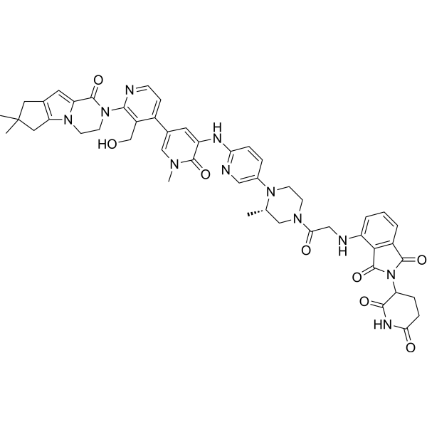
PTD10 is a selective and potent BTK PROTAC degrader (DC50 = 0.5 nM, KD = 2.28 nM). PTD10 can recruit cereblon (CRBN) E3 ligase and form a ternary complex with BTK, thereby mediating the ubiquitination and proteasome-dependent degradation of BTK. PTD10 inhibits cancer cells proliferation, and induces cell apoptosis via activation of the caspase-dependent pathway and mitochondrial pathway. PTD10 potently inhibits the BCR, AKT and NF-κB signaling pathway. PTD10 can be used for researches of B-cell malignancies and autoimmune disease.
(Pink: Btk ligand (HY-19834); Blue: Cereblon ligand (HY-14658); Black: linker (HY-Y0966)).
商品は「研究用試薬」です。人や動物の医療用・臨床診断用・食品用の製品ではありません。
研究用途以外に使用した場合、当社は一切の責任を負いかねます。
- 純度 : 97.38%
- CAS 番号: 2642231-19-2
- 分子式: C49H51N11O8
- 分子量:922.00
-
保管条件:Powder -20°C, 3 years , 4°C, 2 years ; In solvent -80°C, 6 months , -20°C, 1 month

To place orders, for customer services and technical support, please contact: MedChemExpress USA
Tel: 609-228-6898 E-mail: [email protected] [email protected]


-
 生物活性
生物活性
-
化学情報
-
溶剤 & 溶解度
- プロトコル
- 純度とドキュメンテーション
- 参考文献
-
 Help & FAQs




生物活性
製品説明
IC50 & Target
[1]|
Cereblon |
Caspase 3 |
Bcl-2 |
Cellular Effect
|
Cell Line

|
Type | Value | Description | References |
|---|---|---|---|---|
| TMD8 | IC50 |
1.4 nM
Compound: PTD10
|
Cytotoxicity against human TMD8 cells incubated for 48 hrs by celltitre glo 2.0 assay
Cytotoxicity against human TMD8 cells incubated for 48 hrs by celltitre glo 2.0 assay
|
[PMID: 37195170] |
体外実験
PTD10 (0.15-3000 nM; 17 h) potently degrades BTK via the CRBN-dependent ubiquitin-proteasome pathway in Ramos cells (DC50 = 0.5 nM) and JeKo-1 cells (DC50 = 0.6 nM), with maximal degradation exceeding 95% in both cell lines[1].
PTD10 binds to purified BTK with a Kd of 2.28 nM[1].
PTD10 (2 h) engages intracellular BTK (IC50 = 97 nM) and CRBN (IC50 = 107 nM) in HEK293 cells, with positive cooperativity in BTK binding[1].
PTD10 efficiently promotes ternary complex formation between BTK and CRBN in HEK293 cells, achieving 100% maximal BRET signal[1].
PTD10 is a highly selective BTK degrader in TMD8 cells, with no significant off-target degradation of other tested kinases, while degrading neo-substrates IKZF1, IKZF3, and ZNF280D without affecting GSPT1[1].
PTD10 (0.1-10000 nM; 48 h) potently inhibits growth of TMD8 cells (IC50 = 1.4 nM) and Mino cells (IC50 = 2.2 nM)[1].
PTD10 (0.15-3000 nM; 48 h) induces dose-dependent apoptosis in TMD8 and Mino cells via activation of the caspase-dependent and mitochondrial pathways[1].
PTD10 (0.15-3000 nM; 48 h) potently inhibits the BCR signaling pathway in TMD8 and Mino cells[1].
MedChemExpress (MCE) has not independently confirmed the accuracy of these methods. They are for reference only.
-
Cell Line:Ramos, JeKo-1 B-cell lymphoma cell lines
-
Concentration:0.15, 0.45, 1.4, 4, 12, 37, 111, 333, 1000, 3000 nM
-
Incubation Time:17 h
-
Result:Potently degraded BTK in both cell lines, with a DC50 of 0.5 nM (Dmax 97.1%) in Ramos cells and 0.6 nM (Dmax 95.7%) in JeKo-1 cells.
Completely blocked BTK degradation when cotreated with GDC-0853 (HY-19834) or Iberdomide (HY-101291).
Inhibited BTK degradation when pretreated with Bortezomib (HY-10227) or MLN-4924 (HY-70062).
-
Cell Line:TMD8, Mino B-cell lymphoma cell lines
-
Concentration:0.15, 0.45, 1.4, 4, 12, 37, 111, 333, 1000, 3000 nM
-
Incubation Time:48 h
-
Result:Induced apoptosis in a dose-dependent manner in both cell lines, with robust apoptosis detected at 333 nM (a Hook effect was observed at higher concentrations).
-
Cell Line:TMD8, Mino B-cell lymphoma cell lines
-
Concentration:0.15, 0.45, 1.4, 4, 12, 37, 111, 333, 1000, 3000 nM
-
Incubation Time:48 h
-
Result:Caused dose-dependent decreases in full-length PARP, caspase-3, Bcl-2, and MCL-1, with corresponding increases in cleaved-PARP and cleaved-caspase-3.
Caused robust downregulation of phosphorylated BTK, PLCγ2, AKT, and NF-κB, as well as total PLCγ2, AKT, and NF-κB in both cell lines after 48 h.
Activated ERK phosphorylation in TMD8 cells but unaffected in Mino cells.


化学情報
-
CAS 番号 2642231-19-2
-
性状 Solid
-
分子量 922.00
-
分子式 C49H51N11O8
-
Color Light yellow to yellow
-
SMILES
O=C1N(C=C(C=C1NC2=NC=C(N3CCN(C[C@@H]3C)C(CNC4=CC=CC5=C4C(N(C5=O)C6CCC(NC6=O)=O)=O)=O)C=C2)C7=CC=NC(N8CCN9C(C8=O)=CC%10=C9CC(C)(C%10)C)=C7CO)C
-
輸送条件
Room temperature in continental US; may vary elsewhere.
-
保管条件
Powder -20°C 3 years 4°C 2 years In solvent -80°C 6 months -20°C 1 month


溶剤 & 溶解度
体外:
DMSO : 50 mg/mL (54.23 mM; Need ultrasonic; Hygroscopic DMSO has a significant impact on the solubility of product, please use newly opened DMSO)
Please refer to the solubility information to select the appropriate solvent. Once prepared, please aliquot and store the solution to prevent product inactivation from repeated freeze-thaw cycles.
Storage method and period of stock solution: -80°C, 6 months; -20°C, 1 month. When stored at -80°C, please use it within 6 months. When stored at -20°C, please use it within 1 month.
Please refer to the solubility information to select the appropriate solvent. Once prepared, please aliquot and store the solution to prevent product inactivation from repeated freeze-thaw cycles.
Storage method and period of stock solution: -80°C, 6 months; -20°C, 1 month. When stored at -80°C, please use it within 6 months. When stored at -20°C, please use it within 1 month.
濃度 (開始) × 体積 (開始) = 濃度 (終了) × 体積 (終了)


純度とドキュメンテーション
-
データシート (277 KB)
-
SDS (254 KB)
- English - EN (254 KB)
- Français - FR (254 KB)
- Deutsch - DE (254 KB)
- Norwegian - NO (254 KB)
- Español - ES (254 KB)
- Swedish - SV (254 KB)
- Italian - IT (254 KB)
- Korean - KR (254 KB)
- Portuguese - PT (254 KB)
-
取扱説明書 (2659 KB)
参考文献


Complete Stock Solution Preparation Table
Please refer to the solubility information to select the appropriate solvent. Once prepared, please aliquot and store the solution to prevent product inactivation from repeated freeze-thaw cycles.
Storage method and period of stock solution: -80°C, 6 months; -20°C, 1 month. When stored at -80°C, please use it within 6 months. When stored at -20°C, please use it within 1 month.
| Optional Solvent | Concentration Solvent Mass | 1 mg | 5 mg | 10 mg | 25 mg |
|---|---|---|---|---|---|
| DMSO | 1 mM | 1.0846 mL | 5.4230 mL | 10.8460 mL | 27.1150 mL |
| 5 mM | 0.2169 mL | 1.0846 mL | 2.1692 mL | 5.4230 mL | |
| 10 mM | 0.1085 mL | 0.5423 mL | 1.0846 mL | 2.7115 mL | |
| 15 mM | 0.0723 mL | 0.3615 mL | 0.7231 mL | 1.8077 mL | |
| 20 mM | 0.0542 mL | 0.2711 mL | 0.5423 mL | 1.3557 mL | |
| 25 mM | 0.0434 mL | 0.2169 mL | 0.4338 mL | 1.0846 mL | |
| 30 mM | 0.0362 mL | 0.1808 mL | 0.3615 mL | 0.9038 mL | |
| 40 mM | 0.0271 mL | 0.1356 mL | 0.2711 mL | 0.6779 mL | |
| 50 mM | 0.0217 mL | 0.1085 mL | 0.2169 mL | 0.5423 mL |


PTD10 関連分類
HY-154860 関連分類
 Powered by Bioz