



QCA570
Based on 1 Customer Validation
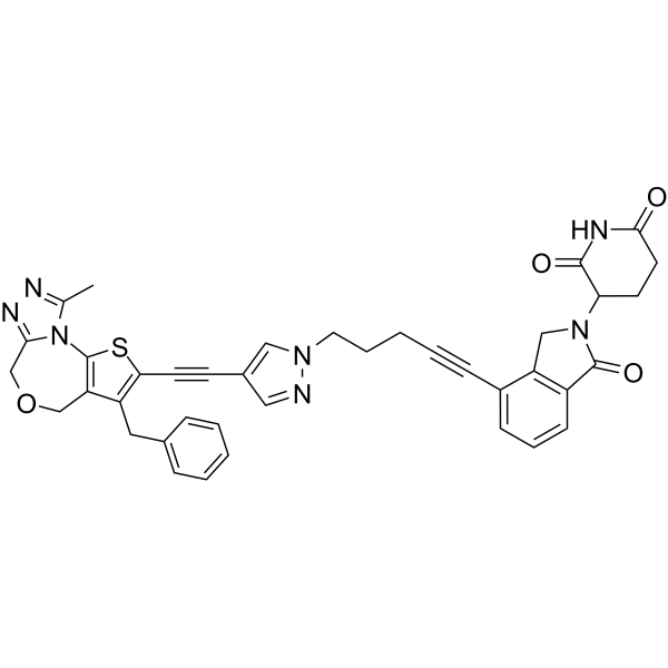
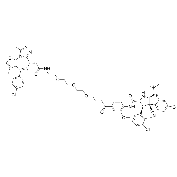
QCA570 is a pan-BET-BRD PROTAC degrader. QCA570 degrades BRD2/3/4 and BRDT via cereblon and the ubiquitin-proteasome pathway, inhibits the expression of c-MYC, EZH2 and Mcl-1, induces tumor cell apoptosis, cycle arrest and regression, and exerts a synergistic inhibitory effect on Osimertinib (HY-15772)-resistant EGFR-mutant non-small cell lung cancer. QCA570 can be used in the research of acute myeloid leukemia, acute lymphoblastic leukemia, bladder cancer and non-small cell lung cancer.
(Pink: BET ligand (HY-44103); Blue: Cereblon ligand (HY-131318); Black: linker (HY-W035972)).
For research use only. We do not sell to patients.
- Purity : 99.30%
- CAS No.: 2207569-08-0
- Formula: C39H33N7O4S
- Molecular Weight:695.79
-
Storage:
-20°C, protect from light
* In solvent : -80°C, 6 months; -20°C, 1 month (protect from light)

To place orders, for customer services and technical support, please contact: MedChemExpress USA
Tel: 609-228-6898 E-mail: [email protected] [email protected]


-
 Biological Activity
-
Chemical Information
-
Solvent & Solubility
- Protocols
- Purity & Documentation
- References
-
 Help & FAQs


-
HY-101519
BETd-260BET PROTAC Degrader -
HY-129937A
GNE-987PROTAC BRD4 Degrader -
HY-100972
ARV-771BET PROTAC Degrader -
HY-114228
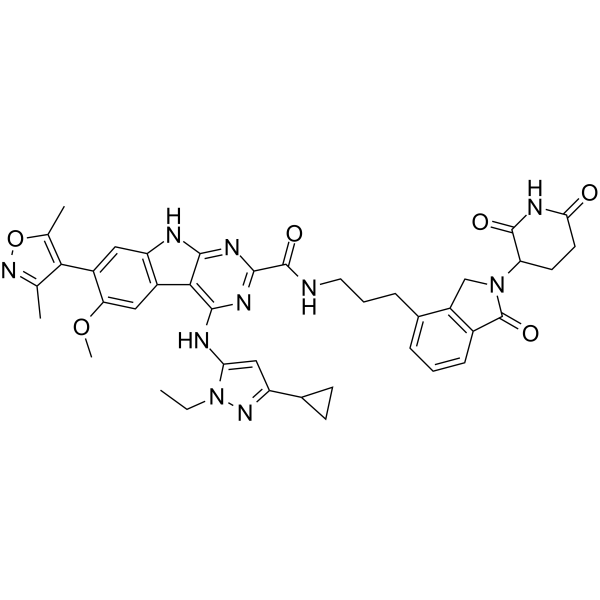
PROTAC BET degrader-2BRD2/BRD3/BRD4 PROTAC Degrader -
HY-112376
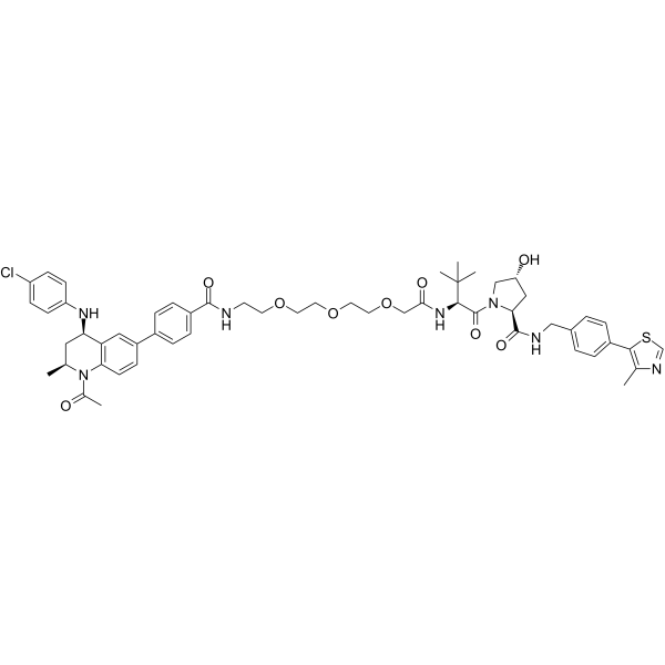
MZP-54BRD3/BRD4 PROTAC Degrader -
HY-16954
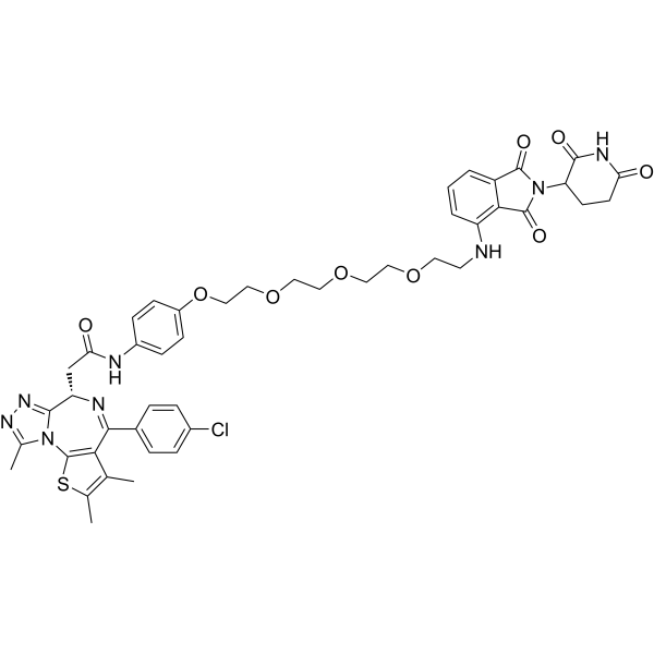
ARV-825PROTAC BRD2/BRD3/BRD4 Degrader -
HY-107425
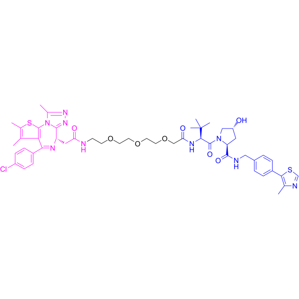
MZ 1PROTAC BRD4 Degrader -
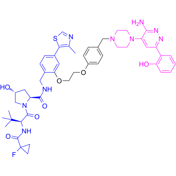
HY-128359
ACBI1SMARCA2/SMARCA4/PBRM1 Degrader -
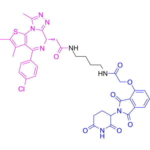
HY-101838
dBET1PROTAC BRD4 Degrader -
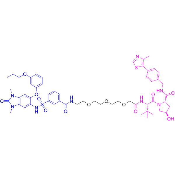
HY-111519
dTRIM24PROTAC TRIM24 Degrader -
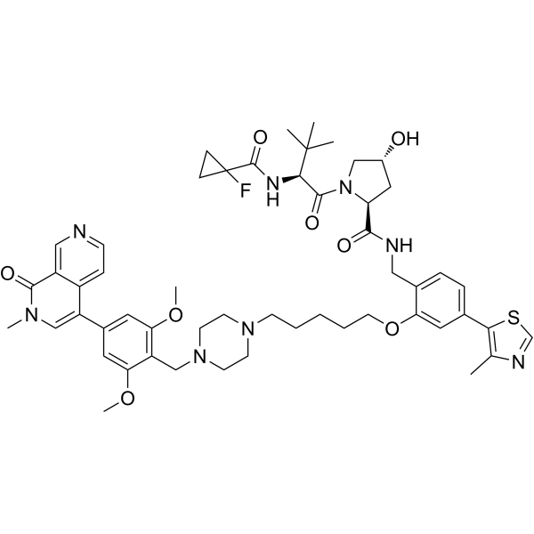
HY-114322
VZ185BRD9/BRD7 PROTAC Degrader -
HY-114305
A1874BRD4 PROTAC Degrader


Biological Activity
Description
|
BRD2 |
BRD3 |
BRD4 |
Mcl-1 |
EZH2 |
Cellular Effect
|
Cell Line

|
Type | Value | Description | References |
|---|---|---|---|---|
| RS4-11 | IC50 |
50 pM
|
Cell growth inhibition against RS4;11 leukemia cells.
Cell growth inhibition against RS4;11 leukemia cells.
|
30019901 |
| Leukemia cell | IC50 |
120 pM
|
Induction of cell death/apoptosis in CD34-enriched leukemia stem cells from primary AML samples measured by flow cytometry for DAPI-Annexin V binding after 3 days of incubation.
Induction of cell death/apoptosis in CD34-enriched leukemia stem cells from primary AML samples measured by flow cytometry for DAPI-Annexin V binding after 3 days of incubation.
|
30019901 |
| MV4-11 | IC50 |
8.3 pM
|
Cell growth inhibition against human acute leukemia MV4;11 cells assessed via lactate dehydrogenase-based WST-8 assay after 4 days of incubation.
Cell growth inhibition against human acute leukemia MV4;11 cells assessed via lactate dehydrogenase-based WST-8 assay after 4 days of incubation.
|
36733715 |
| MOLM-13 | IC50 |
62 pM
|
Cell growth inhibition against human acute leukemia MOLM-13 cells assessed via lactate dehydrogenase-based WST-8 assay after 4 days of incubation.
Cell growth inhibition against human acute leukemia MOLM-13 cells assessed via lactate dehydrogenase-based WST-8 assay after 4 days of incubation.
|
36733715 |
| RS4-11 | IC50 |
32 pM
|
Cell growth inhibition against human acute leukemia RS4;11 cells assessed via lactate dehydrogenase-based WST-8 assay after 4 days of incubation.
Cell growth inhibition against human acute leukemia RS4;11 cells assessed via lactate dehydrogenase-based WST-8 assay after 4 days of incubation.
|
36733715 |
| 5637 | DC50 |
1.184 nM
|
Half-maximal degradation of BRD4 protein in human 5637 bladder cancer cells after 9 h incubation measured via Western blot analysis.
Half-maximal degradation of BRD4 protein in human 5637 bladder cancer cells after 9 h incubation measured via Western blot analysis.
|
35261801 |
| J82 | DC50 |
0.4507 nM
|
Half-maximal degradation of BRD4 protein in human J82 bladder cancer cells after 9 h incubation measured via Western blot analysis.
Half-maximal degradation of BRD4 protein in human J82 bladder cancer cells after 9 h incubation measured via Western blot analysis.
|
35261801 |
| T-24 | DC50 |
0.2313 nM
|
Half-maximal degradation of BRD4 protein in human T24 bladder cancer cells after 9 h incubation measured via Western blot analysis.
Half-maximal degradation of BRD4 protein in human T24 bladder cancer cells after 9 h incubation measured via Western blot analysis.
|
35261801 |
| UMUC3 | DC50 |
1.589 nM
|
Half-maximal degradation of BRD4 protein in human UM-UC-3 bladder cancer cells after 9 h incubation measured via Western blot analysis.
Half-maximal degradation of BRD4 protein in human UM-UC-3 bladder cancer cells after 9 h incubation measured via Western blot analysis.
|
35261801 |
| EJ | DC50 |
1.041 nM
|
Half-maximal degradation of BRD4 protein in human EJ-1 bladder cancer cells after 9 h incubation measured via Western blot analysis.
Half-maximal degradation of BRD4 protein in human EJ-1 bladder cancer cells after 9 h incubation measured via Western blot analysis.
|
35261801 |
| 5637 | IC50 |
2.68 nM
|
Half-maximal inhibition of cell viability in human 5637 bladder cancer cells after 72 h incubation measured via CCK-8 assay.
Half-maximal inhibition of cell viability in human 5637 bladder cancer cells after 72 h incubation measured via CCK-8 assay.
|
35261801 |
| J82 | IC50 |
10.81 nM
|
Half-maximal inhibition of cell viability in human J82 bladder cancer cells after 72 h incubation measured via CCK-8 assay.
Half-maximal inhibition of cell viability in human J82 bladder cancer cells after 72 h incubation measured via CCK-8 assay.
|
35261801 |
| T-24 | IC50 |
30.44 nM
|
Half-maximal inhibition of cell viability in human T24 bladder cancer cells after 72 h incubation measured via CCK-8 assay.
Half-maximal inhibition of cell viability in human T24 bladder cancer cells after 72 h incubation measured via CCK-8 assay.
|
35261801 |
| UMUC3 | IC50 |
21.96 nM
|
Half-maximal inhibition of cell viability in human UM-UC-3 bladder cancer cells after 72 h incubation measured via CCK-8 assay.
Half-maximal inhibition of cell viability in human UM-UC-3 bladder cancer cells after 72 h incubation measured via CCK-8 assay.
|
35261801 |
| EJ | IC50 |
15.59 nM
|
Half-maximal inhibition of cell viability in human EJ-1 bladder cancer cells after 72 h incubation measured via CCK-8 assay.
Half-maximal inhibition of cell viability in human EJ-1 bladder cancer cells after 72 h incubation measured via CCK-8 assay.
|
35261801 |
| H1975 | IC50 |
0.3 nM
|
Cell survival inhibition against human NSCLC H1975 cells assessed by SRB assay following 3-day incubation.
Cell survival inhibition against human NSCLC H1975 cells assessed by SRB assay following 3-day incubation.
|
blood-2019-131759 |
| NCI-H157 | IC50 |
0.3 nM
|
Cell survival inhibition against human NSCLC H157 cells assessed by SRB assay following 3-day incubation.
Cell survival inhibition against human NSCLC H157 cells assessed by SRB assay following 3-day incubation.
|
blood-2019-131759 |
| Calu-1 | IC50 |
0.3 nM
|
Cell survival inhibition against human NSCLC Calu-1 cells assessed by SRB assay following 3-day incubation.
Cell survival inhibition against human NSCLC Calu-1 cells assessed by SRB assay following 3-day incubation.
|
blood-2019-131759 |
| NCI-H1299 | IC50 |
100 nM
|
Cell survival inhibition against human NSCLC H1299 cells assessed by SRB assay following 3-day incubation.
Cell survival inhibition against human NSCLC H1299 cells assessed by SRB assay following 3-day incubation.
|
blood-2019-131759 |
| EKVX | IC50 |
100 nM
|
Cell survival inhibition against human NSCLC EKVX cells assessed by SRB assay following 3-day incubation.
Cell survival inhibition against human NSCLC EKVX cells assessed by SRB assay following 3-day incubation.
|
blood-2019-131759 |
| MV4-11 | IC50 |
0.0083 nM
|
Antiproliferative activity against human MV4;11 cells assessed as inhibition of cell proliferation.
Antiproliferative activity against human MV4;11 cells assessed as inhibition of cell proliferation.
|
34494353 |
In Vitro
QCA570 (30 pM) degrades all BET-BRD proteins in RS4;11 leukemia cells and inhibits the growth of RS4;11 cells with an IC50 of 50 pM[4].
QCA570 (4 h) degrades BRD4 and inhibits c-MYC in CD34-enriched leukemic stem cells from primary AML samples[4].
QCA570 (0.01-10 nM; 3 h) potently induces the degradation of BRD2, BRD3 and BRD4 proteins at picomolar concentrations and suppresses c-Myc protein levels in MV4;11 and RS4;11 human acute leukemia cells after 3 hours of incubation[1].
QCA570 (0.5 nM; 3 h) induces the degradation of BRD2, BRD3 and BRD4 proteins in RS4;11 human acute leukemia cells via cereblon-dependent, neddylation-dependent and proteasome-dependent mechanisms, with no effect on GSPT1 protein levels[1].
QCA570 (0.01-100 nM; 9 h) potently induces proteasome-dependent degradation of BRD2, BRD3 and BRD4 proteins in 5637, J82, T24, UM-UC-3 and EJ-1 human bladder cancer cells[2].
QCA570 (10-100 nM) inhibits the expression of BRD4 target genes c-MYC and EZH2 at both mRNA and protein levels in human bladder cancer J82, T24 and 5637 cells[2].
QCA570 (72 h) potently inhibits the proliferation of 5637, J82, T24, UM-UC-3 and EJ-1 human bladder cancer cells, with IC50 values ranging from 2.68 to 30.44 nM[2].
QCA570 potently reduces the viability of human non-small cell lung cancer cell lines (H1975, H157, Calu-1, H1299, H1650, H460, H1792, PC-9, EKVX, H1944), with IC50 values ranging from 0.3 nM to 100 nM, and its sensitivity correlates with higher baseline BET protein levels[3].
QCA570 potently inhibits the proliferation of MV4;11 cells, with an IC50 of 0.0083 nM[5].
QCA570 (0.1-0.3 nM; 24 h) induces PARP cleavage in human acute leukemia cells MV4;11 and RS4;11 at picomolar concentrations. After 24 h of treatment, caspase 3 activation is detectable in MV4;11 cells, but this phenomenon is not observed in RS4;11 cells at the same time point[1].
QCA570 (10-100 nM; 72 h) induces dose-dependent apoptosis in 5637, J82, T24, UM-UC-3 and EJ-1 human bladder cancer cells, as detected by Annexin V-FITC/PI staining, activated caspase-3 expression assay and TUNEL staining[2].
QCA570 (5-10 nM; 24 h) induces apoptosis in human non-small cell lung cancer cell lines (H157, H1975, H1650, Calu-1), where downregulation of Mcl-1 plays a key mediating role, while Bim is dispensable for this activity[3].
QCA570 (1-10 nM; 7 h) potently and rapidly reduces the levels of BRD4, BRD3, BRD2, Mcl-1 and Bim in human non-small cell lung cancer cell lines (H157, H1975, Calu-1) at concentrations as low as 1 nM, and exerts cell-specific effects on c-Myc levels[3].
QCA570 (0.3125-30 nM; 3 days, 36-72 h, every 3 days for 12 days) acts synergistically with Osimertinib (HY-15772) to reduce cell viability, induce apoptosis, and inhibit colony formation in Osimertinib-resistant human EGFR-mutant non-small cell lung cancer cell lines (PC-9/AR, PC-9/GR/AR, HCC827/AR)[3].
Combination treatment with QCA570 (1 nM; 12 h) and Osimertinib exerts a synergistic apoptosis-inducing effect in Osimertinib-resistant human EGFR-mutant non-small cell lung cancer cell lines (PC-9/AR, PC-9/GR/AR, HCC827/AR), and this effect is mainly mediated by enhanced Mcl-1 degradation[3].
MedChemExpress (MCE) has not independently confirmed the accuracy of these methods. They are for reference only.
-
Cell Line:MV4;11, RS4;11
-
Concentration:0.01, 0.03, 0.1, 0.3, 1, 3, 10 nM
-
Incubation Time:3 h
-
Result:Reduced levels of BRD3 and BRD4 proteins at 10 pM in RS4;11 cells after 3 h.
Reduced BRD2 levels at 30-100 pM in RS4;11 cells after 3 h.
Reduced BRD3 and BRD4 levels at 10 pM in MV4;11 cells after 3 h.
Reduced BRD2 levels at 30 pM in MV4;11 cells after 3 h.
Reduced c-Myc protein levels at 10 pM in RS4;11 cells after 3 h.
Reduced c-Myc protein levels at 30 pM in MV4;11 cells after 3 h.
Induced complete degradation of BRD2, BRD3, and BRD4 at 0.5 nM in RS4;11 cells after 3 h.
-
Cell Line:RS4;11
-
Concentration:0.5 nM
-
Incubation Time:3 h
-
Result:Degradation of BRD2, BRD3, and BRD4 proteins was completely blocked by pre-treatment with cereblon modulator, NEDD8-activating enzyme inhibitor, proteasome inhibitor (peptidyl aldehyde), or proteasome inhibitor (epoxyketone).
Had no effect on GSPT1 protein levels in either MV4;11 or RS4;11 cells.
-
Cell Line:MV4;11, MOLM-13, RS4;11
-
Concentration:0.01, 0.03, 0.1, 0.3, 1, 3, 10 nM
-
Incubation Time:24, 48 h
-
Result:Induced > 60% apoptosis at 1 nM in MV4;11 and MOLM-13 cells after 24 h.
Induced minimal apoptosis at 0.1 nM in RS4;11 cells after 24 h.
Induced 15-30% apoptosis at 0.3-10 nM in RS4;11 cells after 24 h.
Induced > 40% apoptosis at 0.1 nM in RS4;11 cells after 48 h.
-
Cell Line:MV4;11, RS4;11
-
Concentration:0.01, 0.03, 0.1, 0.3, 1, 3, 10 nM
-
Incubation Time:24 h
-
Result:Induced robust cleavage of PARP at 0.1-0.3 nM in both MV4;11 and RS4;11 cells after 24 h.
Induced caspase 3 activation at 0.3 nM in MV4;11 cells after 24 h.
No caspase 3 cleavage was detected in RS4;11 cells after 24 h.
-
Cell Line:5637, J82, T24, UM-UC-3, EJ-1
-
Concentration:0.01, 0.1, 1, 10, 100, 1000 nM (9 h incubation); 30 nM (1-24 h incubation)
-
Incubation Time:9 h (0.01-100 nM); 1, 2, 3, 6, 9, 12, 24 h (30 nM)
-
Result:Induced dose-dependent degradation of BRD2, BRD3, and BRD4 proteins across all five bladder cancer cell lines after 9 h of incubation.
Largely depleted BRD4 at 3 nM in each cell line, with BRD4 degradation half-maximal (DC50) values of 1.184 nM (5637), 0.4507 nM (J82), 0.2313 nM (T24), 1.589 nM (UM-UC-3), and 1.041 nM (EJ-1).
Peaked BRD4 degradation within 1 h in 5637, T24, and UM-UC-3 cells, and within 3 h in J82 and EJ-1 cells at 30 nM, with time-dependent degradation also observed for BRD2 and BRD3.
-
Cell Line:5637
-
Concentration:10 nM
-
Incubation Time:72 h
-
Result:Induced G2/M phase arrest in 5637 cells, resulting in a decrease in the percentage of cells in G1 phase and an increase in the percentage of cells in G2/M phase compared to vehicle control.
-
Cell Line:5637, EJ-1, J82, T24
-
Concentration:100 nM
-
Incubation Time:8, 24, 48 h
-
Result:Significantly suppressed the migration of 5637, EJ-1, J82, and T24 cells, as evidenced by reduced wound closure compared to vehicle control after 24 h (5637, EJ-1, T24) or 48 h (J82) of incubation.
-
Cell Line:5637, J82, T24, UM-UC-3, EJ-1
-
Concentration:10, 100 nM (flow cytometry); 0.001, 0.01, 0.1 μM (Western blot)
-
Incubation Time:72 h
-
Result:Induced dose-dependent cell apoptosis across all five bladder cancer cell lines, with more than 50% of cells undergoing apoptosis in J82 and 5637 lines at the higher concentration.
Increased levels of cleaved caspase-3 (a marker of apoptosis) in 5637, J82, and T24 cells incubated with QCA570.
Confirmed increased apoptotic cells in QCA570-incubated 5637, J82, and T24 cells compared to vehicle control via TUNEL staining.
-
Cell Line:Human NSCLC cell lines (H157, H1975, H1650, Calu-1)
-
Concentration:5, 10 nM
-
Incubation Time:24 h (flow cytometry); 22 h (Western blot)
-
Result:Increased the annexin V-positive cell population in all tested cell lines.
Induced cleavage of PARP, caspase-8, and caspase-3, confirming apoptosis induction.
Had its apoptosis-inducing effect blocked by enforced ectopic expression of Mcl-1 in H157 and H1975 cells.
Showed no change in apoptosis induction in H157 and H1975 cells with Bim knockout.
-
Cell Line:Human NSCLC cell lines (H157, H1975, Calu-1)
-
Concentration:1 nM, 2.5 nM, 5 nM, 10 nM (7 h incubation); 10 nM (time-course incubation)
-
Incubation Time:7 h; 3, 6, 9, 12, 15 h
-
Result:Decreased the levels of BRD4, BRD3, and BRD2 potently and rapidly, with reduction detectable as early as 3 h post-treatment and achievable at 1 nM.
Decreased levels of Mcl-1 and Bim with similar potency, rapidness, and durability.
Decreased c-Myc levels quickly and sustainably in H157 cells, caused transient reduction in H1975 cells, and showed no reduction in Calu-1 cells.
Downregulated Mcl-1 mRNA but not Bim mRNA in H157 cells.
-
Cell Line:Osimertinib-resistant human EGFR-mutant NSCLC cell lines (PC-9/AR, PC-9/GR/AR, HCC827/AR)
-
Concentration:1 nM
-
Incubation Time:12 h
-
Result:Decreased Mcl-1 and c-Myc levels more effectively in combination with Osimertinib than either single agent in all three tested cell lines.
Did not enhance Bim elevation in combination with Osimertinib, and alone decreased Bim levels in HCC827/AR cells.
Had its enhanced apoptosis-inducing and mitochondrial Cyt C/Smac release effects abrogated by enforced ectopic Mcl-1 expression in HCC827/AR cells.
In Vivo
QCA570 (0.5 mg/kg; i.p.; twice weekly) alone shows limited efficacy against Osimertinib-resistant EGFR-mutant non-small cell lung cancer xenografts, but when combined with Osimertinib, it significantly enhances tumor growth inhibition with good tolerance[3].
QCA570 completely abrogates tumor growth in a mouse xenograft model of human acute leukemia[6].
MedChemExpress (MCE) has not independently confirmed the accuracy of these methods. They are for reference only.
-
Animal Model:SCID mice (acute leukemia xenograft model, tumors grown to ~100 mm3)[1]
-
Dosage:1, 2.5, 5 mg/kg
-
Administration:i.v.; single dose; 3 times per week for 3 weeks; weekly
-
Result:Dramatically reduced BRD2, BRD3, BRD4, and c-Myc protein levels in tumor tissue at 1 h, 6 h, and 24 h post-treatment.
Achieved more effective reduction of BRD4 levels at 24 h with 5 mg/kg single dose.
Induced robust PARP cleavage (apoptosis) at 6 h and 24 h with 1 mg/kg and 5 mg/kg single doses.
Achieved complete and long-lasting tumor regression with 1, 2.5, and 5 mg/kg doses administered 3 times per week for 3 weeks.
Achieved complete tumor growth inhibition but not regression with 5 mg/kg weekly dose.
Caused minimal animal weight loss across all treatment groups.
-
Animal Model:SCID mice (acute leukemia xenograft model, tumors grown to ~100 mm3)[1]
-
Dosage:1, 5 mg/kg
-
Administration:i.v.; 3 times per week for 2 weeks
-
Result:Achieved complete tumor regression with 1 mg/kg dose administered 3 times per week for 2 weeks.
Achieved complete and long-lasting tumor regression with 5 mg/kg dose administered 3 times per week for 2 weeks.
Caused a maximum weight loss of less than 10%.
-
Animal Model:NU(NCr)-Foxn1nu (4-week-old female, flank injection of 2×10⁶ HCC827/AR osimertinib-resistant EGFR-mutant NSCLC cells)[3]
-
Dosage:0.5 mg/kg
-
Administration:i.p.; twice per week
-
Result:Exerted limited effects on suppressing HCC827/AR xenograft growth.
Significantly inhibited tumor growth when combined with Osimertinib compared to either single agent.
Caused limited body weight reduction.


Chemical Information
-
CAS No. 2207569-08-0
-
Appearance Solid
-
Molecular Weight 695.79
-
Formula C39H33N7O4S
-
Color White to off-white
-
SMILES
O=C(N(C1CCC(NC1=O)=O)C2)C3=C2C(C#CCCCN4N=CC(C#CC(S5)=C(CC6=CC=CC=C6)C(COC7)=C5N8C7=NN=C8C)=C4)=CC=C3
-
Shipping
Room temperature in continental US; may vary elsewhere.
-
Storage
-20°C, protect from light
* In solvent : -80°C, 6 months; -20°C, 1 month (protect from light)


Solvent & Solubility
In Vitro:
DMSO : 40 mg/mL (57.49 mM; Need ultrasonic; Hygroscopic DMSO has a significant impact on the solubility of product, please use newly opened DMSO)
Please refer to the solubility information to select the appropriate solvent. Once prepared, please aliquot and store the solution to prevent product inactivation from repeated freeze-thaw cycles.
Storage method and period of stock solution: -80°C, 6 months; -20°C, 1 month (protect from light). When stored at -80°C, please use it within 6 months. When stored at -20°C, please use it within 1 month.
Please refer to the solubility information to select the appropriate solvent. Once prepared, please aliquot and store the solution to prevent product inactivation from repeated freeze-thaw cycles.
Storage method and period of stock solution: -80°C, 6 months; -20°C, 1 month (protect from light). When stored at -80°C, please use it within 6 months. When stored at -20°C, please use it within 1 month.
Concentration (start) × Volume (start) = Concentration (final) × Volume (final)
In Vivo:
Select the appropriate dissolution method based on your experimental animal and administration route.
- For the following dissolution methods, please ensure to first prepare a clear stock solution using an In Vitro approach and then sequentially add co-solvents:
- To ensure reliable experimental results, the clarified stock solution can be appropriately stored based on storage conditions. As for the working solution for In Vivo experiments, it is recommended to prepare freshly and use it on the same day.
- The percentages shown for the solvents indicate their volumetric ratio in the final prepared solution. If precipitation or phase separation occurs during preparation, heat and/or sonication can be used to aid dissolution.
Add each solvent one by one: 10% DMSO 40% PEG300 5% Tween-80 45% Saline
Solubility: 2 mg/mL (2.87 mM); Suspended solution; Need ultrasonic
This protocol yields a suspended solution of 2 mg/mL. Suspended solution can be used for oral and intraperitoneal injection.
Taking 1 mL working solution as an example, add 100 μL DMSO stock solution (20.0 mg/mL) to 400 μL PEG300, and mix evenly; then add 50 μL Tween-80 and mix evenly; then add 450 μL Saline to adjust the volume to 1 mL.
Preparation of Saline: Dissolve 0.9 g sodium chloride in ddH₂O and dilute to 100 mL to obtain a clear Saline solution.
In Vivo Dissolution Calculator
Please enter the basic information of animal experiments:
-
-
-
-
Recommended: Prepare an additional quantity of animals to account for potential losses during experiments.
Please enter your animal formula composition:
-
%DMSO +
Recommended: Keep the proportion of DMSO in working solution below 2% if your animal is weak.
-
%+
-
+%Tween-80 + +
-
%Saline +
The co-solvents required include: DMSO, . All of co-solvents are available by MedChemExpress (MCE). , Tween 80. All of co-solvents are available by MedChemExpress (MCE).
Working solution concentration: 0.22 mg/mL
Method for preparing stock solution: mg drug dissolved in μL DMSO. Stock solution concentration: mg/mL. * In solvent : -80°C, 6 months; -20°C, 1 month (protect from light)
1. Take μL DMSO stock solution;
2. Add μL .
μL , mix evenly;
3. Then add μL Tween 80, mix evenly;
4. Then add μL
Please ensure that the stock solution in the first step is dissolved to a clear state, and add co-solvents in sequence. You can use ultrasonic heating (ultrasonic cleaner, recommended frequency 20-40 kHz), vortexing, etc. to assist dissolution.


Purity & Documentation
-
Data Sheet (302 KB)
-
SDS (252 KB)
- English - EN (252 KB)
- Français - FR (252 KB)
- Deutsch - DE (252 KB)
- Norwegian - NO (252 KB)
- Español - ES (252 KB)
- Swedish - SV (252 KB)
- Italian - IT (252 KB)
- Korean - KR (252 KB)
- Portuguese - PT (252 KB)
-
Handling Instructions (2659 KB)
References
[1]. Qin C, et al. Discovery of QCA570 as an Exceptionally Potent and Efficacious Proteolysis Targeting Chimera (PROTAC) Degrader of the Bromodomain and Extra-Terminal (BET) Proteins Capable of Inducing Complete and Durable Tumor Regression. Journal of medicinal chemistry. 2018 Aug 09;61(15):6685-6704. [Content Brief]
[3]. Liu C, et al. The novel BET degrader, QCA570, is highly active against the growth of human NSCLC cells and synergizes with osimertinib in suppressing osimertinib-resistant EGFR-mutant NSCLC cells. American journal of cancer research. 2022;12(2):779-792. [Content Brief]
[5]. Hu Z, et al. Recent Developments in PROTAC-Mediated Protein Degradation: From Bench to Clinic. Chembiochem : a European journal of chemical biology. 2022 Jan 19;23(2):e202100270. [Content Brief]


Complete Stock Solution Preparation Table
Please refer to the solubility information to select the appropriate solvent. Once prepared, please aliquot and store the solution to prevent product inactivation from repeated freeze-thaw cycles.
Storage method and period of stock solution: -80°C, 6 months; -20°C, 1 month (protect from light). When stored at -80°C, please use it within 6 months. When stored at -20°C, please use it within 1 month.
| Optional Solvent | Concentration Solvent Mass | 1 mg | 5 mg | 10 mg | 25 mg |
|---|---|---|---|---|---|
| DMSO | 1 mM | 1.4372 mL | 7.1861 mL | 14.3722 mL | 35.9304 mL |
| 5 mM | 0.2874 mL | 1.4372 mL | 2.8744 mL | 7.1861 mL | |
| 10 mM | 0.1437 mL | 0.7186 mL | 1.4372 mL | 3.5930 mL | |
| 15 mM | 0.0958 mL | 0.4791 mL | 0.9581 mL | 2.3954 mL | |
| 20 mM | 0.0719 mL | 0.3593 mL | 0.7186 mL | 1.7965 mL | |
| 25 mM | 0.0575 mL | 0.2874 mL | 0.5749 mL | 1.4372 mL | |
| 30 mM | 0.0479 mL | 0.2395 mL | 0.4791 mL | 1.1977 mL | |
| 40 mM | 0.0359 mL | 0.1797 mL | 0.3593 mL | 0.8983 mL | |
| 50 mM | 0.0287 mL | 0.1437 mL | 0.2874 mL | 0.7186 mL |


QCA570 Related Classifications
HY-112609 Related Classifications
 Powered by Bioz