



FGIN 1-27

Based on 1 Customer Validation
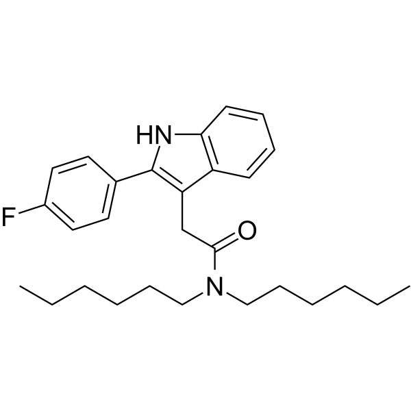
FGIN-1-27 is a blood-brain barrier-penetrant TSPO ligand with a Ki value of 5 nM. FGIN 1-27 inhibits PKC-β, PKA/CREB, p38/ERK MAPK, MITF, tyrosinase, TRP-1, and TRP-2, thereby inhibiting melanogenesis and pigmentation. FGIN-1-27 alleviates X-ray radiation-induced astrocyte mitochondrial hyperfunction, reduces ROS and superoxide production, inhibits excessive activation of A1-type astrocytes, downregulates GFAP and C3 protein expression, and restores astrocyte proliferative capacity. FGIN-1-27 produces anticonvulsant effects in normal mice; in diazepam-withdrawn mice, the brain MDR pathway becomes subsensitive, and the anticonvulsant activity disappears. FGIN-1-27 attenuates pigmentation in zebrafish embryos and ameliorates UVB-induced skin pigmentation in guinea pigs. FGIN-1-27 directly stimulates testicular Leydig cells while upregulating luteinizing hormone levels, causing an acute increase in serum testosterone in male rats. FGIN 1-27 can be used for research related to hyperpigmentation, epilepsy, brain injury, and other diseases.
Nur für Forschungszwecke. Wir verkaufen nicht an Patienten.
- Reinheit : 99.95%
- CAS. Nr.: 142720-24-9
- Formel: C28H37FN2O
- Molecular Weight:436.60
-
Speicherung:Powder -20°C, 3 years , 4°C, 2 years ; In solvent -80°C, 6 months , -20°C, 1 month

To place orders, for customer services and technical support, please contact: MedChemExpress USA
Tel: 609-228-6898 E-mail: [email protected] [email protected]


-
 Biologische Aktivität
-
Chemical Information
-
Lösungsmittel & Löslichkeit
- Protokoll
- Reinheit & Dokumentation
- Verweise
-
 Help & FAQs




Biologische Aktivität
Beschreibung
|
IL-17A |
p38 |
PKA |
PKC |
Cellular Effect
|
Cell Line

|
Type | Value | Description | References |
|---|---|---|---|---|
| C6 | IC50 |
10 nM
Compound: FGIN-1-27
|
Displacement of [3H]PK11195 binding from peripheral benzodiazepine receptor in C6 rat glioma cell line
Displacement of [3H]PK11195 binding from peripheral benzodiazepine receptor in C6 rat glioma cell line
|
[PMID: 8691438] |
In Vitro
FGIN-1-27 (10 min) does not directly inhibit mushroom tyrosinase activity in cell-free assays[1].
FGIN-1-27 (1-16 μM; 48 h) does not reduce the viability of SK-MEL-2 cells or HEM[1].
FGIN-1-27 (1-4 μM; 48 h) inhibits basal and α-MSH (HY-P0252)-, OAG-, or ET-1-induced melanin synthesis in SK-MEL-2 cells and HEM[1].
FGIN-1-27 (0-4 μM; 48 h) inhibits tyrosinase, TRP-1, and TRP-2 expression in SK-MEL-2 cells and HEM, and inhibits OAG-induced cellular tyrosinase activity[1].
FGIN-1-27 (4 μM; 1-12 h) inhibits basal, α-MSH-induced, and ET-1-induced MITF expression in SK-MEL-2 cells and HEM[1].
FGIN-1-27 (4 μM; 0-120 min) reduced PKC-β, p-PKA cat, p-CREB, p-p38, and p-ERK protein levels in SK-MEL-2 cells and HEM[1].
FGIN 1-27 (40 μM; 2 h) stimulates testosterone production in primary rat Leydig cells through a PKA-sensitive mechanism at or downstream of mitochondrial steroidogenesis, without requiring calcium signaling or new protein synthesis, and without altering the mRNA levels of key steroidogenic genes[3].
FGIN-1-27 (1 μM; 24 h) reduced GFAP/C3 expression and restored EdU-positive proliferation in primary C57BL/6 mouse astrocytes exposed to 20 Gy X-rays without altering viability; it also stabilized mitochondrial membrane potential and reduced mitochondrial hyperrespiration and oxidative stress[5].
FGIN-1-27 (10 µM; 6 days) selectively inhibits the proliferation of conventional CD4+ T cells, CD8+ T cells, Th1, and Th17 subsets in phytohemagglutinin-stimulated human PBMCs and reduces IL-17A and IFN-γ production while preserving Treg proliferation[4].
MedChemExpress (MCE) has not independently confirmed the accuracy of these methods. They are for reference only. Further protocols information, click here.
-
Cell Line:SK-MEL-2 cells and human epidermis melanocytes (HEM)
-
Concentration:0, 1, 2, 4, 8, 16 μM
-
Incubation Time:48 h
-
Result:Did not reduce viability of SK-MEL-2 cells or HEM.
-
Cell Line:SK-MEL-2 cells and human epidermis melanocytes (HEM)
-
Concentration:0, 1, 2, 4 μM
-
Incubation Time:48 h
-
Result:Suppressed tyrosinase, TRP-1, and TRP-2 expression and inhibits OAG-induced cellular tyrosinase activity in SK-MEL-2 cells and HEM.
-
Cell Line:SK-MEL-2 cells and human epidermis melanocytes (HEM)
-
Concentration:4 μM
-
Incubation Time:1, 2, 4, 8, 12 h
-
Result:Suppressed basal, α-MSH-induced, and ET-1-induced MITF expression in SK-MEL-2 cells and HEM.
-
Cell Line:SK-MEL-2 cells and human epidermis melanocytes (HEM)
-
Concentration:4 μM
-
Incubation Time:0, 5, 15, 30, 60, 120 min
-
Result:Decreased PKC-β, p-PKA cat, p-CREB, p-p38, and p-ERK protein levels in SK-MEL-2 cells and HEM.
-
Cell Line:primary rat Leydig cells
-
Concentration:40 μM
-
Incubation Time:2 h
-
Result:Did not alter mRNA levels of key steroidogenic genes in primary rat Leydig cells, suggesting its acute stimulatory effect is not mediated by increased expression of these genes.
In Vivo
FGIN-1-27 (20-30 μg/mouse; i.c.v.; single administration 15-30 min before PTZ infusion) increases the pentylenetetrazol seizure threshold in chronically vehicle-treated ddY mice; it does not increase the seizure threshold in benzodiazepine-withdrawn mice[2].
FGIN 1-27 (1 μg/g body weight (1 mg/kg BW); i.p.; single dose) acutely increases serum testosterone in healthy adult male SD rats; it also increases serum LH at 2 h without altering serum FSH[3].
MedChemExpress (MCE) has not independently confirmed the accuracy of these methods. They are for reference only.
-
Animal Model:ddY (male, 20-23 g)[2]
-
Dosage:20 and 30 μg/mouse
-
Administration:i.c.v.; single administration 15 and 30 min before PTZ infusion
-
Result:Increased the pentylenetetrazol seizure threshold in chronically vehicle-treated ddY mice; it does not increase the seizure threshold in benzodiazepine-withdrawn mice.


Chemical Information
-
CAS. Nr. 142720-24-9
-
Appearance Solid
-
Molecular Weight 436.60
-
Formel C28H37FN2O
-
Color Off-white to light yellow
-
SMILES
O=C(N(CCCCCC)CCCCCC)CC1=C(C2=CC=C(F)C=C2)NC3=C1C=CC=C3
-
Versand
Room temperature in continental US; may vary elsewhere.
-
Speicherung
Powder -20°C 3 years 4°C 2 years In solvent -80°C 6 months -20°C 1 month


Lösungsmittel & Löslichkeit
In Vitro:
DMSO : 100 mg/mL (229.04 mM; Need ultrasonic; Hygroscopic DMSO has a significant impact on the solubility of product, please use newly opened DMSO)
Please refer to the solubility information to select the appropriate solvent. Once prepared, please aliquot and store the solution to prevent product inactivation from repeated freeze-thaw cycles.
Storage method and period of stock solution: -80°C, 6 months; -20°C, 1 month. When stored at -80°C, please use it within 6 months. When stored at -20°C, please use it within 1 month.
Please refer to the solubility information to select the appropriate solvent. Once prepared, please aliquot and store the solution to prevent product inactivation from repeated freeze-thaw cycles.
Storage method and period of stock solution: -80°C, 6 months; -20°C, 1 month. When stored at -80°C, please use it within 6 months. When stored at -20°C, please use it within 1 month.
Konzentration (Stammlösung) × Volumen (Stammlösung) = Konzentration (Ziellösung) × Volumen (Ziellösung)
In Vivo:
Select the appropriate dissolution method based on your experimental animal and administration route.
- For the following dissolution methods, please ensure to first prepare a clear stock solution using an In Vitro approach and then sequentially add co-solvents:
- To ensure reliable experimental results, the clarified stock solution can be appropriately stored based on storage conditions. As for the working solution for In Vivo experiments, it is recommended to prepare freshly and use it on the same day.
- The percentages shown for the solvents indicate their volumetric ratio in the final prepared solution. If precipitation or phase separation occurs during preparation, heat and/or sonication can be used to aid dissolution.
Add each solvent one by one: 10% DMSO 40% PEG300 5% Tween-80 45% Saline
Solubility: ≥ 2.5 mg/mL (5.73 mM); Clear solution
This protocol yields a clear solution of ≥ 2.5 mg/mL (saturation unknown).
Taking 1 mL working solution as an example, add 100 μL DMSO stock solution (25.0 mg/mL) to 400 μL PEG300, and mix evenly; then add 50 μL Tween-80 and mix evenly; then add 450 μL Saline to adjust the volume to 1 mL.
Preparation of Saline: Dissolve 0.9 g sodium chloride in ddH₂O and dilute to 100 mL to obtain a clear Saline solution.
Add each solvent one by one: 10% DMSO 90% (20% SBE-β-CD in Saline)
Solubility: 2.5 mg/mL (5.73 mM); Suspended solution; Need ultrasonic
This protocol yields a suspended solution of 2.5 mg/mL. Suspended solution can be used for oral and intraperitoneal injection.
Taking 1 mL working solution as an example, add 100 μL DMSO stock solution (25.0 mg/mL) to 900 μL 20% SBE-β-CD in Saline, and mix evenly.
Preparation of 20% SBE-β-CD in Saline (4°C, storage for one week): 2 g SBE-β-CD powder is dissolved in 10 mL Saline, completely dissolve until clear.
In Vivo Dissolution Calculator
Please enter the basic information of animal experiments:
-
-
-
-
Recommended: Prepare an additional quantity of animals to account for potential losses during experiments.
Please enter your animal formula composition:
-
%DMSO +
Recommended: Keep the proportion of DMSO in working solution below 2% if your animal is weak.
-
%+
-
+%Tween-80 + +
-
%Saline +
The co-solvents required include: DMSO, . All of co-solvents are available by MedChemExpress (MCE). , Tween 80. All of co-solvents are available by MedChemExpress (MCE).
Working solution concentration: 0.22 mg/mL
Method for preparing stock solution: mg drug dissolved in μL DMSO. Stock solution concentration: mg/mL.
1. Take μL DMSO stock solution;
2. Add μL .
μL , mix evenly;
3. Then add μL Tween 80, mix evenly;
4. Then add μL
Please ensure that the stock solution in the first step is dissolved to a clear state, and add co-solvents in sequence. You can use ultrasonic heating (ultrasonic cleaner, recommended frequency 20-40 kHz), vortexing, etc. to assist dissolution.

Protokoll
-
RNA extraction experimental
By lysing cells, releasing RNA, and removing impurities such as proteins and DNA, high-purity RNA products are finally obtained. The commonly used traditional method is the guanidine isothiocyanate/phenol/chloroform method (Trizol), which is suitable for a variety of animal materials including animal tissues, microorganisms, cultured cells, etc., and most plant materials.
-
Western Blot
Western blotting (WB) is a commonly used experimental method in molecular biology, biochemistry, and immunogenetics for identifying and quantifying target proteins. It combines gel electrophoresis with immunoassay, enabling researchers to analyze protein expression, post-translational modifications, and molecular weight.
-
ROS/oxidative-stress fluorescent staining
ROS/oxidative-stress fluorescent staining uses cell-permeant fluorogenic probes that become fluorescent after oxidation inside cells or tissues; commonly used examples include DCFH-DA/DCFDA for broad cellular oxidant detection, DHE for superoxide-related signal detection, MitoSOX for mitochondrial superoxide-related signal detection, and CellROX probes for oxidative-stress-associated fluorescence readouts. The assay detects probe oxidation rather than a single ROS species unless the probe and analysis method have been chemically validated for that species. DCFH-DA enters cells, is deacetylated by intracellular esterases to DCFH, and produces fluorescent DCF after oxidation, so the readout is used as an operational measure of total cellular oxidative stress rather than a species-specific ROS measurement. DHE and MitoSOX can report superoxide-related oxidation, but red fluorescence alone can include non-specific ethidium-like oxidation products; HPLC or optimized spectral approaches are
-
Research Protocol for Endocrine Diseases
Endocrine diseases often arise from disrupted hormone production, hormone signaling, or target-tissue responsiveness; for diabetes-focused endocrine disease models, insulin signaling regulates glucose uptake, hepatic glucose output, lipid metabolism, and β-cell compensation. Type 2 diabetes develops through interacting defects in insulin resistance, β-cell dysfunction, adipose inflammation, hepatic glucose overproduction, altered incretin signaling, and ectopic lipid metabolism. A major unresolved question is whether endocrine dysfunction is driven primarily by target-tissue insulin resistance, intrinsic β-cell failure, immune/inflammatory stress, or combined multi-organ failure that differs by disease stage.
-
Protocol For Protein Expression And Purification
Recombinant protein expression in Escherichia coli followed by purification of a His-tagged soluble protein by immobilized metal affinity chromatography (IMAC), with optional MBP fusion and TEV tag removal when the construct includes these elements. The biological readout is production of the encoded target protein, detected as an inducible band at the expected molecular mass by SDS-PAGE and quantified by total protein assay or chromatographic absorbance; the purification readout is enrichment of the target protein in elution fractions after selective binding of polyhistidine residues to immobilized Ni2+/metal-chelate resin and elution by imidazole-containing buffer. Expression is driven by an inducible bacterial expression system, commonly T7/lac-based, in which IPTG or lactose/auto-induction activates transcription and translation of the cloned gene; lower induction temperature, lower inducer concentration, induction timing, and solubility-enhancing fusion tags can influence the frac


Reinheit & Dokumentation
-
Data Sheet (296 KB)
-
SDS (393 KB)
- English - EN (393 KB)
- Français - FR (393 KB)
- Deutsch - DE (393 KB)
- Norwegian - NO (393 KB)
- Español - ES (393 KB)
- Swedish - SV (393 KB)
- Italian - IT (393 KB)
- Korean - KR (393 KB)
- Portuguese - PT (393 KB)
-
Handling Instructions (2659 KB)
Verweise


Complete Stock Solution Preparation Table
Please refer to the solubility information to select the appropriate solvent. Once prepared, please aliquot and store the solution to prevent product inactivation from repeated freeze-thaw cycles.
Storage method and period of stock solution: -80°C, 6 months; -20°C, 1 month. When stored at -80°C, please use it within 6 months. When stored at -20°C, please use it within 1 month.
| Optional Solvent | Concentration Solvent Mass | 1 mg | 5 mg | 10 mg | 25 mg |
|---|---|---|---|---|---|
| DMSO | 1 mM | 2.2904 mL | 11.4521 mL | 22.9043 mL | 57.2606 mL |
| 5 mM | 0.4581 mL | 2.2904 mL | 4.5809 mL | 11.4521 mL | |
| 10 mM | 0.2290 mL | 1.1452 mL | 2.2904 mL | 5.7261 mL | |
| 15 mM | 0.1527 mL | 0.7635 mL | 1.5270 mL | 3.8174 mL | |
| 20 mM | 0.1145 mL | 0.5726 mL | 1.1452 mL | 2.8630 mL | |
| 25 mM | 0.0916 mL | 0.4581 mL | 0.9162 mL | 2.2904 mL | |
| 30 mM | 0.0763 mL | 0.3817 mL | 0.7635 mL | 1.9087 mL | |
| 40 mM | 0.0573 mL | 0.2863 mL | 0.5726 mL | 1.4315 mL | |
| 50 mM | 0.0458 mL | 0.2290 mL | 0.4581 mL | 1.1452 mL | |
| 60 mM | 0.0382 mL | 0.1909 mL | 0.3817 mL | 0.9543 mL | |
| 80 mM | 0.0286 mL | 0.1432 mL | 0.2863 mL | 0.7158 mL | |
| 100 mM | 0.0229 mL | 0.1145 mL | 0.2290 mL | 0.5726 mL |


FGIN 1-27 Related Classifications
HY-101059 Related Classifications
 Powered by Bioz
Keywords
- FGIN 1-27
- 142720-24-9
- TSPO
- TRP Channel
- Microphthalmia Associated Transcription Factor (MITF)
- PKC
- PKA
- p38 MAPK
- PERK
- Interleukin Related
- Leydig cell steroidogenesis
- Th17
- Treg
- astrocyte
- cholesterol translocation
- melanogenesis
- mitochondrial translocator protein
- neurosteroid biosynthesis
- tyrosinase
- Inhibitor
- inhibitor
- inhibit