



Chondroitin sulfate (from chicken)

Based on 1 Customer Validation
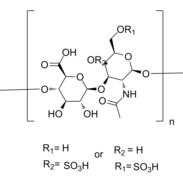
Chondroitin sulfate (from chicken) (Chondroitin polysulfate (from chicken)) is an orally active, sulfated linear polysaccharide extracted from chickens, which belongs to glycosaminoglycans. Chondroitin sulfate (from chicken) reduces the phosphorylation levels of ERK1/2 and p38MAPK, inhibits the expression of inflammatory cytokines and MMP, and downregulates the levels of IL-1β, IL-6, TNF-α and PGE2. Chondroitin sulfate (from chicken) improves motor function, protects cartilage tissue, reverses chondrocyte aggregation, and regulates the structure of intestinal flora. Chondroitin sulfate (from chicken) can be used in research related to osteoarthritis.
For research use only. We do not sell to patients.
- Purity : 99.61%
- CAS No.: 9007-28-7
- Formula: (C14H21NO14S)n
-
Storage:
4°C, protect from light
* In solvent : -80°C, 6 months; -20°C, 1 month (protect from light)

To place orders, for customer services and technical support, please contact: MedChemExpress USA
Tel: 609-228-6898 E-mail: [email protected] [email protected]


-
 Biological Activity
-
Chemical Information
-
Solvent & Solubility
- Protocols
- Purity & Documentation
- References
-
 Help & FAQs


-
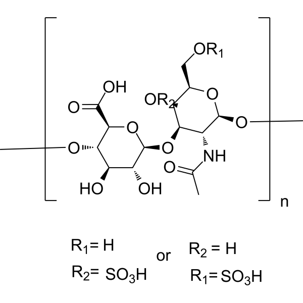
HY-B2162C
Chondroitin sulfate (from pig)Sulfated Linear Polysaccharide -
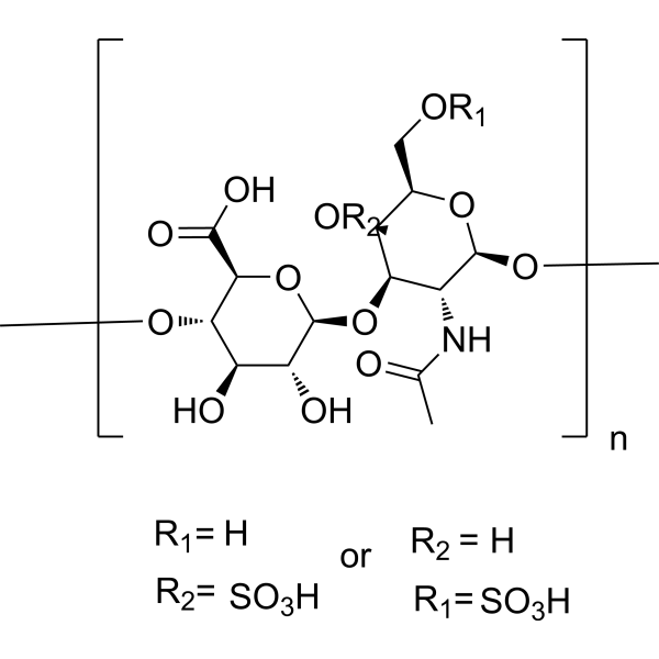
HY-B2162
Chondroitin sulfateGlycosaminoglycan -
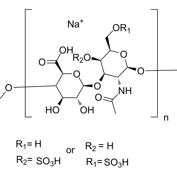
HY-B2162A
Chondroitin sulfate sodium (from shark cartilage)Glycosaminoglycan -
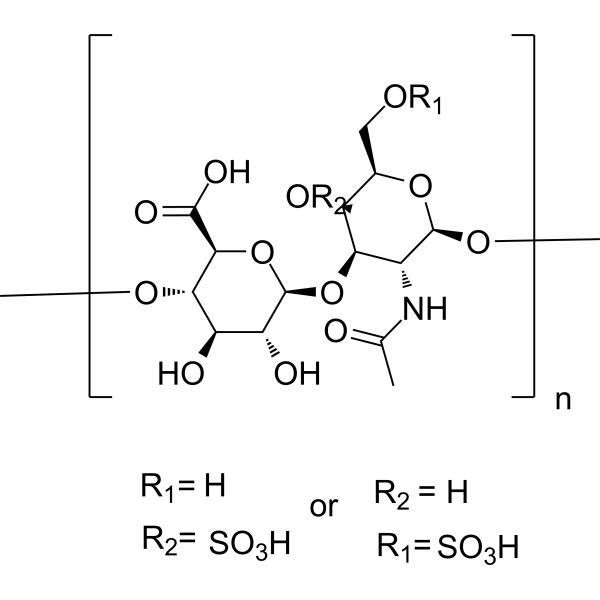
HY-B2162B
Chondroitin sulfate sodium (from bovine)Sulfated Linear Polysaccharide -
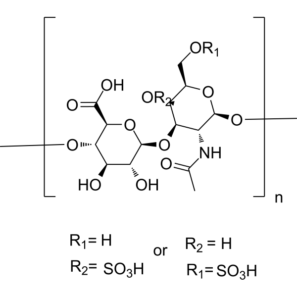
HY-B2162E
Chondroitin sulfate (from shark cartilage)Biochemical Reagent -

HY-B2162H
Chondroitin sulfate (from fish)Glycosaminoglycan -
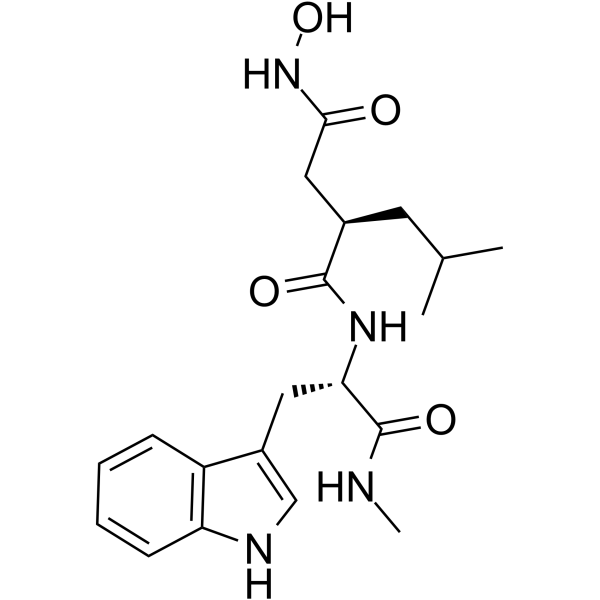
HY-15768
IlomastatMMP Inhibitor -
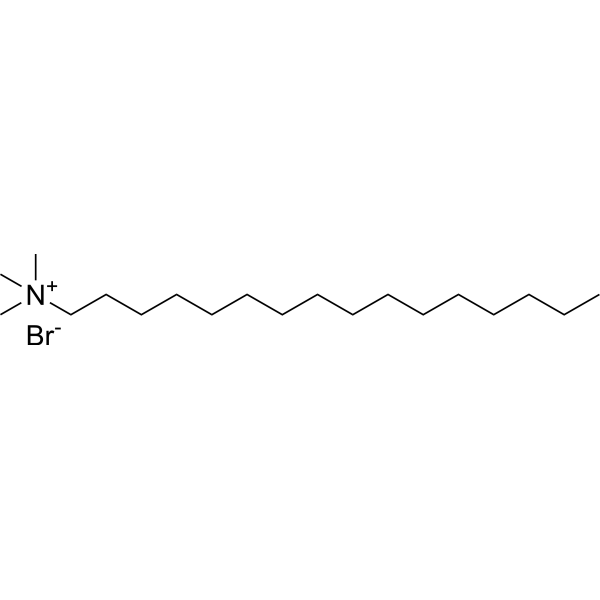
HY-B1260
Cetrimonium bromideCationic Surfactant -
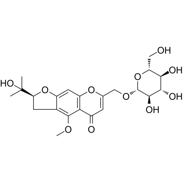
HY-N0635
Prim-O-glucosylcimifuginCOX Inhibitor -
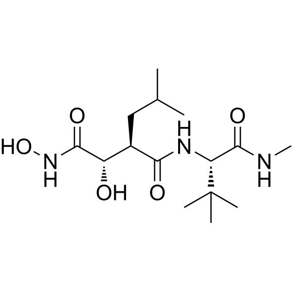
HY-12169
MarimastatMMP Inhibitor -
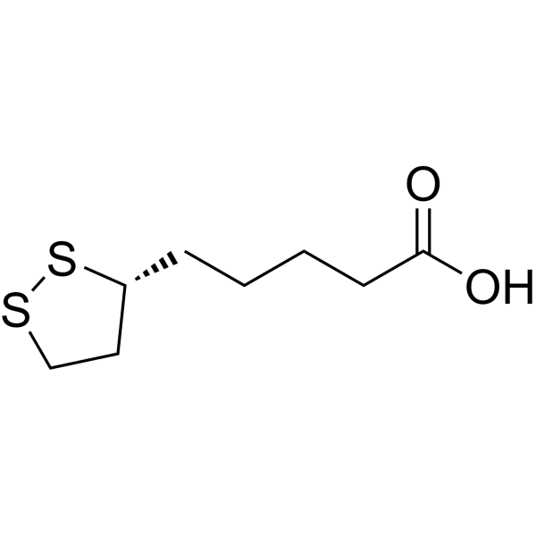
HY-18733
Lipoic acidAntioxidant -
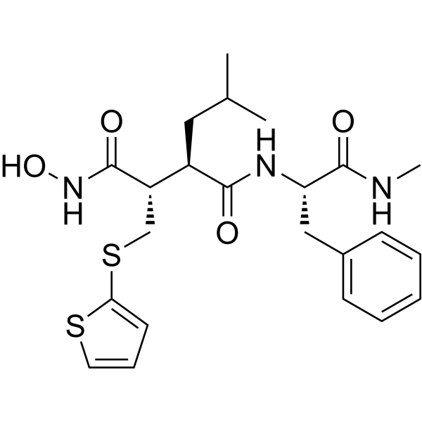
HY-13564
BatimastatMMP Inhibitor
All Endogenous Metabolite Isoforms
More

Biological Activity
Description
IC50 & Target
[1]|
ERK1 |
ERK2 |
IL-1β |
IL-6 |
IL-17 |
In Vivo
MedChemExpress (MCE) has not independently confirmed the accuracy of these methods. They are for reference only.
-
Animal Model:Sprague Dawley (SD) (male, 7-8 weeks old, 220 g, sodium iodoacetate-induced osteoarthritis)[1]
-
Dosage:100 mg/kg/day
-
Administration:p.o.; daily; 28 days
-
Result:Increased rotating rod time and ramp angle with the highest increase rate among tested cartilage extracts, trending closest to the sham group by week 4.
Resulted in intact cartilage surfaces, regular cell arrangement, and intact tide lines via H&E staining; achieved a significantly lower Mankin’s OA score than the osteoarthritis model group and lower than the cartilage powder (CPw) and type II collagen peptides (CP) groups.
Significantly reduced synovial fluid levels of IL-1β, prostaglandin E2 (PGE2), IL-6, and tumor necrosis factor-α (TNF-α) compared to the osteoarthritis model group, with the lowest levels among tested cartilage extracts; modulated serum inflammatory cytokine levels by reducing pro-inflammatory markers relative to the model group.
Altered the gut microbiota structure of osteoarthritis rats, significantly reduced the relative abundance of Lactobacillus compared to the model group; significantly increased ACE index and decreased Simpson index relative to the model group; showed distinct separation from the model group's microbiota profile via principal component analysis.


Chemical Information
-
CAS No. 9007-28-7
-
Appearance Solid
-
Formula (C14H21NO14S)n
-
Color Off-white to light yellow
-
SMILES
O=S(O)=O.O=S(O)=O.OC([C@@H]1[C@@H](OC)[C@H](O)[C@@H](O)[C@H](O[C@@H]2[C@@H](NC(C)=O)[C@H](OC)O[C@H](CO[R])[C@@H]2O[R])O1)=O.[R].[R].[=].[=].[R2=].[R1=].[n].[or]
-
Synonyms
Chondroitin polysulfate (from chicken)
-
Shipping
Room temperature in continental US; may vary elsewhere.
-
Storage
4°C, protect from light
* In solvent : -80°C, 6 months; -20°C, 1 month (protect from light)


Solvent & Solubility
In Vitro:
H2O : 100 mg/mL (Need ultrasonic)

Protocols
-
Kinase activity and phosphorylation assays
Kinase activity assays measure the ability of kinases to transfer phosphate groups from ATP to specific substrates, while phosphorylation assays detect the presence and levels of phosphorylated proteins. Common methods include radiolabeled ATP incorporation (e. g. ,), ADP release detection via bioluminescence (e. g. ,[3]), enzyme-linked immunosorbent assays (ELISA) for phospho-specific epitopes (e. g. ,[6]), and microtiter-based formats for high-throughput screening (e. g. ,[8]). The ADP-Glo assay quantifies kinase activity by measuring ADP produced during phosphorylation using a luciferase-based system. Radiometric assays involve autoradiography or scintillation counting after incorporation of 32P-labeled ATP into substrate proteins. ELISA-based approaches rely on phospho-specific antibodies to detect activated kinases in cell lysates or purified samples.
-
Western Blot
Western blotting (WB) is a commonly used experimental method in molecular biology, biochemistry, and immunogenetics for identifying and quantifying target proteins. It combines gel electrophoresis with immunoassay, enabling researchers to analyze protein expression, post-translational modifications, and molecular weight.
-
RNA extraction experimental
By lysing cells, releasing RNA, and removing impurities such as proteins and DNA, high-purity RNA products are finally obtained. The commonly used traditional method is the guanidine isothiocyanate/phenol/chloroform method (Trizol), which is suitable for a variety of animal materials including animal tissues, microorganisms, cultured cells, etc., and most plant materials.
-
Mitochondrial membrane-potential fluorescent assay
Mitochondrial membrane potential fluorescent assays estimate ΔΨm in living cells using lipophilic cationic dyes such as TMRM, TMRE, rhodamine 123, and JC-1, which accumulate in mitochondria according to membrane polarization; loss of signal after FCCP or CCCP treatment is interpreted as mitochondrial depolarization. TMRM/TMRE and rhodamine 123 are commonly used for semi-quantitative live-cell microscopy or flow cytometry, while JC-1 can report a shift from red aggregate fluorescence to green monomer fluorescence during depolarization; interpretation requires controls because dye concentration, quenching mode, cell type, dye efflux, and mitochondrial mass can affect fluorescence independently of ΔΨm.
-
LPS-Induced Endotoxemia/Systemic Inflammation
Lipopolysaccharide (LPS)-induced endotoxemia is a widely used in vivo model of acute systemic inflammation in which LPS, a Gram-negative bacterial endotoxin, activates innate immune signaling primarily through TLR4, leading to rapid and transient induction of pro-inflammatory cytokines such as TNF-α, IL-6, and IL-1β in circulation and tissues. This cytokine surge is commonly used as a measurable readout of systemic inflammatory activation and immune dysregulation, and is typically assessed within hours after intraperitoneal LPS administration in mouse models of endotoxemia. The model captures key features of systemic inflammatory response syndrome, including cytokine release, immune cell activation, and downstream tissue responses, and has been used to evaluate anti-inflammatory interventions such as cytokine modulation, lipid mediators, and immune cell-targeting therapies.
-
Mitochondrial membrane-potential and mitochondrial mass staining
Mitochondrial membrane potential staining measures the electrochemical polarization across the mitochondrial inner membrane in live cells using lipophilic cationic fluorescent probes; early rhodamine-based work showed that selective mitochondrial dye accumulation is lost when the mitochondrial transmembrane potential is dissipated. JC-1 reports mitochondrial polarization by shifting from green monomer fluorescence to red J-aggregate fluorescence as dye concentration increases within energized mitochondria; therefore, the red/green fluorescence ratio is used as a relative readout of mitochondrial membrane potential. TMRE or TMRM staining provides a single-channel relative readout because these cationic rhodamine esters accumulate in polarized mitochondria, and lower fluorescence indicates reduced mitochondrial polarization when acquisition and dye-loading conditions are controlled. Mitochondrial mass staining is commonly performed with MitoTracker Green FM or related MitoTracker dyes as
-
Protocol for Kinase activity and phosphorylation assays
Kinase activity assays measure transfer of phosphate from ATP to a protein or peptide substrate, generating phosphorylated substrate, ADP, or incorporated radiolabeled phosphate as the readout; phosphorylation assays measure site-specific phosphorylation in cells or tissues as a proxy for kinase-pathway activation, inhibition, or substrate regulation. Phosphorylation can be detected by phospho-specific Western blot, immunoprecipitation kinase assay, phospho-immunofluorescence, phospho-flow cytometry, luminescent ADP detection, radiolabeled ATP incorporation, or reporter-based pathway assays, and these readouts can be applied to cancer cells, primary neurons, mouse tumors, organoids, inflammatory macrophages, ferroptosis studies, and mitophagy studies when the kinase target is biologically relevant.
-
Pyroptosis Solutions
Pyroptosis is a lytic inflammatory cell-death pathway executed by gasdermin pores, most classically through inflammasome-mediated activation of caspase-1, cleavage of gasdermin D, membrane pore formation, LDH release, and secretion of IL-1β and IL-18. The canonical pathway is commonly modeled by priming cells with an inflammatory signal such as LPS to induce pro-IL-1β and inflammasome components, followed by an activation signal such as ATP or nigericin to activate NLRP3, ASC speck formation, caspase-1 cleavage, GSDMD cleavage, cytokine release, and pyroptotic membrane rupture. The non-canonical pathway is triggered when cytosolic LPS activates mouse caspase-11 or human caspase-4/5, leading to GSDMD cleavage and pyroptosis, and this can secondarily activate NLRP3-dependent IL-1β release. Pyroptosis is linked to inflammatory injury, infection, cancer, liver disease, ocular disease, placental inflammation, and other disease phenotypes, but unresolved questions include which gasdermin fam


Purity & Documentation
-
Data Sheet (280 KB)
-
SDS (394 KB)
- English - EN (394 KB)
- Français - FR (394 KB)
- Deutsch - DE (394 KB)
- Norwegian - NO (394 KB)
- Español - ES (394 KB)
- Swedish - SV (394 KB)
- Italian - IT (394 KB)
- Korean - KR (394 KB)
- Portuguese - PT (394 KB)
-
Handling Instructions (2659 KB)
References

Calculators
Concentration (start) × Volume (start) = Concentration (final) × Volume (final)


Chondroitin sulfate (from chicken) Related Classifications
HY-B2162D Related Classifications
 Powered by Bioz
Keywords