



Hydrangenol

Based on 1 Customer Validation
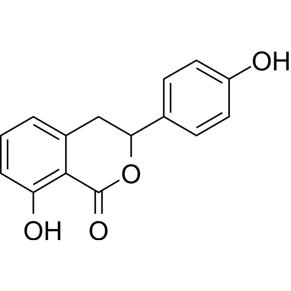
Hydrangenol is an orally active antiphotoaging compound. It can be isolated from Hydrangea serrata leaves. Hydrangenol prevents wrinkle formation by reducing MMP and inflammatory cytokine expression and increasing moisturizing factors and antioxidant genes level.
For research use only. We do not sell to patients.
- Purity : 99.62%
- CAS No.: 480-47-7
- Formula: C15H12O4
- Molecular Weight:256.25
-
Storage:Powder -20°C, 3 years ; In solvent -80°C, 6 months , -20°C, 1 month

To place orders, for customer services and technical support, please contact: MedChemExpress USA
Tel: 609-228-6898 E-mail: [email protected] [email protected]


-
 Biological Activity
-
Chemical Information
-
Solvent & Solubility
- Protocols
- Purity & Documentation
- References
-
 Help & FAQs




Biological Activity
Description
IC50 & Target
[1]|
MMP-1 |
MMP-3 |
COX-2 |
IL-6 |
In Vivo
MedChemExpress (MCE) has not independently confirmed the accuracy of these methods. They are for reference only.
-
Animal Model:UVB-irradiated HR-1 hairless mice[1]
-
Dosage:5, 10, 20, or 40 mg/kg
-
Administration:Oral gavage; daily for 7 weeks
-
Result:Increased the expression of involucrin, filaggrin, and aquaporin-3 (AQP3) as well as hyaluronic acid (HA) production via hyaluronidase (HYAL)-1/-2 downregulation.
Increased the expression of Pro-COL1A1.
Decreased the expression matrix metalloproteinase (MMP)-1/-3, cyclooxygenase-2 (COX-2), and interleukin-6 (IL-6).
Attenuated the phosphorylation of mitogen-activated protein kinases (MAPKs) including ERK and p38, activator protein 1 (AP-1) subunit, and signal transduction and activation of transcription 1 (STAT1).
Upregulated the expression of nuclear factor-E2-related factor 2 (Nrf2), heme oxygenase-1 (HO-1), NAD(P)H quinone dehydrogenase 1 (NQO-1), glutamate cysteine ligase modifier subunit (GCLM), and glutamate cysteine ligase catalysis subunit (GCLC).


Chemical Information
-
CAS No. 480-47-7
-
Appearance Solid
-
Molecular Weight 256.25
-
Formula C15H12O4
-
Color White to off-white
-
SMILES
O=C1C2=C(O)C=CC=C2CC(C3=CC=C(O)C=C3)O1
-
Structure Classification
-
Shipping
Room temperature in continental US; may vary elsewhere.
-
Storage
Powder -20°C 3 years In solvent -80°C 6 months -20°C 1 month


Solvent & Solubility
In Vitro:
DMSO : 125 mg/mL (487.80 mM; Need ultrasonic; Hygroscopic DMSO has a significant impact on the solubility of product, please use newly opened DMSO)
Please refer to the solubility information to select the appropriate solvent. Once prepared, please aliquot and store the solution to prevent product inactivation from repeated freeze-thaw cycles.
Storage method and period of stock solution: -80°C, 6 months; -20°C, 1 month. When stored at -80°C, please use it within 6 months. When stored at -20°C, please use it within 1 month.
Please refer to the solubility information to select the appropriate solvent. Once prepared, please aliquot and store the solution to prevent product inactivation from repeated freeze-thaw cycles.
Storage method and period of stock solution: -80°C, 6 months; -20°C, 1 month. When stored at -80°C, please use it within 6 months. When stored at -20°C, please use it within 1 month.
Concentration (start) × Volume (start) = Concentration (final) × Volume (final)
In Vivo:
Select the appropriate dissolution method based on your experimental animal and administration route.
- For the following dissolution methods, please ensure to first prepare a clear stock solution using an In Vitro approach and then sequentially add co-solvents:
- To ensure reliable experimental results, the clarified stock solution can be appropriately stored based on storage conditions. As for the working solution for In Vivo experiments, it is recommended to prepare freshly and use it on the same day.
- The percentages shown for the solvents indicate their volumetric ratio in the final prepared solution. If precipitation or phase separation occurs during preparation, heat and/or sonication can be used to aid dissolution.
Add each solvent one by one: 10% DMSO 90% (20% SBE-β-CD in Saline)
Solubility: ≥ 2.08 mg/mL (8.12 mM); Clear solution
This protocol yields a clear solution of ≥ 2.08 mg/mL (saturation unknown).
Taking 1 mL working solution as an example, add 100 μL DMSO stock solution (20.8 mg/mL) to 900 μL 20% SBE-β-CD in Saline, and mix evenly.
Preparation of 20% SBE-β-CD in Saline (4°C, storage for one week): 2 g SBE-β-CD powder is dissolved in 10 mL Saline, completely dissolve until clear.
In Vivo Dissolution Calculator
Please enter the basic information of animal experiments:
-
-
-
-
Recommended: Prepare an additional quantity of animals to account for potential losses during experiments.
Please enter your animal formula composition:
-
%DMSO +
Recommended: Keep the proportion of DMSO in working solution below 2% if your animal is weak.
-
%+
-
+%Tween-80 + +
-
%Saline +
The co-solvents required include: DMSO, . All of co-solvents are available by MedChemExpress (MCE). , Tween 80. All of co-solvents are available by MedChemExpress (MCE).
Working solution concentration: 0.22 mg/mL
Method for preparing stock solution: mg drug dissolved in μL DMSO. Stock solution concentration: mg/mL.
1. Take μL DMSO stock solution;
2. Add μL .
μL , mix evenly;
3. Then add μL Tween 80, mix evenly;
4. Then add μL
Please ensure that the stock solution in the first step is dissolved to a clear state, and add co-solvents in sequence. You can use ultrasonic heating (ultrasonic cleaner, recommended frequency 20-40 kHz), vortexing, etc. to assist dissolution.


Purity & Documentation
-
Data Sheet (275 KB)
-
SDS (394 KB)
- English - EN (394 KB)
- Français - FR (394 KB)
- Deutsch - DE (394 KB)
- Norwegian - NO (394 KB)
- Español - ES (394 KB)
- Swedish - SV (394 KB)
- Italian - IT (394 KB)
- Korean - KR (394 KB)
- Portuguese - PT (394 KB)
-
Handling Instructions (2659 KB)
References


Complete Stock Solution Preparation Table
Please refer to the solubility information to select the appropriate solvent. Once prepared, please aliquot and store the solution to prevent product inactivation from repeated freeze-thaw cycles.
Storage method and period of stock solution: -80°C, 6 months; -20°C, 1 month. When stored at -80°C, please use it within 6 months. When stored at -20°C, please use it within 1 month.
| Optional Solvent | Concentration Solvent Mass | 1 mg | 5 mg | 10 mg | 25 mg |
|---|---|---|---|---|---|
| DMSO | 1 mM | 3.9024 mL | 19.5122 mL | 39.0244 mL | 97.5610 mL |
| 5 mM | 0.7805 mL | 3.9024 mL | 7.8049 mL | 19.5122 mL | |
| 10 mM | 0.3902 mL | 1.9512 mL | 3.9024 mL | 9.7561 mL | |
| 15 mM | 0.2602 mL | 1.3008 mL | 2.6016 mL | 6.5041 mL | |
| 20 mM | 0.1951 mL | 0.9756 mL | 1.9512 mL | 4.8780 mL | |
| 25 mM | 0.1561 mL | 0.7805 mL | 1.5610 mL | 3.9024 mL | |
| 30 mM | 0.1301 mL | 0.6504 mL | 1.3008 mL | 3.2520 mL | |
| 40 mM | 0.0976 mL | 0.4878 mL | 0.9756 mL | 2.4390 mL | |
| 50 mM | 0.0780 mL | 0.3902 mL | 0.7805 mL | 1.9512 mL | |
| 60 mM | 0.0650 mL | 0.3252 mL | 0.6504 mL | 1.6260 mL | |
| 80 mM | 0.0488 mL | 0.2439 mL | 0.4878 mL | 1.2195 mL | |
| 100 mM | 0.0390 mL | 0.1951 mL | 0.3902 mL | 0.9756 mL |


Hydrangenol Related Classifications
HY-N4028 Related Classifications
 Powered by Bioz