



Isolinderalactone
Based on 2 publication(s) in Google Scholar
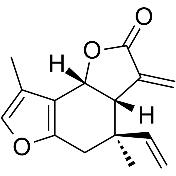
Isolinderalactone is a sesquiterpene that exhibits anti-cancer, anti-inflammatory, and neuroprotective effects. Isolinderalactone inhibits VEGF expression and tyrosine phosphorylation of VEGFR2. Isolinderalactone decreases viability and induces apoptosis in U-87 glioblastoma (GBM) cells and colorectal cancer (CRC) cells. Isolinderalactone induces G2/M phase cell cycle arrest, ROS generation, pJNK/p38 MAPK activation, in colorectal cancer (CRC) cells. Isolinderalactone blocks LPS (HY-D1056)-induced NF-κB activation while activating Nrf2-HMOX1 signaling in RAW264.7 macrophages. Isolinderalactone improves cognitive dysfunction in APP/PS1 mice. Isolinderalactone can be used for the study of Glioblastoma multiforme (GBM), colorectal cancer, Alzheimer’s disease and acute lung injury.
For research use only. We do not sell to patients.
- Purity : 95.62%
- CAS No.: 957-66-4
- Formula: C15H16O3
- Molecular Weight:244.29
-
Storage:
4°C, sealed storage, away from moisture and light
* In solvent : -80°C, 6 months; -20°C, 1 month (sealed storage, away from moisture and light)

To place orders, for customer services and technical support, please contact: MedChemExpress USA
Tel: 609-228-6898 E-mail: [email protected] [email protected]


-
 Biological Activity
-
Chemical Information
-
Solvent & Solubility
- Protocols
- Purity & Documentation
- References
-
 Help & FAQs


Publications Citing Use of MedChemExpress (MCE) Isolinderalactone
MoreAll VEGFR Isoforms
MoreAll Caspase Isoforms
More

Biological Activity
Description
IC50 & Target
[1]|
VEGFR2 |
Bcl-2 |
Cellular Effect
|
Cell Line

|
Type | Value | Description | References |
|---|---|---|---|---|
| RAW264.7 | CC50 |
66.41 μM
Compound: 7
|
Cytotoxicity against mouse RAW264.7 cells assessed as reduction in cell viability after 3 hrs by alamar blue assay
Cytotoxicity against mouse RAW264.7 cells assessed as reduction in cell viability after 3 hrs by alamar blue assay
|
[PMID: 22148193] |
| RAW264.7 | IC50 |
0.3 μM
Compound: 7
|
Inhibition of iNOS-mediated NO production in LPS-stimulated mouse RAW264.7 cells after 24 hrs by Griess reagent method
Inhibition of iNOS-mediated NO production in LPS-stimulated mouse RAW264.7 cells after 24 hrs by Griess reagent method
|
[PMID: 22148193] |
In Vitro
Isolinderalactone (0.5-2.5 μg/mL, 24-72 h) inhibits U-87 glioblastoma cell growth[1].
Isolinderalactone (0.5-2.5 μg/mL, 48 h) activates the apoptotic pathway in U-87 GBM cells by decreasing BCL-2, survivin, and XIAP expression, increasing cleaved caspase-3, and inducing DNA breakage, thereby promoting cell apoptosis[1].
Isolinderalactone (0.5-2 μg/mL, 48 h) suppresses VEGF expression in U-87 GBM cells and inhibits VEGF-induced angiogenesis of human brain microvascular endothelial cells (HBMECs)[2].
Isolinderalactone (2 μg/mL, 8 days) inhibits angiogenic sprouting in a 3D microfluidic chip[2].
Isolinderalactone (2.5-5 μg/mL, 48 h) decreases HIF expression and activity in U-87 cells and VEGFR2 activation in HBMECs[2].
Isolinderalactone (0-9 μM, 24-48 h) suppresses proliferation and colony formation of colorectal cancer cells (HCT116, HCT116-OxR, HT29, and HT29-OxR cells)[3].
Isolinderalactone (0-9 μM, 48 h) induces apoptosis, G2/M cell cycle arrest, ROS generation, and ER stress in colorectal cancer cells[3].
Isolinderalactone (0-9 μM, 48 h) induces apoptosis in CRC cells via mitochondrial and caspase-dependent pathways, potentially by modulating JNK/p38 MAPK activation, with ROS playing a critical role in this process[3].
Isolinderalactone (10 μM, 2-27 h) attenuates Aβ1-42-induced cell damage and reduces neurotoxicity in PC12 cells[4].
Isolinderalactone (10 μM, 27 h) reduces neuronal cell damage by inhibiting JNK in PC12 cells[4].
Isolinderalactone (0.5-10 μM, 19 h) inhibits LPS or TNF-α-induced inflammatory response in RAW264.7 cells, MH-S cells and BMDMs[5].
Isolinderalactone (1-10 μM, 13 h) suppresses the mRNA expression of proinflammatory enzymes and cytokines in LPS-exposed RAW264.7 macrophages[5].
Isolinderalactone (1-10 μM, 2-9 h) blocks LPS-induced NF-κB activation while activating Nrf2-HMOX1 signaling in RAW264.7 macrophages[5].
MedChemExpress (MCE) has not independently confirmed the accuracy of these methods. They are for reference only. Further protocols information, click here.
-
Cell Line:U-87 GBM cells and HBMECs
-
Concentration:0.5, 1, 2, 2.5 μg/mL
-
Incubation Time:24, 48, 72 h
-
Result:Inhibited cell viability in a dose-dependent manner.
Decreased VEGF-induced HBMEC proliferation in a dose-dependent manner.
-
Cell Line:U-87 GBM cells and HBMECs
-
Concentration:0.5, 1, 2.5,5 μg/mL
-
Incubation Time:48 h
-
Result:Decreased BCL-2, survivin, and XIAP expression.
Increased the level of cleaved caspase-3 and cleaved PARP.
Decreased the expression of the potent angiogenic factor, VEGF.
Decreased HIF expression and activity in U-87 cells.
Reduced phosphorylation of VEGFR2 in HBMECs.
-
Cell Line:U-87 GBM cells
-
Concentration:2.5 μg/mL
-
Incubation Time:48 h
-
Result:Showed predominantly bright γ-H2AX nuclear staining.
-
Cell Line:HCT116, HCT116-OxR, HT29, and HT29-OxR cells
-
Concentration:3, 6, 9 μM
-
Incubation Time:24, 48 h
-
Result:Suppressed colorectal cancer cells proliferation with IC50 values of 8.05 μM (HCT116), 5.13 μM (HCT116-OxR), 10.38 μM (HT29) and 9.46 μM (HT29-OxR).
-
Cell Line:HCT116, HCT116-OxR, HT29, and HT29-OxR cells
-
Concentration:3, 6, 9 μM
-
Incubation Time:48 h
-
Result:Contributed to G2/M phase cell cycle arrest.
-
Cell Line:HCT116, HCT116-OxR, HT29, and HT29-OxR cells
-
Concentration:3, 6, 9 μM
-
Incubation Time:48 h
-
Result:Increased the expression levels of ER stress related proteins (GRP78, CHOP, DR4, and DR5).
Activated JNK/p38 MAPK.
Increased the levels of pro-apoptotic proteins (Bim and Bax), apaf-1, and cleaved (c)-PARP.
Decreased expression levels of anti-apoptotic proteins (Mcl-1, Bid, Bcl-xL and Bcl-2), mitochondrial cyto c and caspase 3 in a dose-dependent manner.
-
Cell Line:Aβ1-42-induced PC12 cells
-
Concentration:10 μM
-
Incubation Time:27 h
-
Result:Decreased Bax expression and intracellular levels of caspase-3 activation and increased Bcl2 levels.
Decreased the level of p-JNK, but had no significant effect on ERK and P38MAPK.
-
Cell Line:LPS-exposed RAW264.7 macrophages
-
Concentration:1, 10 μM
-
Incubation Time:13 h
-
Result:Decreased the mRNA levels of iNOS, COX2, IL-1β, IL-6, and TNF-α.
-
Cell Line:LPS-exposed RAW264.7 macrophages
-
Concentration:1, 5, 10 μM
-
Incubation Time:2, 9 h
-
Result:Down-regulated LPS-induced phosphorylation of IKKα/β and the level of NF-κB p65.
Increased the protein level of Nrf2 protein in the cytoplasmic fraction and nuclear fraction.
In Vivo
Isolinderalactone (2.5-5 mg/kg, i.p., every other day, 16 days) reduces tumor growth and vasculature in a human GBM xenograft model [2].
Isolinderalactone (5 mg/kg, i.p., daily, 6 days) inhibits VEGF-mediated angiogenesis in an in vivo Matrigel plug assay in mice[2].
Isolinderalactone (1-10 mg/kg, i.p., daily, 30 days) ameliorates learning and memory deficits in APP/PS1 mice[4].
Isolinderalactone (2.5-10 mg/kg, i.p., daily, 5 days) alleviates LPS-induced lung inflammatory injury in mice[5].
MedChemExpress (MCE) has not independently confirmed the accuracy of these methods. They are for reference only.
-
Animal Model:U-87 GBM cells (3 × 10⁶ cells/100 μL of serum-free DMEM) were subcutaneously implanted into the right flank of the BALB/C nude mice[1]
-
Dosage:1, 2.5, 5 mg/kg
-
Administration:i.p., every other day for 12 days
-
Result:Reduced tumor volume and weight Showed no differences on body weight.
Increased cleaved caspase-3 fluorescence.
Showed a significantly higher number of TUNEL-positive cells.
-
Animal Model:U-87 GBM cells (3 × 10⁶ cells/100 μL of serum-free DMEM) were subcutaneously implanted into the right flank of the BALB/C nude mice[2]
-
Dosage:2.5, 5 mg/kg
-
Administration:i.p., every other day for 16 days
-
Result:Decreased tumor volume and suppressed tumor progression.
Inhibited tumor angiogenesis.
Reduced VEGF immunoreactivity.
-
Animal Model:C57BL/6 mice (6-week-old, male)[2]
-
Dosage:5 mg/kg
-
Administration:i.p. daily for 6 days
-
Result:Decreased hemoglobin concentration and CD31 staining.
Inhibited VEGF-induced new vessel formation in vivo.
-
Animal Model:10-month-old APP/PS1 transgenic mice[4]
-
Dosage:1, 10 mg/kg
-
Administration:i.p. daily for 30 days
-
Result:Enhanced the exploration of the novel object by the mice.
Ameliorated learning and memory deficits in APP/PS1mice.
Reduced the level of APP/β-amyloid.
Increased the levels of synapse-associated proteins, including PSD95 and Map2.
Reduced neuronal apoptosis in the cortex and hippocampus.
Increased the SOD content and decreased MDA levels in the hippocampus.
Increased Bcl2 levels, decreased Bax, p-JNK expression, and decreased caspase-3 activation.
-
Animal Model:Male Institute of Cancer Research (ICR) mice (SPF grade, 20-22 g, 6 weeks old) injected with LPS (15 mg/kg)[5]
-
Dosage:2.5-10 mg/kg
-
Administration:i.p., daily for 5 days
-
Result:Improved the lung injury scores.
Reduced the MPO activity in LPS-exposed lung tissue.
Reduced the levels of PGE2, IL-1β, IL-6, and TNF-α.


Chemical Information
-
CAS No. 957-66-4
-
Appearance Solid
-
Molecular Weight 244.29
-
Formula C15H16O3
-
Color White to off-white
-
SMILES
C=C[C@@]1([C@]2([H])[C@](OC(C2=C)=O)([H])C3=C(OC=C3C)C1)C
-
Structure Classification
-
Initial Source
-
Shipping
Room temperature in continental US; may vary elsewhere.
-
Storage
4°C, sealed storage, away from moisture and light
* In solvent : -80°C, 6 months; -20°C, 1 month (sealed storage, away from moisture and light)


Publications (2)
-
Journal Impact Factor
-
Most Recent
-
Eur J Pharmacol
Isolinderalactone targets TNF-α/STAT3 inflammatory pathways to attenuate psoriasis-like dermatitis. [Abstract]2026 Mar 28:1019:178733. PMID: 41795538 -


Solvent & Solubility
In Vitro:
DMSO : 50 mg/mL (204.67 mM; Need ultrasonic; Hygroscopic DMSO has a significant impact on the solubility of product, please use newly opened DMSO)
Please refer to the solubility information to select the appropriate solvent. Once prepared, please aliquot and store the solution to prevent product inactivation from repeated freeze-thaw cycles.
Storage method and period of stock solution: -80°C, 6 months; -20°C, 1 month (sealed storage, away from moisture and light). When stored at -80°C, please use it within 6 months. When stored at -20°C, please use it within 1 month.
Please refer to the solubility information to select the appropriate solvent. Once prepared, please aliquot and store the solution to prevent product inactivation from repeated freeze-thaw cycles.
Storage method and period of stock solution: -80°C, 6 months; -20°C, 1 month (sealed storage, away from moisture and light). When stored at -80°C, please use it within 6 months. When stored at -20°C, please use it within 1 month.
Concentration (start) × Volume (start) = Concentration (final) × Volume (final)
In Vivo:
Select the appropriate dissolution method based on your experimental animal and administration route.
- For the following dissolution methods, please ensure to first prepare a clear stock solution using an In Vitro approach and then sequentially add co-solvents:
- To ensure reliable experimental results, the clarified stock solution can be appropriately stored based on storage conditions. As for the working solution for In Vivo experiments, it is recommended to prepare freshly and use it on the same day.
- The percentages shown for the solvents indicate their volumetric ratio in the final prepared solution. If precipitation or phase separation occurs during preparation, heat and/or sonication can be used to aid dissolution.
In Vivo Dissolution Calculator
Please enter the basic information of animal experiments:
-
-
-
-
Recommended: Prepare an additional quantity of animals to account for potential losses during experiments.
Please enter your animal formula composition:
-
%DMSO +
Recommended: Keep the proportion of DMSO in working solution below 2% if your animal is weak.
-
%+
-
+%Tween-80 + +
-
%Saline +
The co-solvents required include: DMSO, . All of co-solvents are available by MedChemExpress (MCE). , Tween 80. All of co-solvents are available by MedChemExpress (MCE).
Working solution concentration: 0.22 mg/mL
Method for preparing stock solution: mg drug dissolved in μL DMSO. Stock solution concentration: mg/mL. * In solvent : -80°C, 6 months; -20°C, 1 month (sealed storage, away from moisture and light)
1. Take μL DMSO stock solution;
2. Add μL .
μL , mix evenly;
3. Then add μL Tween 80, mix evenly;
4. Then add μL
Please ensure that the stock solution in the first step is dissolved to a clear state, and add co-solvents in sequence. You can use ultrasonic heating (ultrasonic cleaner, recommended frequency 20-40 kHz), vortexing, etc. to assist dissolution.

Protocols
-
RNA extraction experimental
By lysing cells, releasing RNA, and removing impurities such as proteins and DNA, high-purity RNA products are finally obtained. The commonly used traditional method is the guanidine isothiocyanate/phenol/chloroform method (Trizol), which is suitable for a variety of animal materials including animal tissues, microorganisms, cultured cells, etc., and most plant materials.
-
Western Blot
Western blotting (WB) is a commonly used experimental method in molecular biology, biochemistry, and immunogenetics for identifying and quantifying target proteins. It combines gel electrophoresis with immunoassay, enabling researchers to analyze protein expression, post-translational modifications, and molecular weight.
-
Annexin V plus membrane-impermeant dye apoptosis staining
Annexin V-based apoptosis assays rely on the detection of phosphatidylserine (PS) externalization from the inner leaflet of the plasma membrane to the outer leaflet, an early biochemical hallmark of apoptosis. Fluorescently labeled Annexin V binds PS in a calcium-dependent manner, enabling identification of early apoptotic cells by flow cytometry or fluorescence microscopy. When combined with a membrane-impermeant DNA-binding dye (e. g. , propidium iodide), this approach allows discrimination between viable (Annexin V−/dye−), early apoptotic (Annexin V+/dye−), and late apoptotic or necrotic (Annexin V+/dye+) cell populations by assessing membrane integrity and PS exposure.
-
BrdU Incorporation Assay
Bromodeoxyuridine (BrdU) incorporation assay is based on the principle that BrdU, a thymidine analog, is incorporated into newly synthesized DNA during the S phase of the cell cycle, thereby serving as a marker of DNA replication and cellular proliferation. Incorporated BrdU can be detected using anti-BrdU antibodies following DNA denaturation, enabling visualization or quantification of proliferating cells through immunochemical detection methods such as immunofluorescence or immunohistochemistry.
-
ROS/oxidative-stress fluorescent staining
ROS/oxidative-stress fluorescent staining uses cell-permeant fluorogenic probes that become fluorescent after oxidation inside cells or tissues; commonly used examples include DCFH-DA/DCFDA for broad cellular oxidant detection, DHE for superoxide-related signal detection, MitoSOX for mitochondrial superoxide-related signal detection, and CellROX probes for oxidative-stress-associated fluorescence readouts. The assay detects probe oxidation rather than a single ROS species unless the probe and analysis method have been chemically validated for that species. DCFH-DA enters cells, is deacetylated by intracellular esterases to DCFH, and produces fluorescent DCF after oxidation, so the readout is used as an operational measure of total cellular oxidative stress rather than a species-specific ROS measurement. DHE and MitoSOX can report superoxide-related oxidation, but red fluorescence alone can include non-specific ethidium-like oxidation products; HPLC or optimized spectral approaches are
-
Protocol for Cell Cycle
Cell-cycle analysis by flow cytometry measures DNA content in single cells to estimate the fraction of cells in G0/G1, S, and G2/M phases. Propidium iodide intercalates into DNA, and after RNA removal with RNase, fluorescence intensity reflects cellular DNA content: 2N cells are assigned to G0/G1, cells between 2N and 4N to S phase, and 4N cells to G2/M. DNA-content analysis alone cannot reliably separate G0 from G1 or G2 from M. Ki-67 can distinguish quiescent G0 cells from cycling cells, EdU or BrdU incorporation marks active DNA synthesis in S phase, and phospho-histone H3 staining identifies mitotic cells within the 4N population.
-
Research Protocol for Inflammation-related Diseases
The NLRP3 inflammasome is a cytosolic innate immune signaling platform that integrates priming signals and danger-signal activation to promote caspase-1 activation, maturation of IL-1β and IL-18, and gasdermin D-mediated pyroptotic cell death. The core experimental logic is to determine whether inflammatory disease phenotypes are driven by increased NLRP3 expression, ASC-containing inflammasome assembly, caspase-1 cleavage, GSDMD cleavage, and extracellular release of IL-1β/IL-18 rather than by nonspecific cell injury alone. The pathway is strongly linked to inflammation-related disease phenotypes because monosodium urate crystals activate NALP3/NLRP3 inflammasome signaling in gout-like crystal inflammation, cholesterol crystals activate NLRP3 inflammasomes in atherogenesis models, and DSS-induced intestinal inflammation has been reported to involve NLRP3 inflammasome activity. However, experimental colitis studies also show context-dependent protective effects of NLRP3 inflammasome co
-
Cell Viability Determination by MTT Colorimetric Assay
The following protocol uses the MTT colorimetric assay as a classic literature-established method for assessing cell viability/metabolic activity in cultured mammalian cells. MTT[3-(4,5-dimethylthiazol-2-yl)-2,5-diphenyltetrazolium bromide] is reduced by metabolically active cells to a colored formazan product; the amount of formazan is quantified spectrophotometrically and provides an indirect measure of metabolically active viable cells. Importantly, MTT reduction reflects cellular oxidoreductase/metabolic activity rather than an absolute direct count of living cells, so changes in cellular metabolism can alter the signal independently of cell number.
-
Kinase activity and phosphorylation assays
Kinase activity assays measure the ability of kinases to transfer phosphate groups from ATP to specific substrates, while phosphorylation assays detect the presence and levels of phosphorylated proteins. Common methods include radiolabeled ATP incorporation (e. g. ,), ADP release detection via bioluminescence (e. g. ,[3]), enzyme-linked immunosorbent assays (ELISA) for phospho-specific epitopes (e. g. ,[6]), and microtiter-based formats for high-throughput screening (e. g. ,[8]). The ADP-Glo assay quantifies kinase activity by measuring ADP produced during phosphorylation using a luciferase-based system. Radiometric assays involve autoradiography or scintillation counting after incorporation of 32P-labeled ATP into substrate proteins. ELISA-based approaches rely on phospho-specific antibodies to detect activated kinases in cell lysates or purified samples.
-
Apoptosis
Apoptosis, also called programmed cell death, is generally characterized by distinct morphological characteristics.
-
TUNEL staining for apoptotic DNA fragmentation
TUNEL staining detects DNA strand breaks by using terminal deoxynucleotidyl transferase to add labeled nucleotides to exposed 3′-OH DNA termini, generating either microscopic staining in fixed cells or tissue sections, or fluorescence/cytometric signal in cell suspensions. TUNEL positivity reflects DNA fragmentation but should not be interpreted alone as definitive apoptosis, because TUNEL can also label necrotic, autolytic, mechanically damaged, or DNA-repair-associated DNA breaks.
-
Flow cytometric DNA-content cell-cycle staining
Flow cytometric DNA-content cell-cycle staining measures the fluorescence intensity of DNA-bound fluorochromes in single cells or nuclei to estimate DNA content distributions, allowing assignment of populations to G0/G1, S, and G2/M phases by DNA histogram deconvolution. Propidium iodide (PI) intercalates into DNA, and PI fluorescence is proportional to cellular DNA content when staining is performed under conditions that make DNA accessible and minimize non-DNA signal. Cells with G2/M DNA content are expected to show approximately twice the fluorescence intensity of G0/G1 cells, while S-phase cells occupy intermediate fluorescence values. PI-based DNA-content analysis can also detect cells with fractional DNA content, often reported as sub-G1, when DNA fragmentation and extraction during staining reduce retained DNA signal in apoptotic cells. DAPI is an alternative DNA fluorochrome for univariate DNA-content analysis, while bivariate approaches combining DNA content with proliferation
-
Apoptosis Solutions
Apoptosis is a regulated, generally non-lytic cell-death pathway that removes unwanted, damaged, infected, or abnormal cells through coordinated morphological changes, caspase activation, DNA fragmentation, and membrane remodeling. The intrinsic apoptosis pathway is controlled mainly by mitochondrial outer membrane permeabilization, BCL-2 family proteins, cytochrome c release, apoptosome formation, caspase-9 activation, and downstream executioner caspase-3/7 activation. The extrinsic apoptosis pathway is initiated by death receptors such as Fas, TNFR, and TRAIL receptors, which recruit adaptor proteins and activate caspase-8 before engaging executioner caspases or mitochondrial amplification through BID cleavage. Apoptosis is linked to many phenotypes, including cancer cell killing, tissue homeostasis, immune regulation, neurodegeneration, infection response, and treatment-induced cytotoxicity; unresolved questions include how apoptosis interacts with necroptosis, pyroptosis, ferroptos
-
Protocol for Kinase activity and phosphorylation assays
Kinase activity assays measure transfer of phosphate from ATP to a protein or peptide substrate, generating phosphorylated substrate, ADP, or incorporated radiolabeled phosphate as the readout; phosphorylation assays measure site-specific phosphorylation in cells or tissues as a proxy for kinase-pathway activation, inhibition, or substrate regulation. Phosphorylation can be detected by phospho-specific Western blot, immunoprecipitation kinase assay, phospho-immunofluorescence, phospho-flow cytometry, luminescent ADP detection, radiolabeled ATP incorporation, or reporter-based pathway assays, and these readouts can be applied to cancer cells, primary neurons, mouse tumors, organoids, inflammatory macrophages, ferroptosis studies, and mitophagy studies when the kinase target is biologically relevant.
-
Inhalation Toxicity Study
Inhalation toxicity studies expose rodents to a controlled aerosol, vapor, gas, or smoke atmosphere and assess respiratory and systemic toxicity using exposure-atmosphere characterization, clinical observations, body and organ weights, bronchoalveolar lavage fluid, histopathology, blood chemistry, hematology, and, when included, molecular endpoints such as transcriptomics, proteomics, lipidomics, or tissue burden analysis. The primary biological readouts are airway irritation, pulmonary inflammation, cytotoxicity, altered surfactant or lipid homeostasis, impaired particle clearance, and tissue remodeling, reflected by BALF cell differentials, BALF protein, LDH, phosphatase activities, cytokines, lung weight, microscopic respiratory-tract lesions, and retained lung burden.


Purity & Documentation
-
Data Sheet (302 KB)
-
SDS (393 KB)
- English - EN (393 KB)
- Français - FR (393 KB)
- Deutsch - DE (393 KB)
- Norwegian - NO (393 KB)
- Español - ES (393 KB)
- Swedish - SV (393 KB)
- Italian - IT (393 KB)
- Korean - KR (393 KB)
- Portuguese - PT (393 KB)
-
Handling Instructions (2659 KB)
References
[1]. Hwang JY, et al. Isolinderalactone regulates the BCL-2/caspase-3/PARP pathway and suppresses tumor growth in a human glioblastoma multiforme xenograft mouse model. Cancer Lett. 2019 Feb 28;443:25-33. [Content Brief]
[2]. Park JH, et al. Isolinderalactone suppresses human glioblastoma growth and angiogenic activity in 3D microfluidic chip and in vivo mouse models. [Content Brief]
[3]. Kwak AW, et al. Isolinderalactone sensitizes oxaliplatin-resistance colorectal cancer cells through JNK/p38 MAPK signaling pathways. Phytomedicine. 2022 Oct;105:154383. [Content Brief]
[4]. Xiong L, et al. Isolinderalactone Ameliorates the Pathology of Alzheimer's Disease by Inhibiting the JNK Signaling Pathway. J Nat Prod. 2023 Dec 22;86(12):2718-2729. [Content Brief]
[5]. Shen X, et al. A natural sesquiterpene lactone isolinderalactone attenuates lipopolysaccharide-induced inflammatory response and acute lung injury through inhibition of NF-κB pathway and activation Nrf2 pathway in macrophages. Int Immunopharmacol. 2023 Nov;124(Pt B):110965. [Content Brief]


Complete Stock Solution Preparation Table
Please refer to the solubility information to select the appropriate solvent. Once prepared, please aliquot and store the solution to prevent product inactivation from repeated freeze-thaw cycles.
Storage method and period of stock solution: -80°C, 6 months; -20°C, 1 month (sealed storage, away from moisture and light). When stored at -80°C, please use it within 6 months. When stored at -20°C, please use it within 1 month.
| Optional Solvent | Concentration Solvent Mass | 1 mg | 5 mg | 10 mg | 25 mg |
|---|---|---|---|---|---|
| DMSO | 1 mM | 4.0935 mL | 20.4675 mL | 40.9350 mL | 102.3374 mL |
| 5 mM | 0.8187 mL | 4.0935 mL | 8.1870 mL | 20.4675 mL | |
| 10 mM | 0.4093 mL | 2.0467 mL | 4.0935 mL | 10.2337 mL | |
| 15 mM | 0.2729 mL | 1.3645 mL | 2.7290 mL | 6.8225 mL | |
| 20 mM | 0.2047 mL | 1.0234 mL | 2.0467 mL | 5.1169 mL | |
| 25 mM | 0.1637 mL | 0.8187 mL | 1.6374 mL | 4.0935 mL | |
| 30 mM | 0.1364 mL | 0.6822 mL | 1.3645 mL | 3.4112 mL | |
| 40 mM | 0.1023 mL | 0.5117 mL | 1.0234 mL | 2.5584 mL | |
| 50 mM | 0.0819 mL | 0.4093 mL | 0.8187 mL | 2.0467 mL | |
| 60 mM | 0.0682 mL | 0.3411 mL | 0.6822 mL | 1.7056 mL | |
| 80 mM | 0.0512 mL | 0.2558 mL | 0.5117 mL | 1.2792 mL | |
| 100 mM | 0.0409 mL | 0.2047 mL | 0.4093 mL | 1.0234 mL |


Isolinderalactone Related Classifications
HY-N3001 Related Classifications
 Powered by Bioz