



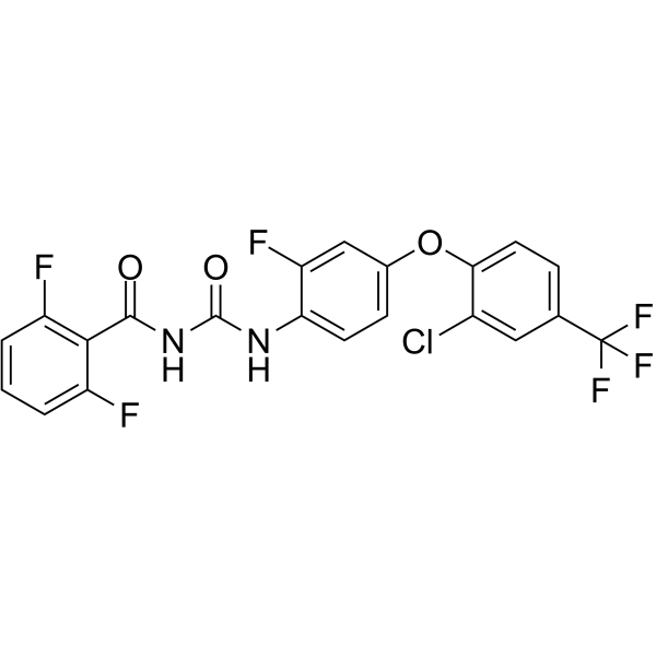
Flufenoxuron
Based on 1 publication(s) in Google Scholar
Flufenoxuron is an orally active chitin synthesis inhibitor. Flufenoxuron inhibits PI3K/AKT/p70S6K/S6 and MAPK/ERK1/2/JNK/p38 phosphorylation, reduces VEGFA/B/C and Flt1/Flt4 mRNA, and induces cell cycle dysregulation, apoptosis, DNA fragmentation, ROS production, mitochondrial calcium overload, ER stress/UPR, and autophagy, while inhibiting proliferation, migration, angiogenesis, and embryonic development. Flufenoxuron disrupts thyroid hormone regulation and induces reproductive toxicity. Flufenoxuron can be used in research on early embryo loss and infertility, testicular dysfunction, and anemia.
商品は「研究用試薬」です。人や動物の医療用・臨床診断用・食品用の製品ではありません。
研究用途以外に使用した場合、当社は一切の責任を負いかねます。
- 純度 : 99.37%
- CAS 番号: 101463-69-8
- 分子式: C21H11ClF6N2O3
- 分子量:488.77
-
保管条件:
4°C, protect from light
* In solvent : -80°C, 6 months; -20°C, 1 month (protect from light)

To place orders, for customer services and technical support, please contact: MedChemExpress USA
Tel: 609-228-6898 E-mail: [email protected] [email protected]


-
 生物活性
-
化学情報
-
溶剤 & 溶解度
- プロトコル
- 純度とドキュメンテーション
- 参考文献
-
 Help & FAQs




生物活性
製品説明
|
Akt |
S6 |
p70S6K |
ERK1 |
ERK2 |
JNK |
p38 |
VEGFA |
VEGFB |
VEGFC |
Flt1 |
Flt4 |
体外実験
Flufenoxuron (50-200 μM; 48 h) induces late apoptosis in pLE and pTr cells, with relative late apoptosis reaching 675.0% and 2900.0%, respectively[1].
Flufenoxuron (5-20 μM; 6 h) reduces phosphorylated Bad to below 50% in TM3 and TM4 cells at 20 μM, while slightly increasing Bak and decreasing Bcl-xL in both cell lines[2].
Flufenoxuron (5-20 μM; 6 h) induces the ER stress markers Ire1α, p-Eif2α, and Chop in TM4 cells, and increases Chop in TM3 cells at 20 μM[2].
Flufenoxuron (5-20 μM; 6 h) increases Vapb, Fam82a2, and p-p62 in TM3 and TM4 cells, and upregulates Grp75/Vdac in TM4 cells[2].
Flufenoxuron (5-20 μM; 6 h) decreases p-Rps6ka1 by 51% in TM3 cells and by 53% in TM4 cells at 20 μM, while increasing p-Erk1/2, p-Jnk, and p-Akt[2].
Flufenoxuron (15-30 µM; 48 h) increases late apoptosis in HUVECs at 30 µM, reported as a 188% increase relative to the vehicle group[3].
Flufenoxuron (20 μM; 24 h) reduces nuclear PCNA expression in TM3 and TM4 cells[2].
Flufenoxuron (2-20 μM; 24 h at 37 °C) induces G0/G1 phase arrest in TM4 Sertoli cells but does not significantly alter the cell cycle phases of TM3 Leydig cells[2].
Flufenoxuron (50-200 μM; 48 h) increases mitochondrial Ca2+ in pLE and pTr cells to 242.2% and 236.6%, respectively, whereas co-application of 2-APB, BAPTA, or ruthenium red attenuates this increase[1].
Flufenoxuron (2-20 μM) increases mitochondrial matrix calcium up to 7.8-fold in TM3 cells and 18-fold in TM4 cells at 20 μM, and increases mitochondrial membrane potential loss in both cell lines[2].
Flufenoxuron (30-300 µM; 7 d) induces leaf ROS accumulation and lipid peroxidation in barley, with leaf MDA increasing at 100 and 300 µM and root MDA decreasing at 300 µM[7].
Flufenoxuron (30-300 µM; 7 d) increases POD in leaves and roots, decreases SOD in leaves, and increases SOD in roots of barley[7].
Flufenoxuron (50-200 μM; 48 h) increases cytosolic Ca2+ in pLE and pTr cells to 214.1% and 227.0%, respectively, whereas co-application of 2-APB, BAPTA, or ruthenium red attenuates this increase[1].
Flufenoxuron (200 μM; 12 h) inhibits pLE-pTr cell migration and prevents closure of the 500-μm gap within 12 h[1].
Flufenoxuron (50-200 μM) reduces the expression of pregnancy-related genes in pLE and pTr cells, with the lowest expression reaching 0.13-fold for EPHA2 in pLE cells and 0.33-fold for EPHA2 in pTr cells[1].
Flufenoxuron (1-30 µM; 48 h) reduces HUVEC viability, with inhibition occurring at 5 µM and a 27% reduction at 30 µM[3].
Flufenoxuron (30-300 μM; 7 days) inhibits barley seedling growth and biomass, reducing root length by 29.57% and root fresh weight by 50.86%[5].
Flufenoxuron (300 µM; 7 d) induces chloroplast swelling, thylakoid disorganization, loss of starch granules, and proliferation of plastoglobuli in barley mesophyll cells[7].
MedChemExpress (MCE) has not independently confirmed the accuracy of these methods. They are for reference only.
-
Cell Line:pLE and pTr cells
-
Concentration:5, 10, 25, 50, 100, 150, 200 μM
-
Incubation Time:48 h
-
Result:Inhibited cell proliferation in a dose-dependent manner by up to 64.7% in pLE cells and up to 56.3% in pTr cells compared with vehicle-treated cells.
-
Cell Line:pLE and pTr cells
-
Concentration:50, 100, 200 μM
-
Incubation Time:48 h
-
Result:Increased the sub-G1 fraction in pLE and pTr cells.
Decreased the G2/M fraction in pTr cells.
-
Cell Line:pLE and pTr cells
-
Concentration:200 μM
-
Incubation Time:48 h
-
Result:Induced DNA fragmentation in both pLE and pTr cells.
-
Cell Line:pLE and pTr cells
-
Concentration:50, 100, 200 μM
-
Incubation Time:48 h
-
Result:Late-apoptotic populations at 0, 50, 100, and 200 μM were 0.4%, 0.8%, 2.1%, and 2.7% in pLE cells and 0.1%, 0.6%, 2.1%, and 2.9% in pTr cells.
Relative late apoptosis increased up to 675.0% in pLE cells and up to 2900.0% in pTr cells.
-
Cell Line:pLE and pTr cells
-
Concentration:200 μM
-
Incubation Time:12 h
-
Result:In vehicle-treated dishes, the area between pLE and pTr cells decreased from 100% to 10%.
Inhibited cell migration between pLE and pTr cells.
-
Cell Line:mouse Leydig TM3 and Sertoli TM4 cells
-
Concentration:20 μM
-
Incubation Time:24 h
-
Result:Relative nuclear PCNA expression was reduced in both TM3 and TM4 cells at 20 μM.
-
Cell Line:mouse Leydig TM3 and Sertoli TM4 cells
-
Concentration:2, 5, 10, 20 μM
-
Incubation Time:24 h at 37 °C
-
Result:Sub-G1 and G0/G1 populations increased and S and G2/M populations decreased in TM4 cells, indicating G0/G1 arrest.
TM3 cells showed no significant changes in cell-cycle phase.
-
Cell Line:mouse Leydig TM3 and Sertoli TM4 cells
-
Concentration:5, 10, 20 μM
-
Incubation Time:6 h
-
Result:Bak increased slightly, and Bcl-xL was slightly reduced in both TM3 and TM4 cells.
Phosphorylation of Bad decreased to under 50% at 20 μM in both TM3 and TM4 cells.\nGrp78 was not significantly changed in either cell line.
In TM3 cells, Ire1α and p-Eif2α were slightly increased in a dose-dependent manner, and Chop steeply increased at 20 μM.
In TM4 cells, Ire1α, p-Eif2α, and Chop gradually increased.\nGrp75 and Vdac were gradually upregulated in TM4 cells but not in TM3 cells.
Vapb and Fam82a2 increased in both TM3 and TM4 cells.
Phosphorylation of p62 increased in both cell lines.\nPhosphorylation of Erk1/2 and Jnk gradually increased, and p-Akt was upregulated in a dose-dependent manner in both TM3 and TM4 cells.
p-Rps6ka1 decreased by 51% in TM3 cells and 53% in TM4 cells at 20 μM.
p-Rps6kb1 was slightly decreased at 20 μM in both cell lines.
-
Cell Line:human umbilical vein endothelial cells (HUVECs)
-
Concentration:1, 5, 10, 15, 30 µM
-
Incubation Time:48 h
-
Result:At 5 µM, HUVEC viability was significantly suppressed.
Viability decreased by 27% at 30 µM.
体内実験
Flufenoxuron reduces hemolymph and testicular carbohydrate, lipid, and protein levels in sixth-instar male S. mauritia and disrupts testicular spermatogenesis[6].
Flufenoxuron is effective against sixth-instar S. mauritia larvae and impairs male reproductive development, reducing testis weight, volume, GSI, adult emergence rate, fecundity, and fertility[9].
MedChemExpress (MCE) has not independently confirmed the accuracy of these methods. They are for reference only.
-
Animal Model:C57BL/6 (male, 3 weeks old at start)[2]
-
Dosage:100 mg/kg body weight
-
Administration:p.o. (oral gavage); once daily; 28 days
-
Result:In testes, relative mRNA expression of Ar, Jun, and Jund was significantly reduced.
Relative mRNA expression of Star, Hsd17b3, Cyp17a1, and Cyp19a1 was slightly increased by up to 10-30%.
Hsd17b1 mRNA was decreased.
- Reproductive and Developmental Toxicity Study
- Western Blot
- RNA extraction experimental
- Human pluripotent stem cell endothelial-cell differentiation
- Vascular/Branching Fractal Analysis
- Annexin V plus membrane-impermeant dye apoptosis staining
- BrdU Incorporation Assay
- ROS/oxidative-stress fluorescent staining
- Cell migration
- Protocol for Cell Cycle
- Cell Viability Determination by MTT Colorimetric Assay
- Kinase activity and phosphorylation assays
- Totipotent stem cell culture
- Detection of Apoptosis
- TUNEL staining for apoptotic DNA fragmentation
- Flow cytometric DNA-content cell-cycle staining
- Research Protocol for Endocrine Diseases
- Autophagy
- Endothelial Tube Formation Assay
- Apoptosis Solutions
- Protocol for Kinase activity and phosphorylation assays
- Lysosome and acidic-vesicle live-cell staining
- Macroautophagy Solutions
- Endothelial Cell Migration/Angiogenic Sprouting Assay


化学情報
-
CAS 番号 101463-69-8
-
性状 Solid
-
分子量 488.77
-
分子式 C21H11ClF6N2O3
-
Color Off-white to light yellow
-
SMILES
O=C(NC(NC1=CC=C(OC2=CC=C(C(F)(F)F)C=C2Cl)C=C1F)=O)C3=C(F)C=CC=C3F
-
別名
フルフェノクスロン
-
輸送条件
Room temperature in continental US; may vary elsewhere.
-
保管条件
4°C, protect from light
* In solvent : -80°C, 6 months; -20°C, 1 month (protect from light)


Publications (1)
-
Journal Impact Factor
-
Most Recent
-
Int J Biol Macromol
Reticuline isomerase AKR1B1 with aldo-keto reductase activity and detoxification function from the insect Blaps rhynchopetera. [Abstract]2026 Mar:352:151224. PMID: 41791543


溶剤 & 溶解度
体外:
DMSO : 250 mg/mL (511.49 mM; Need ultrasonic; Hygroscopic DMSO has a significant impact on the solubility of product, please use newly opened DMSO)
Please refer to the solubility information to select the appropriate solvent. Once prepared, please aliquot and store the solution to prevent product inactivation from repeated freeze-thaw cycles.
Storage method and period of stock solution: -80°C, 6 months; -20°C, 1 month (protect from light). When stored at -80°C, please use it within 6 months. When stored at -20°C, please use it within 1 month.
Please refer to the solubility information to select the appropriate solvent. Once prepared, please aliquot and store the solution to prevent product inactivation from repeated freeze-thaw cycles.
Storage method and period of stock solution: -80°C, 6 months; -20°C, 1 month (protect from light). When stored at -80°C, please use it within 6 months. When stored at -20°C, please use it within 1 month.
濃度 (開始) × 体積 (開始) = 濃度 (終了) × 体積 (終了)
体内:
Select the appropriate dissolution method based on your experimental animal and administration route.
- For the following dissolution methods, please ensure to first prepare a clear stock solution using an In Vitro approach and then sequentially add co-solvents:
- To ensure reliable experimental results, the clarified stock solution can be appropriately stored based on storage conditions. As for the working solution for In Vivo experiments, it is recommended to prepare freshly and use it on the same day.
- The percentages shown for the solvents indicate their volumetric ratio in the final prepared solution. If precipitation or phase separation occurs during preparation, heat and/or sonication can be used to aid dissolution.
Add each solvent one by one: 10% DMSO 40% PEG300 5% Tween-80 45% Saline
Solubility: ≥ 2.08 mg/mL (4.26 mM); Clear solution
This protocol yields a clear solution of ≥ 2.08 mg/mL (saturation unknown).
Taking 1 mL working solution as an example, add 100 μL DMSO stock solution (20.8 mg/mL) to 400 μL PEG300, and mix evenly; then add 50 μL Tween-80 and mix evenly; then add 450 μL Saline to adjust the volume to 1 mL.
Preparation of Saline: Dissolve 0.9 g sodium chloride in ddH₂O and dilute to 100 mL to obtain a clear Saline solution.
In Vivo Dissolution Calculator
Please enter the basic information of animal experiments:
-
-
-
-
Recommended: Prepare an additional quantity of animals to account for potential losses during experiments.
Please enter your animal formula composition:
-
%DMSO +
Recommended: Keep the proportion of DMSO in working solution below 2% if your animal is weak.
-
%+
-
+%Tween-80 + +
-
%Saline +
The co-solvents required include: DMSO, . All of co-solvents are available by MedChemExpress (MCE). , Tween 80. All of co-solvents are available by MedChemExpress (MCE).
Working solution concentration: 0.22 mg/mL
Method for preparing stock solution: mg drug dissolved in μL DMSO. Stock solution concentration: mg/mL. * In solvent : -80°C, 6 months; -20°C, 1 month (protect from light)
1. Take μL DMSO stock solution;
2. Add μL .
μL , mix evenly;
3. Then add μL Tween 80, mix evenly;
4. Then add μL
Please ensure that the stock solution in the first step is dissolved to a clear state, and add co-solvents in sequence. You can use ultrasonic heating (ultrasonic cleaner, recommended frequency 20-40 kHz), vortexing, etc. to assist dissolution.


純度とドキュメンテーション


参考文献

Complete Stock Solution Preparation Table
Please refer to the solubility information to select the appropriate solvent. Once prepared, please aliquot and store the solution to prevent product inactivation from repeated freeze-thaw cycles.
Storage method and period of stock solution: -80°C, 6 months; -20°C, 1 month (protect from light). When stored at -80°C, please use it within 6 months. When stored at -20°C, please use it within 1 month.
| Optional Solvent | Concentration Solvent Mass | 1 mg | 5 mg | 10 mg | 25 mg |
|---|---|---|---|---|---|
| DMSO | 1 mM | 2.0460 mL | 10.2298 mL | 20.4595 mL | 51.1488 mL |
| 5 mM | 0.4092 mL | 2.0460 mL | 4.0919 mL | 10.2298 mL | |
| 10 mM | 0.2046 mL | 1.0230 mL | 2.0460 mL | 5.1149 mL | |
| 15 mM | 0.1364 mL | 0.6820 mL | 1.3640 mL | 3.4099 mL | |
| 20 mM | 0.1023 mL | 0.5115 mL | 1.0230 mL | 2.5574 mL | |
| 25 mM | 0.0818 mL | 0.4092 mL | 0.8184 mL | 2.0460 mL | |
| 30 mM | 0.0682 mL | 0.3410 mL | 0.6820 mL | 1.7050 mL | |
| 40 mM | 0.0511 mL | 0.2557 mL | 0.5115 mL | 1.2787 mL | |
| 50 mM | 0.0409 mL | 0.2046 mL | 0.4092 mL | 1.0230 mL | |
| 60 mM | 0.0341 mL | 0.1705 mL | 0.3410 mL | 0.8525 mL | |
| 80 mM | 0.0256 mL | 0.1279 mL | 0.2557 mL | 0.6394 mL | |
| 100 mM | 0.0205 mL | 0.1023 mL | 0.2046 mL | 0.5115 mL |


Flufenoxuron 関連分類
HY-B2009 関連分類
 Powered by Bioz
Keywords
- Flufenoxuron
- 101463-69-8
- Environmental Pollutants
- PI3K
- Akt
- Ribosomal S6 Kinase (RSK)
- p38 MAPK
- ERK
- JNK
- VEGFR
- Apoptosis
- Reactive Oxygen Species (ROS)
- Autophagy
- Environmental Contaminants
- Phosphoinositide 3-kinase
- PKB
- Protein kinase B
- S6K
- Extracellular signal regulated kinases
- c-Jun N-terminal kinase
- Vascular endothelial growth factor receptor
- Flt1/Flt4
- MAPK/ERK1/2/JNK/p38
- PI3K/AKT/p70S6K/S6
- VEGFA/B/C
- angiogenesis
- apoptosis
- autophagy
- chitin synthesis inhibitor
- embryonic development
- reproductive toxicity
- Inhibitor
- inhibitor
- inhibit