



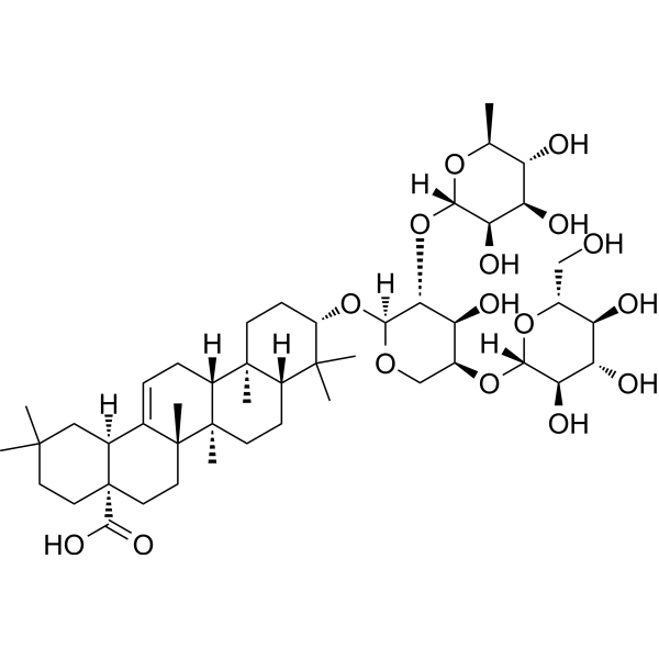
Macranthoside B

Based on 1 Customer Validation
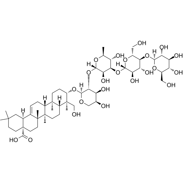
Macranthoside B is an apoptosis inducer with in vitro and in vivo anticancer activity. Macranthoside B induces autophagy and ferroptosis in cancer cells. Macranthoside B induces intracellular ROS accumulation, activates AMPK, inhibits mTOR and P70S6 kinase phosphorylation, and modulates Bcl-2/Bax expression. Macranthoside B activates caspase-3, caspase-9, and the intrinsic caspase cascade, induces PARP cleavage/degradation, and inhibits NRF2 signaling. Macranthoside B disrupts iron homeostasis via NCOA4-dependent ferritinophagy, up-regulates Lip-ROS, reduces mitochondrial membrane potential, and activates the JNK pathway. Macranthoside B can be used for the research of ovarian cancer, hepatoma, gastric carcinoma, breast carcinoma, colon carcinoma, glioma, melanoma, adenocarcinoma of the esophagogastric junction, and cervical cancer.
For research use only. We do not sell to patients.
- Purity : 99.95%
- CAS No.: 146100-02-9
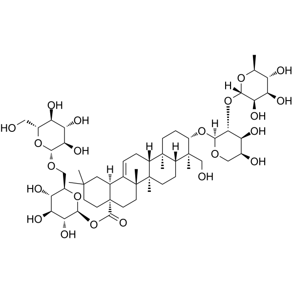
- Formula: C53H86O22
- Molecular Weight:1075.24
-
Storage:
4°C, sealed storage, away from moisture and light
* In solvent : -80°C, 6 months; -20°C, 1 month (sealed storage, away from moisture and light)

To place orders, for customer services and technical support, please contact: MedChemExpress USA
Tel: 609-228-6898 E-mail: [email protected] [email protected]


-
 Biological Activity
-
Chemical Information
-
Solvent & Solubility
- Protocols
- Purity & Documentation
- References
-
 Help & FAQs


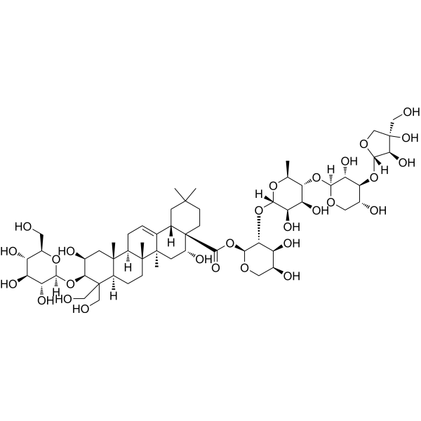
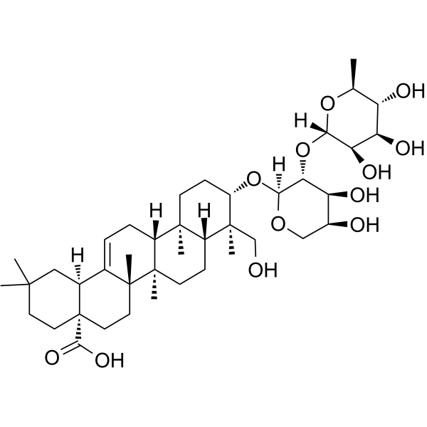
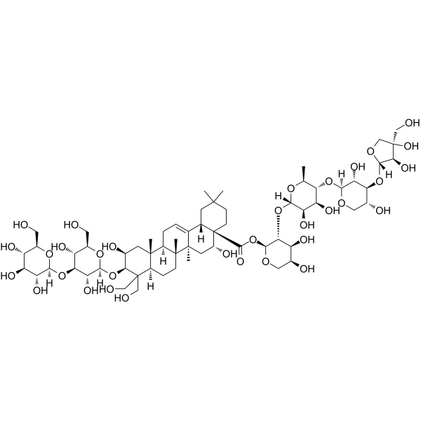


Biological Activity
Description
|
mTOR |
AMPK |
Caspase-9 |
Caspase 3 |
Bcl-2 |
Bax |
In Vitro
Macranthoside B (1.25-20 μM; 24-72 h) inhibits proliferation of human ovarian carcinoma A2780 cells in a time- and concentration-dependent manner, with IC50 values ranging from 7.52 μM to 14.66 μM, and exhibits lower cytotoxicity toward normal human embryonic lung fibroblast HFL1 cells[1].
Macranthoside B (5-20 μM; 6 h treatment, followed by 15 days of growth) dose-dependently inhibits clonogenicity of human ovarian carcinoma A2780 cells, with an IC50 of 8.46 μM[1].
Macranthoside B (5-20 μM; 48 h) dose-dependently activates AMPK and inhibits the mTOR/P70S6K pathway in human ovarian carcinoma A2780 cells[1].
Macranthoside B (2.55-80 μM; 48 h) potently inhibits the proliferation of human HepG2, MGC-803, MCF-7, SW1116, U251, rat C6, and mouse B16F1, B16F10 cancer cells in vitro, with IC50 values ranging from 10 μM to 18 μM[2].
Macranthoside B (0-40 μM; 24, 48 h) inhibits the viability of SKGT-4, OE-19, and OE-33 AEG cells in a concentration-dependent manner, with 48 h IC50 values ranging from 4.74 to 8.59 μM, and is less potent against normal NGEC cells[3].
Macranthoside B (8-10 μM; 24 h) inhibits the colony-forming ability of SKGT-4 and OE-19 AEG cells in a concentration-dependent manner, with significant suppression at 8 μM and 10 μM[3].
Macranthoside B (10-15 μM; 48 h) inhibits the proliferation of SKGT-4 and OE-19 AEG cells in a concentration-dependent manner, with significant reduction in EDU-positive cells at 10 μM and 15 μM[3].
Macranthoside B (8-10 μM; 36 h) inhibits the migration and invasion of SKGT-4 and OE-19 AEG cells in a concentration-dependent manner, with significant suppression at 8 μM and 10 μM[3].
Macranthoside B (5-20 μM; 24-48 h) dose-dependently induces apoptosis in human ovarian carcinoma A2780 cells, and this apoptosis is reduced by pretreatment with the autophagy inhibitor 3-Methyladenine (HY-19312) or the pan-caspase inhibitor Z-VAD-FMK (HY-16658B)[1].
Macranthoside B (5-20 μM; 48 h) dose-dependently upregulates pro-apoptotic markers (cleaved caspase-3, cleaved caspase-9, cleaved PARP) and the autophagy marker LC3-II in human ovarian carcinoma A2780 cells; inhibition of autophagy reduces apoptosis, while inhibition of apoptosis enhances autophagy, and inhibition of ROS or AMPK reduces both apoptosis and autophagy[1].
Macranthoside B (20 μM; 48 h) induces apoptotic nuclear morphological changes in human ovarian carcinoma A2780 cells after 48 h of incubation[1].
Macranthoside B (10-20 μM) dose-dependently induces autophagic puncta formation in GFP-LC3-expressing human ovarian carcinoma A2780 cells[1].
Macranthoside B (15 μM; 48 h) activates autophagy in SKGT-4 and OE-19 AEG cells, increasing LC3B puncta, autophagic vacuoles, and LC3B II/P62 protein levels, while impairing autophagic flux[3].
Macranthoside B (10 μM; 24-48 h) increases intracellular ROS levels in human ovarian carcinoma A2780 cells[1].
Macranthoside B (30-40 μM; 4 h) induces morphological changes characteristic of early apoptosis, including cell rounding, chromatin condensation, and karyopyknosis, in human hepatoma HepG2 cells[2].
Macranthoside B (40 μM; 4 h) induces apoptosis in human hepatoma HepG2 cells, with 39.02% of cells entering early apoptosis and 13.45% entering late apoptosis/necrosis[2].
Macranthoside B (30-40 μM; 4 h) induces apoptosis in human hepatoma HepG2 cells via a mitochondrion-mediated pathway, activating caspase-9 and caspase-3, cleaving PARP, increasing Bax protein expression, decreasing Bcl-2 protein expression, and elevating the Bax/Bcl-2 ratio[2].
Macranthoside B (15 μM; 48 h) treatment of SKGT-4 AEG cells alters the expression of 1800+ genes, upregulating pathways related to oxidoreductase activity, ferroptosis, and autophagy[3].
Macranthoside B (10-15 μM; 48 h) induces concentration-dependent accumulation of lipid reactive oxygen species in SKGT-4 and OE-19 AEG cells, with significant increases at 10 μM and 15 μM[3].
Macranthoside B (0-15 μM; 0-48 h) downregulates GPX4 protein levels in SKGT-4 and OE-19 AEG cells in both concentration- and time-dependent manners, reducing levels to as low as 0.1 and 0.5 of control, respectively[3].
Macranthoside B (0-15 μM; 0-48 h) upregulates NCOA4 and FTH1 protein levels in SKGT-4 and OE-19 AEG cells in both concentration- and time-dependent manners, activating NCOA4-mediated ferritinophagy[3].
Macranthoside B (0-15 μM; 48 h) inhibits NRF2 in SKGT-4 and OE-19 AEG cells, downregulating NRF2 protein levels and its downstream target genes HERC2 and VAMP8, disrupting iron homeostasis[3].
Macranthoside B (48 h) disrupts iron homeostasis in SKGT-4 and OE-19 AEG cells, significantly increasing intracellular Fe2+ levels[3].
Macranthoside B enhances Paclitaxel (HY-B0015)-mediated apoptosis in human cervical adenocarcinoma HeLa cells via ROS overgeneration and activation of the MAPKs/JNK pathway[4].
MedChemExpress (MCE) has not independently confirmed the accuracy of these methods. They are for reference only. Further protocols information, click here.
-
Cell Line:Human ovarian carcinoma A2780 cells, human embryonic lung fibroblast HFL1 cells
-
Concentration:1.25 μM, 2.5 μM, 5 μM, 1 μM, 20 μM
-
Incubation Time:24 h, 48 h, 72 h
-
Result:Inhibited A2780 cell viability in a concentration- and time-dependent manner, with IC50 values of 14.66 μM (24 h), 11.23 μM (48 h), 7.52 μM (72 h).
Exhibited IC50 values of 59.58 μM (24 h), 42.34 μM (48 h), 38.44 μM (72 h) in HFL1 cells, showing greater cytotoxicity toward tumor cells than normal cells.
-
Cell Line:human ovarian carcinoma A2780 cells
-
Concentration:5 μM, 10 μM, 20 μM (48 h incubation); 10 μM (48 h incubation with inhibitor pretreatment or beclin 1 siRNA transfection)
-
Incubation Time:48 h
-
Result:Increased levels of cleaved caspase-3, cleaved caspase-9, cleaved PARP, and LC3-II in a concentration-dependent manner at 5, 10, 20 μM for 48 h (cleaved caspase-3 levels relative to β-actin: 4.4, 20.0, 24.7; cleaved caspase-9: 10.5, 14.9, 23.5; cleaved PARP: 2.2, 3.4, 4.7; LC3-II: 3.4, 12.4, 24.9).
Decreased levels of cleaved caspase-3 (from 10.0 to 8.5), cleaved caspase-9 (from 2.0 to 1.8), cleaved PARP (from 1.0 to 0.5), and LC3-II (from 1.0 to 0.23) when cells were pretreated with 5 mM 3-methyladenine prior to 10 μM treatment.
Decreased LC3-II levels (from 1.0 to 0.19) and cleaved PARP levels (from 1.0 to 0.05) when cells were transfected with beclin 1 siRNA prior to 10 μM treatment.
Decreased levels of cleaved caspase-3 (from 2.1 to 0.2), cleaved caspase-9 (from 6.5 to 2.7), cleaved PARP (from 4.8 to 1.9), but increased LC3-II levels (from 2.0 to 3.8) when cells were pretreated with 10 μM Z-VAD-FMK prior to 10 μM treatment.
Decreased levels of cleaved caspase-3 (from 2.2 to 0.8), cleaved caspase-9 (from 1.7 to 1.5), cleaved PARP (from 1.6 to 0.7), and LC3-II (from 5.0 to 2.5) when cells were pretreated with 5 mM NAC prior to 10 μM treatment.
Decreased levels of cleaved caspase-3 (from 1.8 to 0.7) and LC3-II (from 1.6 to 0.7) when cells were pretreated with 10 μM Compound C prior to 10 μM treatment.
-
Cell Line:human ovarian carcinoma A2780 cells
-
Concentration:5 μM, 10 μM, 20 μM (48 h incubation); 10 μM (48 h incubation with inhibitor pretreatment)
-
Incubation Time:48 h
-
Result:Increased p-AMPK levels relative to β-actin (13.0, 18.9, 23.9) and decreased p-mTOR (0.9, 0.8, 0.6) and p-P70S6K (0.6, 0.4, 0.1) levels in a concentration-dependent manner at 5, 10, 20 μM for 48 h.
Decreased p-AMPK (from 2.4 to 1.1), p-mTOR (from 0.4 to 0.9), and p-P70S6K (from 0.5 to 1.0) levels when cells were pretreated with 5 mM NAC prior to 10 μM treatment.
Decreased p-AMPK (from 5.1 to 0.3), p-mTOR (from 0.3 to 0.9), and p-P70S6K (from 0.4 to 0.6) levels when cells were pretreated with 10 μM Compound C prior to 10 μM treatment.
-
Cell Line:human hepatoma HepG2, human gastric cancer MGC-803, human mammary adenocarcinoma MCF-7, human colonic carcinoma SW1116, human glioblastoma U251, rat glioma C6, mouse melanoma B16F1, mouse melanoma B16F10
-
Concentration:2.55-80 μM; 4.25-48 μM (HepG2 cells)
-
Incubation Time:48 h
-
Result:Inhibited HepG2 cell viability with inhibitory rates of 2.58%, 23.21%, 55.89%, 86.55%, and 98.14% at concentrations of 4.25, 7.08, 14.75, 23.04, and 48 μM respectively, with an IC50 of 10 μM.
Inhibited viability of all tested cell lines with IC50 values: MGC-803 (16 μM), MCF-7 (12 μM), SW1116 (14 μM), U251 (18 μM), C6 (14 μM), B16F1 (15 μM), and B16F10 (16 μM).
-
Cell Line:Human hepatoma HepG2
-
Concentration:40 μM
-
Incubation Time:4 h
-
Result:Increased the percentage of early apoptotic cells to 39.02% and the percentage of late apoptotic/necrotic cells to 13.45%, compared to control cells.
-
Cell Line:human hepatoma HepG2
-
Concentration:30 μM, 40 μM
-
Incubation Time:4 h
-
Result:Increased cleaved PARP levels in a concentration-dependent manner.
Decreased procaspase-9 expression significantly, decreased procaspase-3 expression slightly, and increased levels of activated caspase-3 p17 and p12 subunits.
Increased Bax protein levels by 82% and 113% at 30 μM and 40 μM respectively, decreased Bcl-2 protein levels slightly, resulting in a notable increase in the Bax/Bcl-2 ratio.
-
Cell Line:normal gastric epithelial cell line (NGEC), adenocarcinoma of the esophagogastric junction (AEG) cell lines SKGT-4, OE-19, OE-33
-
Concentration:0, 5, 10, 20, 30, 40 μM
-
Incubation Time:24 h; 48 h
-
Result:Inhibited AEG cell viability in a concentration-dependent manner, with normal NGEC cells showing lower sensitivity.
Exhibited 24 h IC50 values of 9.51 μM (SKGT-4), 12.70 μM (OE-19), 10.98 μM (OE-33), and 22.80 μM (NGEC).
Exhibited 48 h IC50 values of 8.59 μM (SKGT-4), 8.47 μM (OE-19), 4.74 μM (OE-33), and 11.69 μM (NGEC).
-
Cell Line:adenocarcinoma of the esophagogastric junction (AEG) cell lines SKGT-4, OE-19
-
Concentration:0, 8, 10 μM
-
Incubation Time:24 h, followed by 15-20 days of drug-free growth
-
Result:Reduced the number of colonies in SKGT-4 and OE-19 cells in a concentration-dependent manner.
Caused statistically significant decreases (***p < 0.001) at both 8 μM and 10 μM compared to the control.
-
Cell Line:adenocarcinoma of the esophagogastric junction (AEG) cell lines SKGT-4, OE-19
-
Concentration:0, 10, 15 μM
-
Incubation Time:48 h
-
Result:Reduced the EDU-positive cell rate in SKGT-4 and OE-19 cells in a concentration-dependent manner.
Caused statistically significant decreases (**p < 0.01, ***p < 0.001) at 10 μM and 15 μM compared to the control.
-
Cell Line:adenocarcinoma of the esophagogastric junction (AEG) cell lines SKGT-4, OE-19
-
Concentration:0, 5, 10, 15 μM
-
Incubation Time:0, 12, 24, 36, 48 h
-
Result:Reduced GPX4 protein levels in SKGT-4 and OE-19 cells in both concentration- and time-dependent manners.
Reduced GPX4 levels to 0.1 of control at 15 μM, and to 0.3 of control after 48 h in SKGT-4 cells.
Reduced GPX4 levels to 0.8 of control at 15 μM, and to 0.5 of control after 48 h in OE-19 cells.\nUpregulated NCOA4 and FTH1 protein levels in SKGT-4 and OE-19 cells in both concentration- and time-dependent manners.
Increased NCOA4 levels to 1.09 of control at 15 μM and 1.1 of control after 48 h; increased FTH1 levels to 1.1 of control at 15 μM and 1.3 of control after 48 h in SKGT-4 cells.
Increased NCOA4 levels to 2.5 of control at 15 μM and 1.1 of control after 48 h; increased FTH1 levels to 1.1 of control at 15 μM and 1.4 of control after 48 h in OE-19 cells.
In Vivo
MedChemExpress (MCE) has not independently confirmed the accuracy of these methods. They are for reference only.
-
Animal Model:Athymic BALB/cA nude mice (female, 35-40 days old, 18-22 g, subcutaneous implantation of HepG2 cells)[2]
-
Dosage:5 mg/kg
-
Administration:i.v.; every two days; 14 days
-
Result:Achieved 53.29% inhibition of tumor growth compared to vehicle control.
Significantly reduced relative tumor volume.
Lowered excised tumor weights compared to vehicle control.


Chemical Information
-
CAS No. 146100-02-9
-
Appearance Solid
-
Molecular Weight 1075.24
-
Formula C53H86O22
-
Color White to off-white
-
SMILES
C[C@@]12C([C@@]3([H])[C@](C(O)=O)(CCC(C)(C)C3)CC2)=CC[C@@]4([H])[C@]1(CC[C@]5([H])[C@@]4(CC[C@H](O[C@@]6([H])[C@@H]([C@H]([C@@H](O)CO6)O)O[C@@]7([H])[C@@H]([C@@H]([C@@H](O)[C@H](C)O7)O[C@]8([H])O[C@@H]([C@@H](O[C@]9([H])O[C@@H]([C@@H](O)[C@H](O)[C@H]9O)CO)[C@H](O)[C@H]8O)CO)O)[C@@]5(C)CO)C)C
-
Structure Classification
-
Initial Source
-
Shipping
Room temperature in continental US; may vary elsewhere.
-
Storage
4°C, sealed storage, away from moisture and light
* In solvent : -80°C, 6 months; -20°C, 1 month (sealed storage, away from moisture and light)


Solvent & Solubility
In Vitro:
DMSO : 100 mg/mL (93.00 mM; Need ultrasonic; Hygroscopic DMSO has a significant impact on the solubility of product, please use newly opened DMSO)
Please refer to the solubility information to select the appropriate solvent. Once prepared, please aliquot and store the solution to prevent product inactivation from repeated freeze-thaw cycles.
Storage method and period of stock solution: -80°C, 6 months; -20°C, 1 month (sealed storage, away from moisture and light). When stored at -80°C, please use it within 6 months. When stored at -20°C, please use it within 1 month.
Please refer to the solubility information to select the appropriate solvent. Once prepared, please aliquot and store the solution to prevent product inactivation from repeated freeze-thaw cycles.
Storage method and period of stock solution: -80°C, 6 months; -20°C, 1 month (sealed storage, away from moisture and light). When stored at -80°C, please use it within 6 months. When stored at -20°C, please use it within 1 month.
Concentration (start) × Volume (start) = Concentration (final) × Volume (final)

Protocols
-
Western Blot
Western blotting (WB) is a commonly used experimental method in molecular biology, biochemistry, and immunogenetics for identifying and quantifying target proteins. It combines gel electrophoresis with immunoassay, enabling researchers to analyze protein expression, post-translational modifications, and molecular weight.
-
RNA extraction experimental
By lysing cells, releasing RNA, and removing impurities such as proteins and DNA, high-purity RNA products are finally obtained. The commonly used traditional method is the guanidine isothiocyanate/phenol/chloroform method (Trizol), which is suitable for a variety of animal materials including animal tissues, microorganisms, cultured cells, etc., and most plant materials.
-
Mitochondrial membrane-potential fluorescent assay
Mitochondrial membrane potential fluorescent assays estimate ΔΨm in living cells using lipophilic cationic dyes such as TMRM, TMRE, rhodamine 123, and JC-1, which accumulate in mitochondria according to membrane polarization; loss of signal after FCCP or CCCP treatment is interpreted as mitochondrial depolarization. TMRM/TMRE and rhodamine 123 are commonly used for semi-quantitative live-cell microscopy or flow cytometry, while JC-1 can report a shift from red aggregate fluorescence to green monomer fluorescence during depolarization; interpretation requires controls because dye concentration, quenching mode, cell type, dye efflux, and mitochondrial mass can affect fluorescence independently of ΔΨm.
-
Annexin V plus membrane-impermeant dye apoptosis staining
Annexin V-based apoptosis assays rely on the detection of phosphatidylserine (PS) externalization from the inner leaflet of the plasma membrane to the outer leaflet, an early biochemical hallmark of apoptosis. Fluorescently labeled Annexin V binds PS in a calcium-dependent manner, enabling identification of early apoptotic cells by flow cytometry or fluorescence microscopy. When combined with a membrane-impermeant DNA-binding dye (e. g. , propidium iodide), this approach allows discrimination between viable (Annexin V−/dye−), early apoptotic (Annexin V+/dye−), and late apoptotic or necrotic (Annexin V+/dye+) cell populations by assessing membrane integrity and PS exposure.
-
ROS/oxidative-stress fluorescent staining
ROS/oxidative-stress fluorescent staining uses cell-permeant fluorogenic probes that become fluorescent after oxidation inside cells or tissues; commonly used examples include DCFH-DA/DCFDA for broad cellular oxidant detection, DHE for superoxide-related signal detection, MitoSOX for mitochondrial superoxide-related signal detection, and CellROX probes for oxidative-stress-associated fluorescence readouts. The assay detects probe oxidation rather than a single ROS species unless the probe and analysis method have been chemically validated for that species. DCFH-DA enters cells, is deacetylated by intracellular esterases to DCFH, and produces fluorescent DCF after oxidation, so the readout is used as an operational measure of total cellular oxidative stress rather than a species-specific ROS measurement. DHE and MitoSOX can report superoxide-related oxidation, but red fluorescence alone can include non-specific ethidium-like oxidation products; HPLC or optimized spectral approaches are
-
Liver Cancer Modeling
Liver cancer can be classified into primary liver cancer and secondary liver cancer. Secondary liver cancer is the metastatic liver cancer. Primary liver cancer includes hepatocellular carcinoma (HCC), intrahepatic cholangiocarcinoma (ICC) and fibrolamellar HCC, of which HCC is the most common form, accounting for approximately 90% of primary liver cancers[1]. HCC mouse models include chemical agent-induced models, transplanted tumor models, and genetic engineered models.
-
Breast Cancer Modeling
Breast cancer is a heterogeneous cancer, and it has been distinguished into four subtypes: luminal A, luminal B, HER2-positive and basal-like. Molecular mutations, epigenetic alterations, hormone exposure and immune microenvironment are related to the progression of breast cancer.
-
Kinase activity and phosphorylation assays
Kinase activity assays measure the ability of kinases to transfer phosphate groups from ATP to specific substrates, while phosphorylation assays detect the presence and levels of phosphorylated proteins. Common methods include radiolabeled ATP incorporation (e. g. ,), ADP release detection via bioluminescence (e. g. ,[3]), enzyme-linked immunosorbent assays (ELISA) for phospho-specific epitopes (e. g. ,[6]), and microtiter-based formats for high-throughput screening (e. g. ,[8]). The ADP-Glo assay quantifies kinase activity by measuring ADP produced during phosphorylation using a luciferase-based system. Radiometric assays involve autoradiography or scintillation counting after incorporation of 32P-labeled ATP into substrate proteins. ELISA-based approaches rely on phospho-specific antibodies to detect activated kinases in cell lysates or purified samples.
-
Detection of Apoptosis
Apoptosis, also called programmed cell death, is generally characterized by distinct morphological characteristics.
-
TUNEL staining for apoptotic DNA fragmentation
TUNEL staining detects DNA strand breaks by using terminal deoxynucleotidyl transferase to add labeled nucleotides to exposed 3′-OH DNA termini, generating either microscopic staining in fixed cells or tissue sections, or fluorescence/cytometric signal in cell suspensions. TUNEL positivity reflects DNA fragmentation but should not be interpreted alone as definitive apoptosis, because TUNEL can also label necrotic, autolytic, mechanically damaged, or DNA-repair-associated DNA breaks.
-
Fluorescent plasma-membrane potential dye assay
Fluorescent plasma-membrane potential dye assays measure changes in cell membrane potential using voltage-sensitive dyes whose fluorescence changes when cells depolarize or hyperpolarize. Anionic bis-oxonol dyes such as DiBAC4(3) enter depolarized cells more readily and show increased fluorescence after intracellular binding, while hyperpolarization reduces dye accumulation and fluorescence. FMP/FLIPR membrane-potential dyes are used for faster, homogeneous microplate assays of ion-channel or receptor-mediated membrane-potential changes.
-
Autophagy
Autophagy is a process in which eukaryotic cells use lysosomes to degrade their own cytoplasmic proteins and damaged organelles under the regulation of autophagy related gene (Atg). Microtubule-associated proteins light chain 3 (LC3) is recognized as autophagy marker, which transfers from cytoplasmic LC3 (LC3-I) to membrane type (LC3-II). LC3-II/I ratio could be detected by Western Blot and fluorescence microscopy.
-
Mitochondrial membrane-potential and mitochondrial mass staining
Mitochondrial membrane potential staining measures the electrochemical polarization across the mitochondrial inner membrane in live cells using lipophilic cationic fluorescent probes; early rhodamine-based work showed that selective mitochondrial dye accumulation is lost when the mitochondrial transmembrane potential is dissipated. JC-1 reports mitochondrial polarization by shifting from green monomer fluorescence to red J-aggregate fluorescence as dye concentration increases within energized mitochondria; therefore, the red/green fluorescence ratio is used as a relative readout of mitochondrial membrane potential. TMRE or TMRM staining provides a single-channel relative readout because these cationic rhodamine esters accumulate in polarized mitochondria, and lower fluorescence indicates reduced mitochondrial polarization when acquisition and dye-loading conditions are controlled. Mitochondrial mass staining is commonly performed with MitoTracker Green FM or related MitoTracker dyes as
-
Apoptosis Solutions
Apoptosis is a regulated, generally non-lytic cell-death pathway that removes unwanted, damaged, infected, or abnormal cells through coordinated morphological changes, caspase activation, DNA fragmentation, and membrane remodeling. The intrinsic apoptosis pathway is controlled mainly by mitochondrial outer membrane permeabilization, BCL-2 family proteins, cytochrome c release, apoptosome formation, caspase-9 activation, and downstream executioner caspase-3/7 activation. The extrinsic apoptosis pathway is initiated by death receptors such as Fas, TNFR, and TRAIL receptors, which recruit adaptor proteins and activate caspase-8 before engaging executioner caspases or mitochondrial amplification through BID cleavage. Apoptosis is linked to many phenotypes, including cancer cell killing, tissue homeostasis, immune regulation, neurodegeneration, infection response, and treatment-induced cytotoxicity; unresolved questions include how apoptosis interacts with necroptosis, pyroptosis, ferroptos
-
Lysosome and acidic-vesicle live-cell staining
Lysosome and acidic-vesicle live-cell staining detects acidic intracellular compartments by using membrane-permeant acidotropic probes that accumulate in low-pH vesicles, including lysosomes, late endosomes, autolysosomes, and acidic phagosomes. LysoTracker staining is commonly used as an intensity-based readout of acidic lysosomal compartment abundance or enlargement, while acridine orange produces green fluorescence in less concentrated compartments and red fluorescence after concentration-dependent accumulation in acidic vesicular organelles. Loss or reduction of acridine-orange red signal can be used as a readout of lysosomal membrane permeabilization or reduced acidic-vesicle integrity. This protocol is designed for live cultured cells and can be adapted for fluorescence microscopy, high-content imaging, plate-reader readout, or flow cytometry when the selected literature supports the readout. Because these dyes report acidotropic accumulation rather than lysosome identity alone,
-
Ferroptosis Solutions
Ferroptosis is an iron-dependent, non-apoptotic form of regulated cell death characterized by lethal lipid peroxidation and sensitivity to suppression by iron chelators or lipophilic radical-trapping antioxidants. The core pathway links cystine uptake through system Xc−, glutathione availability, GPX4-dependent detoxification of phospholipid hydroperoxides, iron-dependent oxidative reactions, and polyunsaturated-phospholipid metabolism into a cell-death program that is biochemically and morphologically distinct from apoptosis, necrosis, and autophagy. The ferroptosis pathway is experimentally linked to phenotype through chemical and genetic perturbation. Erastin induces ferroptosis by inhibiting cystine uptake through system Xc− and weakening antioxidant defenses, while GPX4 inhibition or depletion causes lipid peroxide accumulation and ferroptotic cancer-cell death. ACSL4 and oxidizable arachidonoyl- or adrenoyl-containing phosphatidylethanolamines shape ferroptosis sensitivity by con
-
Protocol for Kinase activity and phosphorylation assays
Kinase activity assays measure transfer of phosphate from ATP to a protein or peptide substrate, generating phosphorylated substrate, ADP, or incorporated radiolabeled phosphate as the readout; phosphorylation assays measure site-specific phosphorylation in cells or tissues as a proxy for kinase-pathway activation, inhibition, or substrate regulation. Phosphorylation can be detected by phospho-specific Western blot, immunoprecipitation kinase assay, phospho-immunofluorescence, phospho-flow cytometry, luminescent ADP detection, radiolabeled ATP incorporation, or reporter-based pathway assays, and these readouts can be applied to cancer cells, primary neurons, mouse tumors, organoids, inflammatory macrophages, ferroptosis studies, and mitophagy studies when the kinase target is biologically relevant.
-
Macroautophagy Solutions
Macroautophagy is a conserved lysosome-dependent degradation pathway in which cytoplasmic material is sequestered into double-membrane autophagosomes and delivered to lysosomes for degradation and recycling. The pathway supports cellular homeostasis during nutrient limitation, organelle stress, protein-aggregate accumulation, infection, differentiation, and tissue remodeling by coupling cargo sequestration, autophagosome maturation, lysosomal fusion, and degradation of cargo-derived macromolecules. The core molecular sequence includes initiation by nutrient- and stress-regulated autophagy machinery, autophagosome nucleation, LC3/ATG8-family conjugation to autophagosomal membranes, cargo selection through receptors such as SQSTM1/p62, autophagosome-lysosome fusion, and lysosomal degradation. LC3 was identified as a mammalian homolog of yeast Atg8 that localizes to autophagosomal membranes after processing, and p62/SQSTM1 was shown to connect ubiquitinated cargo with autophagic degradati


Purity & Documentation
-
Data Sheet (311 KB)
-
SDS (252 KB)
- English - EN (252 KB)
- Français - FR (252 KB)
- Deutsch - DE (252 KB)
- Norwegian - NO (252 KB)
- Español - ES (252 KB)
- Swedish - SV (252 KB)
- Italian - IT (252 KB)
- Korean - KR (252 KB)
- Portuguese - PT (252 KB)
-
Handling Instructions (2659 KB)
References
[1]. Shan Y, et al. Macranthoside B Induces Apoptosis and Autophagy Via Reactive Oxygen Species Accumulation in Human Ovarian Cancer A2780 Cells. Nutrition and cancer. 2016;68(2):280-9. [Content Brief]
[2]. Wang J, et al. Macranthoside B, a hederagenin saponin extracted from Lonicera macranthoides and its anti-tumor activities in vitro and in vivo. Food and chemical toxicology : an international journal published for the British Industrial Biological Research Association. 2009 Jul;47(7):1716-21. [Content Brief]
[3]. Wang L, et al. Macranthoside B Suppresses the Growth of Adenocarcinoma of Esophagogastric Junction by Regulating Iron Homeostasis and Ferroptosis through NRF2 Inhibition. Current cancer drug targets. 2025;25(8):1013-1027. [Content Brief]
[4]. Li M, et al. Macranthoside B Enhances Paclitaxel-induced Human Cervical Cancer Cell Apoptosis Through ROS-JNK Pathway. Anticancer research. 2025 Jul;45(7):2859-2870. [Content Brief]


Complete Stock Solution Preparation Table
Please refer to the solubility information to select the appropriate solvent. Once prepared, please aliquot and store the solution to prevent product inactivation from repeated freeze-thaw cycles.
Storage method and period of stock solution: -80°C, 6 months; -20°C, 1 month (sealed storage, away from moisture and light). When stored at -80°C, please use it within 6 months. When stored at -20°C, please use it within 1 month.
| Optional Solvent | Concentration Solvent Mass | 1 mg | 5 mg | 10 mg | 25 mg |
|---|---|---|---|---|---|
| DMSO | 1 mM | 0.9300 mL | 4.6501 mL | 9.3002 mL | 23.2506 mL |
| 5 mM | 0.1860 mL | 0.9300 mL | 1.8600 mL | 4.6501 mL | |
| 10 mM | 0.0930 mL | 0.4650 mL | 0.9300 mL | 2.3251 mL | |
| 15 mM | 0.0620 mL | 0.3100 mL | 0.6200 mL | 1.5500 mL | |
| 20 mM | 0.0465 mL | 0.2325 mL | 0.4650 mL | 1.1625 mL | |
| 25 mM | 0.0372 mL | 0.1860 mL | 0.3720 mL | 0.9300 mL | |
| 30 mM | 0.0310 mL | 0.1550 mL | 0.3100 mL | 0.7750 mL | |
| 40 mM | 0.0233 mL | 0.1163 mL | 0.2325 mL | 0.5813 mL | |
| 50 mM | 0.0186 mL | 0.0930 mL | 0.1860 mL | 0.4650 mL | |
| 60 mM | 0.0155 mL | 0.0775 mL | 0.1550 mL | 0.3875 mL | |
| 80 mM | 0.0116 mL | 0.0581 mL | 0.1163 mL | 0.2906 mL |


Macranthoside B Related Classifications
HY-N5008 Related Classifications
 Powered by Bioz