



(+)-Magnoflorine chloride
Based on 2 publication(s) in Google Scholar
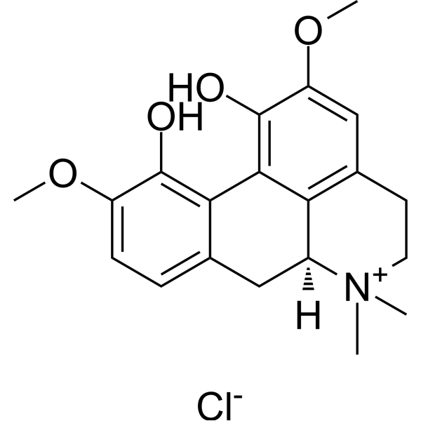
(+)-Magnoflorine (α-Magnoflorine) chloride is an orally active aporphine alkaloid with multiple biological activities. (+)-Magnoflorine chloride promotes Parkin/PINK1-mediated mitochondrial autophagy, inhibits the activation of NLRP3/Caspase-1 pathway, regulates the intestinal microbiota, and exhibits significant anti-inflammatory and immunomodulatory activities. (+)-Magnoflorine chloride inhibits JNK and TLR4/NF-κB signaling pathways, activates Sirt1/AMPK pathway, alleviates neuronal oxidative stress and apoptosis. Magnoflorine chloride upregulates miR-410-3p, inhibits HMGB1/NF-κB pathway, and has anti-tumor activity. (+)-Magnoflorine chloride also has significant antifungal activity.
For research use only. We do not sell to patients.
- Purity : 99.10%
- CAS No.: 6681-18-1
- Formula: C20H24ClNO4
- Molecular Weight:377.86
-
Storage:
4°C, sealed storage, away from moisture and light
* In solvent : -80°C, 6 months; -20°C, 1 month (sealed storage, away from moisture and light)

To place orders, for customer services and technical support, please contact: MedChemExpress USA
Tel: 609-228-6898 E-mail: [email protected] [email protected]


-
 Biological Activity
-
Chemical Information
-
Solvent & Solubility
- Protocols
- Purity & Documentation
- References
-
 Help & FAQs


-
HY-18258
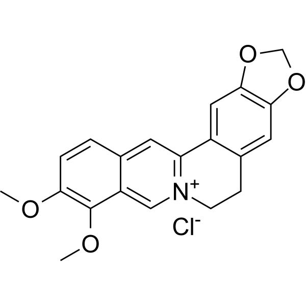
Berberine chlorideAntibiotic -
HY-N0740
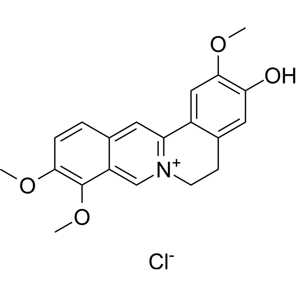
Jatrorrhizine chlorideBioactive Alkaloid -
HY-N0519
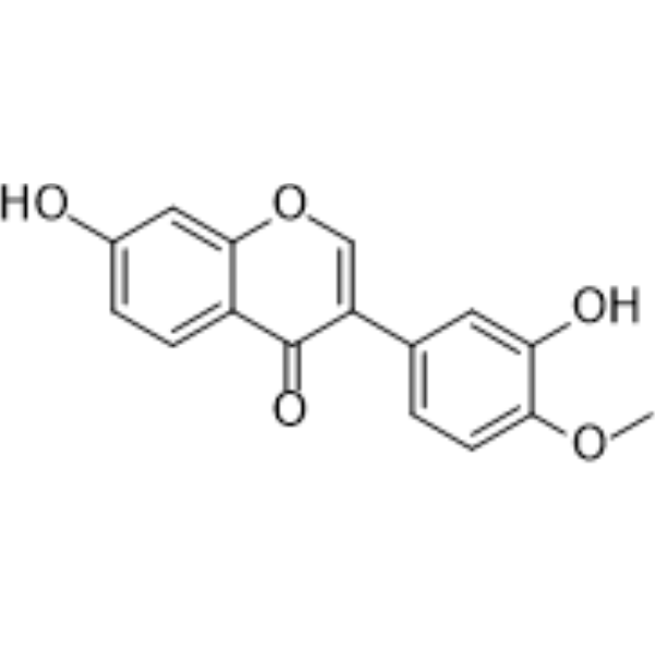
CalycosinPhytoestrogen -
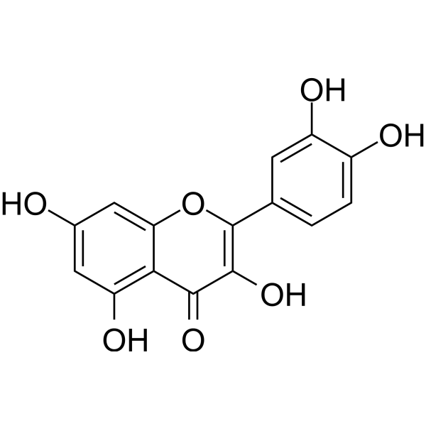
HY-18085
QuercetinFlavonoid Antioxidant -
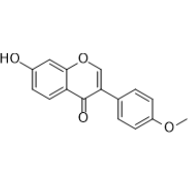
HY-N0183
FormononetinFGFR2 Inhibitor -
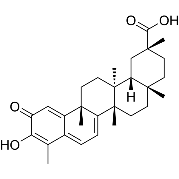
HY-13067
CelastrolAutophagy Inducer -
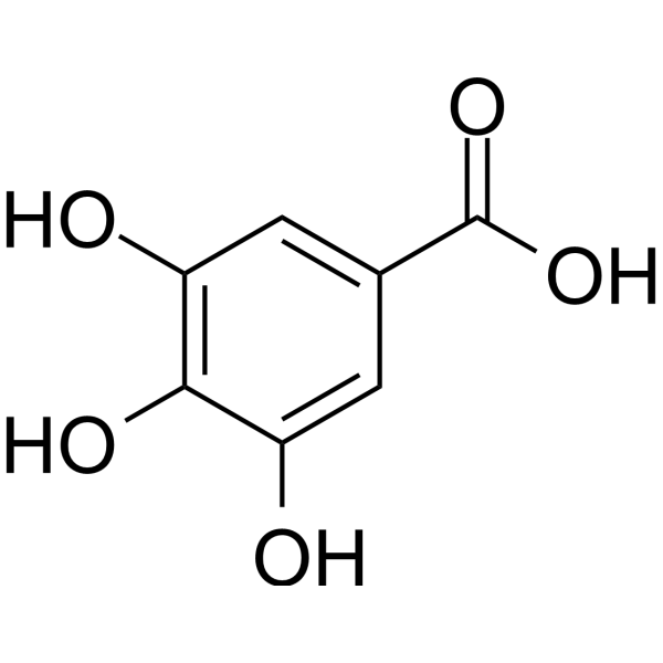
HY-N0523
Gallic acidBioactive Polyhydroxyphenolic Compound -
HY-N0110
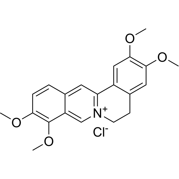
Palmatine chlorideIDO-1/NS2B-NS3 Inhibitor -
HY-N2072
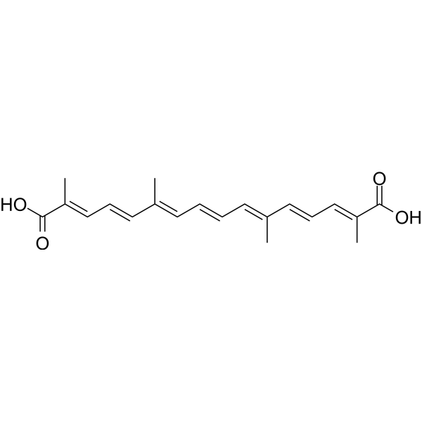
CrocetinAnticancer/Neuroprotective Agent -
HY-N0736
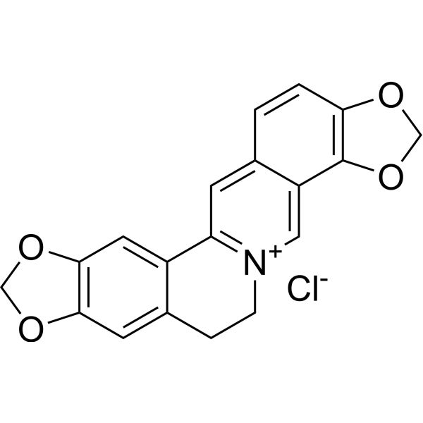
Coptisine chlorideBacterial Inhibitor -
HY-N0055
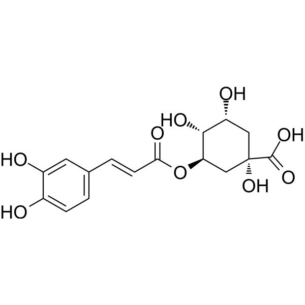
Chlorogenic acidAntioxidant -
HY-N0226A
Epiberberine chlorideAChE/BChE Inhibitor
Publications Citing Use of MedChemExpress (MCE) (+)-Magnoflorine chloride
MoreAll Caspase Isoforms
MoreAll AMPK Isoforms
More

Biological Activity
Description
In Vitro
(+)-Magnoflorine chloride has significant antifungal activities against Penicillium avellaneum UC-4376, T. rubrum and T. mentagrophyte with MIC values of 5 μg/disc, 62.5 μg/mL and 62.5 μg/mL, respectively[1][2].
(+)-Magnoflorine (5-80 μM, 24 h) chloride inhibit the proliferation, invasion and epithelial-mesenchymal transition process of MG-63 and U-2 OS cells[3].
(+)-Magnoflorine (10-40 μM, 24 h) chloride upregulates miR-410-3p and thereby inhibits the HMGB1/NF-κB pathway[3].
(+)-Magnoflorine (10 μM, 27 h) chloride significantly reverses the decline in PC12 cell viability caused by Aβ1-42and reduces the apoptosis rate[4].
(+)-Magnoflorine (10 μM, 27 h) chloride significantly reduces the increase in intracellular ROS levels caused by Aβ1-42, and restores the damaged mitochondrial membrane potential[4].
(+)-Magnoflorine (0-50 μM, 24 h) chloride inhibits the activation of the NLRP3 inflammasome in HK-2 cells by promoting Parkin-dependent mitochondrial autophagy[7].
MedChemExpress (MCE) has not independently confirmed the accuracy of these methods. They are for reference only. Further protocols information, click here.
-
Cell Line:MG-63 and U-2 OS cells
-
Concentration:10, 20 and 40 μM
-
Incubation Time:24 h
-
Result:Upregulated the mRNA expression of E-cadherin in a dose-dependent manner, and downregulate the expressions of N-cadherin and Vimentin.
Dose-dependently down-regulates the mRNA expression of HMGB1.
Inhibited the activation of the NF-κB pathway resulting in a decrease in the levels of phosphorylated-p65 and phosphorylated-IκBα.
Significantly upregulated the expression of miR-410-3p in osteosarcoma cells.
-
Cell Line:MG-63 and U-2 OS cells
-
Concentration:10, 20 and 40 μM
-
Incubation Time:24 h
-
Result:Significantly reduced the number of MG-63 and U-2 OS cells passing through the matrix gel.
-
Cell Line:MG-63 and U-2 OS cells
-
Concentration:10, 20 and 40 μM
-
Incubation Time:24 h
-
Result:Upregulated the protein expression of E-cadherin in a dose-dependent manner, and downregulate the expressions of N-cadherin and Vimentin.
Dose-dependently down-regulates the protein expression of HMGB1.
Inhibited the activation of the NF-κB pathway resulting in a decrease in the levels of phosphorylated-p65 and phosphorylated-IκBα.
Significantly upregulated the expression of miR-410-3p in osteosarcoma cells.
-
Cell Line:PC12 cells
-
Concentration:10 μM
-
Incubation Time:pre-treated for 3 h, and then add Aβ1-42 for co-culture for 24 h
-
Result:Significantly reduced the apoptosis rate caused by Aβ1-42.
Reduced the expression of pro-apoptotic proteins Bax and Cleaved Caspase-3, and increased the expression of anti-apoptotic protein Bcl-2.
In Vivo
(+)-Magnoflorine (10-20 mg/kg, i.g., once daily for 15 days) chloride has a protective effect on cerebral ischemia-reperfusion injury in rats[5].
(+)-Magnoflorine (10 mg/kg, i.g., once daily for 10 days) chloride relieves colitis induced by DDS (HY-116282C) in mice and the concurrent anxiety-like behaviors[6].
(+)-Magnoflorine (5-10 mg/kg, i.g., once daily for 12 weeks) chloride exhibits renal protective effects in a mouse model of chronic kidney disease induced by a high-fat and high-fructose diet[7].
MedChemExpress (MCE) has not independently confirmed the accuracy of these methods. They are for reference only.
-
Animal Model:AD model established in APP/PS1 (APPswe/PSEN1dE9) double-transgenic mice (8 months, 30-32 g, female)[4]
-
Dosage:1 and 10 mg/kg
-
Administration:i.p., once daily for a month
-
Result:Improved the learning and memory abilities of mice. Reduced the core pathological changes in the brain, including Aβ deposition, excessive phosphorylation of Tau protein, neuroinflammation and neuronal loss.
-
Animal Model:Middle cerebral artery occlusion (MCAO) models model established in male Sprague-Dawley rats (weighing 220 g)[5]
-
Dosage:10 and 20 mg/kg
-
Administration:i.g., once daily for 15 days
-
Result:Significantly reduced the neurological deficit score, indicating that it improved motor, balance and sensory functions.
Significantly reduced the volume of cerebral infarction and decreased the brain water content. Reduced the number of damaged neurons shown by Nissl staining and increased the density of NeuN-positive neurons.
Reduced the upregulation of LC3 fluorescence intensity and the LC3-II/LC3-I ratio caused by cerebral ischemia, and increased the expression of p62, Sirt1 and p-AMPK proteins.
-
Animal Model:Colitis models model established in female C57BL/6 J mice (6-8 weeks old)[6]
-
Dosage:10 mg/kg
-
Administration:i.g., once daily for 10 days
-
Result:Reduced the disease activity index, the length of the colon, the tissue pathological damage, and decreased the mRNA levels of pro-inflammatory cytokines (TNF-α, IL-1β, IL-6) in the colon tissue.
Significantly improved the anxiety-like behaviors induced by DSS. Inhibited the activation of microglia (IBA1+) in the hippocampus caused by DSS.
Reduced the mRNA levels of TNF-α, IL-1β, and IL-6 in the hippocampal tissue, and upregulated the expression of tight junction proteins ZO-1 and PV1 in the choroid plexus and blood-brain barrier.
Promoted the production of secondary bile acid HDCA by the microbiota.
-
Animal Model:Chronic kidney disease model established in male C57BL/6 J mice (20 g)[6]
-
Dosage:5 and 10 mg/kg
-
Administration:i.g., once daily for 12 weeks
-
Result:Significantly reduced the neurological deficit score, indicating that it improved motor, balance and sensory functions.
Significantly reduced the levels of serum creatinine, urea nitrogen, uric acid and elevated urine protein.
Reduced the fatty degeneration of renal tubules, lipid deposition and tubular dilation. Reduced the collagen deposition in the kidneys, and downregulated the mRNA and protein expressions of fibrosis-related markers (TGF-β, α-SMA, COL-I, COL-III).
Reduced the levels of reactive oxygen species (ROS) and malondialdehyde (MDA) in the kidneys, and increased the activity of total superoxide dismutase (T-SOD).
Inhibited the activation of the NLRP3 inflammasome (reduced the expression of NLRP3, ASC, Cleaved Caspase-1, GSDMD-N and IL-1β).


Chemical Information
-
CAS No. 6681-18-1
-
Appearance Solid
-
Molecular Weight 377.86
-
Formula C20H24ClNO4
-
Color Yellow to brown
-
SMILES
OC1=C(C2=C3O)C([C@]([N+]4(C)C)([H])CC2=CC=C3OC)=C(CC4)C=C1OC.[Cl-]
-
Synonyms
Magnoflorine chloride; α-Magnoflorine chloride; Thalictrine chloride
-
Structure Classification
-
Shipping
Room temperature in continental US; may vary elsewhere.
-
Storage
4°C, sealed storage, away from moisture and light
* In solvent : -80°C, 6 months; -20°C, 1 month (sealed storage, away from moisture and light)


Publications (2)


Solvent & Solubility
In Vitro:
H2O : 41.67 mg/mL (110.28 mM; ultrasonic and warming and heat to 60°C)
Please refer to the solubility information to select the appropriate solvent. Once prepared, please aliquot and store the solution to prevent product inactivation from repeated freeze-thaw cycles.
Storage method and period of stock solution: -80°C, 6 months; -20°C, 1 month (sealed storage, away from moisture and light). When stored at -80°C, please use it within 6 months. When stored at -20°C, please use it within 1 month.
* Note: If you choose water as the stock solution, please dilute it to the working solution, then filter and sterilize it with a 0.22 μm filter before use.
Please refer to the solubility information to select the appropriate solvent. Once prepared, please aliquot and store the solution to prevent product inactivation from repeated freeze-thaw cycles.
Storage method and period of stock solution: -80°C, 6 months; -20°C, 1 month (sealed storage, away from moisture and light). When stored at -80°C, please use it within 6 months. When stored at -20°C, please use it within 1 month.
* Note: If you choose water as the stock solution, please dilute it to the working solution, then filter and sterilize it with a 0.22 μm filter before use.
Concentration (start) × Volume (start) = Concentration (final) × Volume (final)
In Vivo:
For the following dissolution methods, please prepare the working solution directly:
It is recommended to prepare fresh solutions and use them promptly within a short period of time.
The percentages shown for the solvents indicate their volumetric ratio in the final prepared solution. If precipitation or phase separation occurs during preparation, heat and/or sonication can be used to aid dissolution.
Add each solvent one by one: PBS
Solubility: 100 mg/mL (264.65 mM); Clear solution; Need ultrasonic

Protocols
-
Apoptosis
Apoptosis, also called programmed cell death, is generally characterized by distinct morphological characteristics.
-
TUNEL staining for apoptotic DNA fragmentation
TUNEL staining detects DNA strand breaks by using terminal deoxynucleotidyl transferase to add labeled nucleotides to exposed 3′-OH DNA termini, generating either microscopic staining in fixed cells or tissue sections, or fluorescence/cytometric signal in cell suspensions. TUNEL positivity reflects DNA fragmentation but should not be interpreted alone as definitive apoptosis, because TUNEL can also label necrotic, autolytic, mechanically damaged, or DNA-repair-associated DNA breaks.
-
Annexin V plus membrane-impermeant dye apoptosis staining
Annexin V-based apoptosis assays rely on the detection of phosphatidylserine (PS) externalization from the inner leaflet of the plasma membrane to the outer leaflet, an early biochemical hallmark of apoptosis. Fluorescently labeled Annexin V binds PS in a calcium-dependent manner, enabling identification of early apoptotic cells by flow cytometry or fluorescence microscopy. When combined with a membrane-impermeant DNA-binding dye (e. g. , propidium iodide), this approach allows discrimination between viable (Annexin V−/dye−), early apoptotic (Annexin V+/dye−), and late apoptotic or necrotic (Annexin V+/dye+) cell populations by assessing membrane integrity and PS exposure.
-
ROS/oxidative-stress fluorescent staining
ROS/oxidative-stress fluorescent staining uses cell-permeant fluorogenic probes that become fluorescent after oxidation inside cells or tissues; commonly used examples include DCFH-DA/DCFDA for broad cellular oxidant detection, DHE for superoxide-related signal detection, MitoSOX for mitochondrial superoxide-related signal detection, and CellROX probes for oxidative-stress-associated fluorescence readouts. The assay detects probe oxidation rather than a single ROS species unless the probe and analysis method have been chemically validated for that species. DCFH-DA enters cells, is deacetylated by intracellular esterases to DCFH, and produces fluorescent DCF after oxidation, so the readout is used as an operational measure of total cellular oxidative stress rather than a species-specific ROS measurement. DHE and MitoSOX can report superoxide-related oxidation, but red fluorescence alone can include non-specific ethidium-like oxidation products; HPLC or optimized spectral approaches are
-
Autophagy
Autophagy is a process in which eukaryotic cells use lysosomes to degrade their own cytoplasmic proteins and damaged organelles under the regulation of autophagy related gene (Atg). Microtubule-associated proteins light chain 3 (LC3) is recognized as autophagy marker, which transfers from cytoplasmic LC3 (LC3-I) to membrane type (LC3-II). LC3-II/I ratio could be detected by Western Blot and fluorescence microscopy.
-
Apoptosis Solutions
Apoptosis is a regulated, generally non-lytic cell-death pathway that removes unwanted, damaged, infected, or abnormal cells through coordinated morphological changes, caspase activation, DNA fragmentation, and membrane remodeling. The intrinsic apoptosis pathway is controlled mainly by mitochondrial outer membrane permeabilization, BCL-2 family proteins, cytochrome c release, apoptosome formation, caspase-9 activation, and downstream executioner caspase-3/7 activation. The extrinsic apoptosis pathway is initiated by death receptors such as Fas, TNFR, and TRAIL receptors, which recruit adaptor proteins and activate caspase-8 before engaging executioner caspases or mitochondrial amplification through BID cleavage. Apoptosis is linked to many phenotypes, including cancer cell killing, tissue homeostasis, immune regulation, neurodegeneration, infection response, and treatment-induced cytotoxicity; unresolved questions include how apoptosis interacts with necroptosis, pyroptosis, ferroptos
-
Research Protocol for Microbiome Analysis
Microbiome analysis characterizes microbial communities in biological or environmental samples by measuring community composition, diversity, taxonomic structure, functional potential, and associations with host or environmental phenotypes. 16S rRNA gene amplicon sequencing is commonly used for bacterial and archaeal taxonomic profiling, while shotgun metagenomics provides higher taxonomic resolution and direct functional information, including microbial genes, pathways, viruses, fungi, and antimicrobial-resistance genes when sequencing depth and host-DNA contamination are adequately controlled. Microbiome results are strongly affected by sample collection, storage, DNA extraction, contamination, sequencing method, reference database, and bioinformatic pipeline; therefore, standardized protocols, negative controls, mock communities, and transparent analysis workflows are required. Unresolved issues include low-biomass contamination, compositional-data bias, inconsistent species-level c
-
Lysosome and acidic-vesicle live-cell staining
Lysosome and acidic-vesicle live-cell staining detects acidic intracellular compartments by using membrane-permeant acidotropic probes that accumulate in low-pH vesicles, including lysosomes, late endosomes, autolysosomes, and acidic phagosomes. LysoTracker staining is commonly used as an intensity-based readout of acidic lysosomal compartment abundance or enlargement, while acridine orange produces green fluorescence in less concentrated compartments and red fluorescence after concentration-dependent accumulation in acidic vesicular organelles. Loss or reduction of acridine-orange red signal can be used as a readout of lysosomal membrane permeabilization or reduced acidic-vesicle integrity. This protocol is designed for live cultured cells and can be adapted for fluorescence microscopy, high-content imaging, plate-reader readout, or flow cytometry when the selected literature supports the readout. Because these dyes report acidotropic accumulation rather than lysosome identity alone,
-
Research Protocol for Inflammation-related Diseases
The NLRP3 inflammasome is a cytosolic innate immune signaling platform that integrates priming signals and danger-signal activation to promote caspase-1 activation, maturation of IL-1β and IL-18, and gasdermin D-mediated pyroptotic cell death. The core experimental logic is to determine whether inflammatory disease phenotypes are driven by increased NLRP3 expression, ASC-containing inflammasome assembly, caspase-1 cleavage, GSDMD cleavage, and extracellular release of IL-1β/IL-18 rather than by nonspecific cell injury alone. The pathway is strongly linked to inflammation-related disease phenotypes because monosodium urate crystals activate NALP3/NLRP3 inflammasome signaling in gout-like crystal inflammation, cholesterol crystals activate NLRP3 inflammasomes in atherogenesis models, and DSS-induced intestinal inflammation has been reported to involve NLRP3 inflammasome activity. However, experimental colitis studies also show context-dependent protective effects of NLRP3 inflammasome co
-
Mitophagy Solutions
Mitophagy is the selective autophagic degradation of mitochondria and functions as a mitochondrial quality-control pathway that removes damaged, depolarized, excess, or developmentally programmed mitochondria. The pathway links mitochondrial damage recognition, autophagosome recruitment, lysosomal delivery, and mitochondrial turnover to phenotypes such as mitochondrial homeostasis, oxidative-stress control, metabolic remodeling, differentiation, and neurodegeneration-related mitochondrial fidelity. The best-characterized damage-induced pathway is the PINK1-Parkin axis. Parkin is recruited selectively to impaired mitochondria and promotes their autophagic elimination, while mitochondrial depolarization stabilizes PINK1 on damaged mitochondria, recruits Parkin, and activates Parkin-dependent mitophagy. PINK1 also phosphorylates ubiquitin to activate Parkin E3 ubiquitin ligase activity, and PINK1-driven ubiquitin phosphorylation creates a feed-forward signal for recruiting autophagy machi
-
Macroautophagy Solutions
Macroautophagy is a conserved lysosome-dependent degradation pathway in which cytoplasmic material is sequestered into double-membrane autophagosomes and delivered to lysosomes for degradation and recycling. The pathway supports cellular homeostasis during nutrient limitation, organelle stress, protein-aggregate accumulation, infection, differentiation, and tissue remodeling by coupling cargo sequestration, autophagosome maturation, lysosomal fusion, and degradation of cargo-derived macromolecules. The core molecular sequence includes initiation by nutrient- and stress-regulated autophagy machinery, autophagosome nucleation, LC3/ATG8-family conjugation to autophagosomal membranes, cargo selection through receptors such as SQSTM1/p62, autophagosome-lysosome fusion, and lysosomal degradation. LC3 was identified as a mammalian homolog of yeast Atg8 that localizes to autophagosomal membranes after processing, and p62/SQSTM1 was shown to connect ubiquitinated cargo with autophagic degradati
-
Pyroptosis Solutions
Pyroptosis is a lytic inflammatory cell-death pathway executed by gasdermin pores, most classically through inflammasome-mediated activation of caspase-1, cleavage of gasdermin D, membrane pore formation, LDH release, and secretion of IL-1β and IL-18. The canonical pathway is commonly modeled by priming cells with an inflammatory signal such as LPS to induce pro-IL-1β and inflammasome components, followed by an activation signal such as ATP or nigericin to activate NLRP3, ASC speck formation, caspase-1 cleavage, GSDMD cleavage, cytokine release, and pyroptotic membrane rupture. The non-canonical pathway is triggered when cytosolic LPS activates mouse caspase-11 or human caspase-4/5, leading to GSDMD cleavage and pyroptosis, and this can secondarily activate NLRP3-dependent IL-1β release. Pyroptosis is linked to inflammatory injury, infection, cancer, liver disease, ocular disease, placental inflammation, and other disease phenotypes, but unresolved questions include which gasdermin fam


Purity & Documentation
-
Data Sheet (295 KB)
-
SDS (393 KB)
- English - EN (393 KB)
- Français - FR (393 KB)
- Deutsch - DE (393 KB)
- Norwegian - NO (393 KB)
- Español - ES (393 KB)
- Swedish - SV (393 KB)
- Italian - IT (393 KB)
- Korean - KR (393 KB)
- Portuguese - PT (393 KB)
-
Handling Instructions (2659 KB)
References
[1]. Chen JH, et al. Aporphine alkaloids from Clematis parviloba and their antifungal activity. Arch Pharm Res. 2009 Jan;32(1):3-5. [Content Brief]
[2]. Luo N, et al. Antifungal activity and potential mechanism of magnoflorine against Trichophyton rubrum. J Antibiot (Tokyo). 2021 Mar;74(3):206-214. [Content Brief]
[3]. Wang Y, et al. Magnoflorine inhibits the malignant phenotypes and increases cisplatin sensitivity of osteosarcoma cells via regulating miR-410-3p/HMGB1/NF-κB pathway. Life Sci. 2020 Sep 1;256:117967. [Content Brief]
[4]. Zhong L, et al. Magnoflorine improves cognitive deficits and pathology of Alzheimer's disease via inhibiting of JNK signaling pathway. Phytomedicine. 2023 Apr;112:154714. [Content Brief]
[5]. Liang H, et al. Magnoflorine Attenuates Cerebral Ischemia-Induced Neuronal Injury via Autophagy/Sirt1/AMPK Signaling Pathway. Evid Based Complement Alternat Med. 2022 Sep 10;2022:2131561. [Content Brief]
[6]. Wang L, et al. Magnoflorine alleviates colitis-induced anxiety-like behaviors by regulating gut microbiota and microglia-mediated neuroinflammation. Microbiome. 2025 Jul 26;13(1):172. [Content Brief]
[7]. Cheng Y, et al. Magnoflorine Ameliorates Chronic Kidney Disease in High-Fat and High-Fructose-Fed Mice by Promoting Parkin/PINK1-Dependent Mitophagy to Inhibit NLRP3/Caspase-1-Mediated Pyroptosis. J Agric Food Chem. 2024 Jun 5;72(22):12775-12787. [Content Brief]


Complete Stock Solution Preparation Table
Please refer to the solubility information to select the appropriate solvent. Once prepared, please aliquot and store the solution to prevent product inactivation from repeated freeze-thaw cycles.
Storage method and period of stock solution: -80°C, 6 months; -20°C, 1 month (sealed storage, away from moisture and light). When stored at -80°C, please use it within 6 months. When stored at -20°C, please use it within 1 month.
| Optional Solvent | Concentration Solvent Mass | 1 mg | 5 mg | 10 mg | 25 mg |
|---|---|---|---|---|---|
| H2O | 1 mM | 2.6465 mL | 13.2324 mL | 26.4648 mL | 66.1621 mL |
| 5 mM | 0.5293 mL | 2.6465 mL | 5.2930 mL | 13.2324 mL | |
| 10 mM | 0.2646 mL | 1.3232 mL | 2.6465 mL | 6.6162 mL | |
| 15 mM | 0.1764 mL | 0.8822 mL | 1.7643 mL | 4.4108 mL | |
| 20 mM | 0.1323 mL | 0.6616 mL | 1.3232 mL | 3.3081 mL | |
| 25 mM | 0.1059 mL | 0.5293 mL | 1.0586 mL | 2.6465 mL | |
| 30 mM | 0.0882 mL | 0.4411 mL | 0.8822 mL | 2.2054 mL | |
| 40 mM | 0.0662 mL | 0.3308 mL | 0.6616 mL | 1.6541 mL | |
| 50 mM | 0.0529 mL | 0.2646 mL | 0.5293 mL | 1.3232 mL | |
| 60 mM | 0.0441 mL | 0.2205 mL | 0.4411 mL | 1.1027 mL | |
| 80 mM | 0.0331 mL | 0.1654 mL | 0.3308 mL | 0.8270 mL | |
| 100 mM | 0.0265 mL | 0.1323 mL | 0.2646 mL | 0.6616 mL |
* Note: If you choose water as the stock solution, please dilute it to the working solution, then filter and sterilize it with a 0.22 μm filter before use.


(+)-Magnoflorine chloride Related Classifications
HY-N0535 Related Classifications
 Powered by Bioz