



5-O-Methylvisammioside

Based on 1 Customer Validation
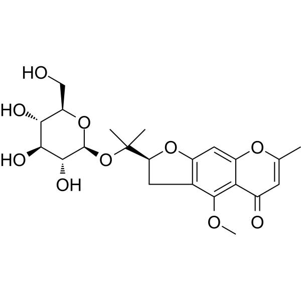
5-O-Methylvisammioside (4'-O-β-D-Glucosyl-5-O-methylvisamminol) is an orally active natural chromone glycoside and multiple biological activities. 5-O-Methylvisammioside inhibits ferroptosis by activating the Nrf2/HO-1 signaling axis. 5-O-Methylvisammioside alleviates intestinal barrier damage by inhibiting the ROS/NF-κB/NLRP3 pathway. 5-O-Methylvisammioside exerts a protective effect against acute liver injury by reducing ALT/AST, decreasing inflammatory infiltration, and inhibiting IκB-α phosphorylation and NF-κB nuclear translocation. 5-O-Methylvisammioside blocks the HMGB1/RAGE/MEK/ERK signaling axis to exert anti-tumor and anti-angiogenic effects. 5-O-Methylvisammioside improves depression-like behaviors by inhibiting Src kinase and the NF-κB pathway.
For research use only. We do not sell to patients.
- Purity : 99.90%
- CAS No.: 84272-85-5
- Formula: C22H28O10
- Molecular Weight:452.45
-
Storage:Powder -20°C, 3 years , 4°C, 2 years ; In solvent -80°C, 2 years , -20°C, 1 year

To place orders, for customer services and technical support, please contact: MedChemExpress USA
Tel: 609-228-6898 E-mail: [email protected] [email protected]


-
 Biological Activity
-
Chemical Information
-
Solvent & Solubility
- Protocols
- Purity & Documentation
- References
-
 Help & FAQs




Biological Activity
Description
In Vitro
5-O-Methylvisammioside effectively blocks the production of inflammatory cytokines induced by lipopolysaccharide in BV-2 microglial cells[1].
5-O-Methylvisammioside (50-500 μg/mL; 24 h) shows no toxicity to human umbilical vein endothelial cells at concentrations up to 500 μg/mL, and inhibits HMGB1-induced proliferation of human umbilical vein endothelial cells in a dose-dependent manner[2].
5-O-Methylvisammioside (100-500 μg/mL; 24 h) dose-dependently inhibits HMGB1-induced migration of human umbilical vein endothelial cells in wound healing assays[2].
5-O-Methylvisammioside (100-500 μg/mL; 24 h) dose-dependently inhibits HMGB1-induced migration of human umbilical vein endothelial cells in Transwell assays[2].
5-O-Methylvisammioside (100-500 μg/mL; 16 h) dose-dependently inhibits HMGB1-induced tube formation in human umbilical vein endothelial cells[2].
5-O-Methylvisammioside (100-500 μg/mL) dose-dependently inhibits HMGB1-induced upregulation of RAGE, phosphorylated MEK, and phosphorylated ERK in human umbilical vein endothelial cells[2].
5-O-Methylvisammioside (250 μg/mL) inhibits HMGB1-induced upregulation of RAGE in human umbilical vein endothelial cells[2].
5-O-Methylvisammioside (100-500 μg/mL) dose-dependently inhibits HMGB1-induced VEGF production in human umbilical vein endothelial cells[2].
5-O-Methylvisammioside (40 μM; 48 h) significantly reduces oxidative stress levels, corrects iron metabolism disorders, restores the expression levels of ferroptosis-related proteins, and decreases proinflammatory cytokine levels in Erastin (HY-15763)-treated Caco-2 cells. These protective effects strictly depend on the Nrf2/HO-1 pathway, as Nrf2 knockdown significantly attenuates the aforementioned effects[3].
5-O-Methylvisammioside (10 μM; 45 min) potently inhibits CNT2-mediated adenosine uptake in HEK293 cells overexpressing CNT2, with an IC50 of 11.22 μM, and exhibits stronger activity than other tested compounds at the concentration of 10 μM[4].
5-O-Methylvisammioside (10 μM; 24 h) inhibits ROS production in human intestinal epithelial cells stimulated with 720 μM uric acid (UA) (HY-B2130) for 24 h, which is confirmed by reduced DCFH-DA fluorescence intensity and regulated mRNA expression of SOD/NOX4[4].
5-O-Methylvisammioside (10 μM; 24 h) inhibits the activation of the ROS/NF-κB/NLRP3 pathway and restores the expression of tight junction proteins in human intestinal epithelial cells stimulated with 720 μM UA for 24 h[4].
MedChemExpress (MCE) has not independently confirmed the accuracy of these methods. They are for reference only. Further protocols information, click here.
-
Cell Line:human umbilical vein endothelial cells (HUVECs)
-
Concentration:100, 250, 500 μg/mL
-
Incubation Time:24 h
-
Result:Dose-dependently reduced HMGB1-enhanced wound closure at 100, 250, 500 μg/mL, with significant inhibition observed at all tested concentrations.\n
Significantly reduced the number of HMGB1-increased migrated cells at 100, 250, 500 μg/mL, with dose-dependent inhibition observed.
-
Cell Line:UA-stimulated human intestinal epithelial cells (HIEC)
-
Concentration:10 μM
-
Incubation Time:24 h
-
Result:Significantly reduced phosphorylation of NF-κB p65, and decreased protein expression of NLRP3, ASC, and caspase-1 in UA-stimulated HIEC.
Increased protein expression of tight junction proteins ZO-1 and Occludin in UA-stimulated HIEC.
In Vivo
5-O-Methylvisammioside (12.5-50 mg/kg; once daily; 14 days) dose-dependently inhibits orthotopic hepatocellular carcinoma growth in C57BL/6 mice, with a maximum tumor inhibition rate of 62.64% at 50 mg/kg, by suppressing tumor angiogenesis and downregulating the HMGB1/RAGE/MEK/ERK signaling pathway[2].
5-O-Methylvisammioside (3.5-7 mg/kg/day; i.p.; once daily; 14 days) dose-dependently alleviates DSS (HY-116282C)-induced ulcerative colitis in male Sprague-Dawley rats[3].
5-O-Methylvisammioside (5-20 mg/kg; i.g.; single dose) reduces peak serum uric acid levels in single-dose HUA mice by up to 39.1% (10 mg/kg dose) and accelerates the return to baseline uric acid levels[4].
5-O-Methylvisammioside (5-20 mg/kg; i.g.; once daily; 4 weeks) reduces serum uric acid and adenosine levels, improves liver/kidney function, alleviates intestinal inflammation and oxidative stress, restores intestinal barrier function, and normalizes intestinal CNT2 expression in chronic HUA mice[4].
MedChemExpress (MCE) has not independently confirmed the accuracy of these methods. They are for reference only.
-
Animal Model:ICR (male, adult, 20-22 g, acetaminophen-induced acute liver injury)[1]
-
Dosage:5 mg/kg; 10 mg/kg
-
Administration:i.p.; daily; 7 days
-
Result:Significantly reduced serum alanine aminotransferase and aspartate aminotransferase levels compared to the acetaminophen-only group.
Significantly reduced liver inflammatory cell infiltration and hepatocyte vacuolation compared to the acetaminophen-only group.
Significantly decreased serum TNF-α and IL-1β levels compared to the acetaminophen-only group.
Significantly suppressed hepatic mRNA expression of TNF-α, IL-1β, Ptges, p38, and RelB compared to the acetaminophen-only group.
Significantly inhibited IκB-α phosphorylation and NF-κB nuclear translocation compared to the acetaminophen-only group.
Significantly suppressed phosphorylation of JNK and p38 in the MAPK pathway compared to the acetaminophen-only group, with no significant effect on Erk1/2 phosphorylation.
Regulated 4675 differentially expressed hepatic genes compared to the acetaminophen-only group, with enrichment in TNF, MAPK, NF-κB, and arachidonic acid pathways at 10 mg/kg dose.
Regulated 249 hepatic metabolites compared to the acetaminophen-only group, including significant reduction in pro-inflammatory prostaglandins (D2, B2, A2) linked to the arachidonic acid pathway at 10 mg/kg dose.
-
Animal Model:C57BL/6 (male, 8 weeks old, 20g)[2]
-
Dosage:12.5 mg/kg; 25 mg/kg; 50 mg/kg
-
Administration:daily; 14 days
-
Result:Reduced tumor weight with tumor inhibition rates of 42.24%, 55.48%, and 62.64% for the 12.5 mg/kg, 25 mg/kg, and 50 mg/kg doses, respectively.
Decreased CD31 H-scores in tumor tissues across all three doses.
Exhibited dose-dependent reductions in HMGB1, RAGE, p-MEK, and p-ERK protein levels compared to the model group.
Reduced tumor tissue HMGB1 levels and VEGF levels across all three doses.
Caused no significant changes in body weight during the 14-day period.
-
Animal Model:Sprague-Dawley (male, 6 weeks old, 200 g, specific pathogen-free, ulcerative colitis induced by 3% dextran sulfate sodium)[3]
-
Dosage:3.5 mg/kg/day; 7 mg/kg/day
-
Administration:i.p.; daily; 14 days
-
Result:Reduced disease activity index scores compared to the DSS model group, with the high dose showing greater efficacy.
Significantly increased colon length, reduced colon damage index scores, lowered spleen index, and increased thymus index.
Partially ameliorated mucosal ulceration, crypt atrophy, and inflammatory infiltration at low dose; showed improved gland/crypt organization and reduced inflammation, with near-normal mucosa similar to mesalazine at high dose.
Reduced colon tissue levels of IL-6, IL-8, and TNF-α.
Restored goblet cell numbers, upregulated protein and mRNA levels of tight junction proteins occludin and ZO-1.
Reduced oxidative stress markers MDA, 4-HNE, and MPO activity, and increased GSH content.
Reduced total iron content, upregulated FTH1 expression, and downregulated Tfrc and Slc11a2 levels to restore iron homeostasis.
Improved mitochondrial morphology (reduced swelling, increased cristae organization) compared to the DSS group, with high-dose rats showing discernible mitochondrial double membranes.
Upregulated protein and mRNA levels of ferroptosis markers GPX4 and SLC7A11, and downregulated ACSL4.
Dose-dependently upregulated protein and mRNA levels of Nrf2 and HO-1, with high-dose levels exceeding normal control levels.
-
Animal Model:C57BL/6J (6-week-old, SPF-grade, male, hyperuricemia induced by daily intraperitoneal injection of potassium oxonate plus intragastric gavage of yeast paste for single-dose study)[4]
-
Dosage:5 mg/kg; 10 mg/kg; 20 mg/kg
-
Administration:i.g.; single dose
-
Result:Reduced peak serum uric acid levels to 194.44 μM (5 mg/kg), 174.76 μM (10 mg/kg), 176.93 μM (20 mg/kg).
Lowered peak serum uric acid levels by up to 39.1% (10 mg/kg dose).
Accelerated serum uric acid return to pre-dose levels by 4 h post-treatment.
-
Animal Model:C57BL/6J (6-week-old, SPF-grade, male, hyperuricemia induced by daily intraperitoneal injection of potassium oxonate plus intragastric gavage of yeast paste for 4 weeks)[4]
-
Dosage:5 mg/kg; 10 mg/kg; 20 mg/kg
-
Administration:i.g.; daily; 4 weeks
-
Result:Significantly reduced serum uric acid levels in all treated groups, with the 10 mg/kg dose showing the greatest reduction.
Significantly decreased serum and urine adenosine levels; 5 and 10 mg/kg doses showed better inhibitory effects on adenosine absorption than positive control drugs.
Significantly reduced elevated CNT2 protein expression in the duodenum, jejunum, and ileum induced by HUA.
Ameliorated liver pathology (bile duct hyperplasia, inflammatory cell infiltration, steatosis) and kidney pathology (renal tubular epithelial cell edema, tubular/corpuscle dilation, inflammatory cell infiltration), and reduced serum ALT, AST, and Cr levels compared to the model group.
Significantly decreased serum and intestinal levels of inflammatory factors (IL-1β, IL-6, TNF-α) and oxidative stress marker MDA, while increased serum SOD levels compared to the model group.
Reduced intestinal permeability (measured via FITC-dextran), and restored intestinal tight junction protein expression (ZO-1, Occludin) in the duodenum, jejunum, and ileum compared to the model group.
Improved intestinal histopathology (epithelial detachment, shortened/sparse villi, inflammatory cell infiltration) and preserved villi structure.


Chemical Information
-
CAS No. 84272-85-5
-
Appearance Solid
-
Molecular Weight 452.45
-
Formula C22H28O10
-
Color White to off-white
-
SMILES
O[C@H]1[C@H](O)[C@@H](CO)O[C@@H](OC(C)([C@H]2OC3=C(C(OC)=C(C(C=C(C)O4)=O)C4=C3)C2)C)[C@@H]1O
-
Synonyms
4'-O-β-D-Glucosyl-5-O-methylvisamminol
-
Structure Classification
-
Initial Source
-
Shipping
Room temperature in continental US; may vary elsewhere.
-
Storage
Powder -20°C 3 years 4°C 2 years In solvent -80°C 2 years -20°C 1 year


Solvent & Solubility
In Vitro:
DMSO : 50 mg/mL (110.51 mM; Need ultrasonic; Hygroscopic DMSO has a significant impact on the solubility of product, please use newly opened DMSO)
H2O : ≥ 25 mg/mL (55.25 mM)
* "≥" means soluble, but saturation unknown.
Please refer to the solubility information to select the appropriate solvent. Once prepared, please aliquot and store the solution to prevent product inactivation from repeated freeze-thaw cycles.
Storage method and period of stock solution: -80°C, 2 years; -20°C, 1 year. When stored at -80°C, please use it within 2 years. When stored at -20°C, please use it within 1 year.
* Note: If you choose water as the stock solution, please dilute it to the working solution, then filter and sterilize it with a 0.22 μm filter before use.
Please refer to the solubility information to select the appropriate solvent. Once prepared, please aliquot and store the solution to prevent product inactivation from repeated freeze-thaw cycles.
Storage method and period of stock solution: -80°C, 2 years; -20°C, 1 year. When stored at -80°C, please use it within 2 years. When stored at -20°C, please use it within 1 year.
* Note: If you choose water as the stock solution, please dilute it to the working solution, then filter and sterilize it with a 0.22 μm filter before use.
Concentration (start) × Volume (start) = Concentration (final) × Volume (final)
In Vivo:
Select the appropriate dissolution method based on your experimental animal and administration route.
- For the following dissolution methods, please ensure to first prepare a clear stock solution using an In Vitro approach and then sequentially add co-solvents:
- To ensure reliable experimental results, the clarified stock solution can be appropriately stored based on storage conditions. As for the working solution for In Vivo experiments, it is recommended to prepare freshly and use it on the same day.
- The percentages shown for the solvents indicate their volumetric ratio in the final prepared solution. If precipitation or phase separation occurs during preparation, heat and/or sonication can be used to aid dissolution.
Add each solvent one by one: 10% DMSO 40% PEG300 5% Tween-80 45% Saline
Solubility: ≥ 2.5 mg/mL (5.53 mM); Clear solution
This protocol yields a clear solution of ≥ 2.5 mg/mL (saturation unknown).
Taking 1 mL working solution as an example, add 100 μL DMSO stock solution (25.0 mg/mL) to 400 μL PEG300, and mix evenly; then add 50 μL Tween-80 and mix evenly; then add 450 μL Saline to adjust the volume to 1 mL.
Preparation of Saline: Dissolve 0.9 g sodium chloride in ddH₂O and dilute to 100 mL to obtain a clear Saline solution.
Add each solvent one by one: 10% DMSO 90% (20% SBE-β-CD in Saline)
Solubility: ≥ 2.5 mg/mL (5.53 mM); Clear solution
This protocol yields a clear solution of ≥ 2.5 mg/mL (saturation unknown).
Taking 1 mL working solution as an example, add 100 μL DMSO stock solution (25.0 mg/mL) to 900 μL 20% SBE-β-CD in Saline, and mix evenly.
Preparation of 20% SBE-β-CD in Saline (4°C, storage for one week): 2 g SBE-β-CD powder is dissolved in 10 mL Saline, completely dissolve until clear.
In Vivo Dissolution Calculator
Please enter the basic information of animal experiments:
-
-
-
-
Recommended: Prepare an additional quantity of animals to account for potential losses during experiments.
Working solution concentration: 0.22 mg/mL
This product has good water solubility, please refer to the measured solubility data in water/PBS/Saline for details.

Protocols
-
Kinase activity and phosphorylation assays
Kinase activity assays measure the ability of kinases to transfer phosphate groups from ATP to specific substrates, while phosphorylation assays detect the presence and levels of phosphorylated proteins. Common methods include radiolabeled ATP incorporation (e. g. ,), ADP release detection via bioluminescence (e. g. ,[3]), enzyme-linked immunosorbent assays (ELISA) for phospho-specific epitopes (e. g. ,[6]), and microtiter-based formats for high-throughput screening (e. g. ,[8]). The ADP-Glo assay quantifies kinase activity by measuring ADP produced during phosphorylation using a luciferase-based system. Radiometric assays involve autoradiography or scintillation counting after incorporation of 32P-labeled ATP into substrate proteins. ELISA-based approaches rely on phospho-specific antibodies to detect activated kinases in cell lysates or purified samples.
-
Western Blot
Western blotting (WB) is a commonly used experimental method in molecular biology, biochemistry, and immunogenetics for identifying and quantifying target proteins. It combines gel electrophoresis with immunoassay, enabling researchers to analyze protein expression, post-translational modifications, and molecular weight.
-
Cytoplasmic-Nuclear Fractionated Protein Extraction
Cytoplasmic-nuclear fractionated protein extraction separates soluble cytoplasmic proteins from nuclear-enriched proteins by mild plasma-membrane permeabilization, differential centrifugation, washing of nuclei, and extraction of nuclear proteins for downstream immunoblotting or related molecular analysis. The readout is the relative abundance of a protein in cytoplasmic and nuclear fractions, commonly assessed by western blotting together with compartment markers such as tubulin or pyruvate kinase for cytoplasm and lamin, nucleoporin, hnRNP, H2AX, or Lamin B for nuclear fractions.
-
ROS/oxidative-stress fluorescent staining
ROS/oxidative-stress fluorescent staining uses cell-permeant fluorogenic probes that become fluorescent after oxidation inside cells or tissues; commonly used examples include DCFH-DA/DCFDA for broad cellular oxidant detection, DHE for superoxide-related signal detection, MitoSOX for mitochondrial superoxide-related signal detection, and CellROX probes for oxidative-stress-associated fluorescence readouts. The assay detects probe oxidation rather than a single ROS species unless the probe and analysis method have been chemically validated for that species. DCFH-DA enters cells, is deacetylated by intracellular esterases to DCFH, and produces fluorescent DCF after oxidation, so the readout is used as an operational measure of total cellular oxidative stress rather than a species-specific ROS measurement. DHE and MitoSOX can report superoxide-related oxidation, but red fluorescence alone can include non-specific ethidium-like oxidation products; HPLC or optimized spectral approaches are
-
Ferroptosis Solutions
Ferroptosis is an iron-dependent, non-apoptotic form of regulated cell death characterized by lethal lipid peroxidation and sensitivity to suppression by iron chelators or lipophilic radical-trapping antioxidants. The core pathway links cystine uptake through system Xc−, glutathione availability, GPX4-dependent detoxification of phospholipid hydroperoxides, iron-dependent oxidative reactions, and polyunsaturated-phospholipid metabolism into a cell-death program that is biochemically and morphologically distinct from apoptosis, necrosis, and autophagy. The ferroptosis pathway is experimentally linked to phenotype through chemical and genetic perturbation. Erastin induces ferroptosis by inhibiting cystine uptake through system Xc− and weakening antioxidant defenses, while GPX4 inhibition or depletion causes lipid peroxide accumulation and ferroptotic cancer-cell death. ACSL4 and oxidizable arachidonoyl- or adrenoyl-containing phosphatidylethanolamines shape ferroptosis sensitivity by con
-
Research Protocol for Inflammation-related Diseases
The NLRP3 inflammasome is a cytosolic innate immune signaling platform that integrates priming signals and danger-signal activation to promote caspase-1 activation, maturation of IL-1β and IL-18, and gasdermin D-mediated pyroptotic cell death. The core experimental logic is to determine whether inflammatory disease phenotypes are driven by increased NLRP3 expression, ASC-containing inflammasome assembly, caspase-1 cleavage, GSDMD cleavage, and extracellular release of IL-1β/IL-18 rather than by nonspecific cell injury alone. The pathway is strongly linked to inflammation-related disease phenotypes because monosodium urate crystals activate NALP3/NLRP3 inflammasome signaling in gout-like crystal inflammation, cholesterol crystals activate NLRP3 inflammasomes in atherogenesis models, and DSS-induced intestinal inflammation has been reported to involve NLRP3 inflammasome activity. However, experimental colitis studies also show context-dependent protective effects of NLRP3 inflammasome co
-
Hepatotoxicity Study
This protocol evaluates hepatotoxicity using complementary in vivo mouse APAP acute liver injury and in vitro hepatocyte-based cytotoxicity readouts. In vivo APAP injury is assessed by serum ALT/AST, liver histology, hepatic glutathione, protein adducts, necrosis, inflammation, and regeneration-related endpoints. In vitro hepatotoxicity is assessed by loss of viability, leakage of ALT/AST/LDH, oxidative-stress markers, mitochondrial function, nuclear morphology, intracellular calcium, and high-content imaging endpoints.
-
Protocol for Kinase activity and phosphorylation assays
Kinase activity assays measure transfer of phosphate from ATP to a protein or peptide substrate, generating phosphorylated substrate, ADP, or incorporated radiolabeled phosphate as the readout; phosphorylation assays measure site-specific phosphorylation in cells or tissues as a proxy for kinase-pathway activation, inhibition, or substrate regulation. Phosphorylation can be detected by phospho-specific Western blot, immunoprecipitation kinase assay, phospho-immunofluorescence, phospho-flow cytometry, luminescent ADP detection, radiolabeled ATP incorporation, or reporter-based pathway assays, and these readouts can be applied to cancer cells, primary neurons, mouse tumors, organoids, inflammatory macrophages, ferroptosis studies, and mitophagy studies when the kinase target is biologically relevant.


Purity & Documentation
-
Data Sheet (292 KB)
-
SDS (394 KB)
- English - EN (394 KB)
- Français - FR (394 KB)
- Deutsch - DE (394 KB)
- Norwegian - NO (394 KB)
- Español - ES (394 KB)
- Swedish - SV (394 KB)
- Italian - IT (394 KB)
- Korean - KR (394 KB)
- Portuguese - PT (394 KB)
-
Handling Instructions (2659 KB)
References
[1].
Zhang T, et al. Multi-omics reveals that 5-O-methylvisammioside prevention acute liver injury in mice by regulating the TNF/MAPK/NF-κB/arachidonic acid pathway. Phytomedicine. 2024 Jun;128:155550.
[Content Brief]
[2]. Hou W, et al. 5-O-Methylvisammioside inhibits HMGB1-induced Angiogenesis of hepatocellular carcinoma through RAGE/MEK/ERK signaling pathway. PLoS One. 2025;20(5):e0322056. Published 2025 May 5. [Content Brief]
[3]. Li D, et al. Nrf2/HO-1-dependent inhibition of ferroptosis underlies the antioxidant effects of 5-O-methylvisammioside in colitis. Front Immunol. 2026;16:1738408. Published 2026 Jan 20. [Content Brief]
[4]. Mai S, et al. 5-O-Methylvisammioside, a novel inhibitor of CNT2, improves hyperuricemia by inhibiting intestinal purine nucleoside absorption with a safe profile. Biochem Pharmacol. Published online August 13, 2025. [Content Brief]


Complete Stock Solution Preparation Table
Please refer to the solubility information to select the appropriate solvent. Once prepared, please aliquot and store the solution to prevent product inactivation from repeated freeze-thaw cycles.
Storage method and period of stock solution: -80°C, 2 years; -20°C, 1 year. When stored at -80°C, please use it within 2 years. When stored at -20°C, please use it within 1 year.
| Optional Solvent | Concentration Solvent Mass | 1 mg | 5 mg | 10 mg | 25 mg |
|---|---|---|---|---|---|
| H2O / DMSO | 1 mM | 2.2102 mL | 11.0509 mL | 22.1019 mL | 55.2547 mL |
| 5 mM | 0.4420 mL | 2.2102 mL | 4.4204 mL | 11.0509 mL | |
| 10 mM | 0.2210 mL | 1.1051 mL | 2.2102 mL | 5.5255 mL | |
| 15 mM | 0.1473 mL | 0.7367 mL | 1.4735 mL | 3.6836 mL | |
| 20 mM | 0.1105 mL | 0.5525 mL | 1.1051 mL | 2.7627 mL | |
| 25 mM | 0.0884 mL | 0.4420 mL | 0.8841 mL | 2.2102 mL | |
| 30 mM | 0.0737 mL | 0.3684 mL | 0.7367 mL | 1.8418 mL | |
| 40 mM | 0.0553 mL | 0.2763 mL | 0.5525 mL | 1.3814 mL | |
| 50 mM | 0.0442 mL | 0.2210 mL | 0.4420 mL | 1.1051 mL | |
| DMSO | 60 mM | 0.0368 mL | 0.1842 mL | 0.3684 mL | 0.9209 mL |
| 80 mM | 0.0276 mL | 0.1381 mL | 0.2763 mL | 0.6907 mL | |
| 100 mM | 0.0221 mL | 0.1105 mL | 0.2210 mL | 0.5525 mL |
* Note: If you choose water as the stock solution, please dilute it to the working solution, then filter and sterilize it with a 0.22 μm filter before use.


5-O-Methylvisammioside Related Classifications
HY-N0442 Related Classifications
 Powered by Bioz
Keywords
- 5-O-Methylvisammioside
- 84272-85-5
- 4'-O-β-D-Glucosyl-5-O-methylvisamminol
- NF-κB
- p38 MAPK
- JNK
- Src
- TNF Receptor
- NOD-like Receptor (NLR)
- Amyloid-β
- MEK
- ERK
- Ferroptosis
- VEGFR
- Anaplastic lymphoma kinase (ALK)
- Reactive Oxygen Species (ROS)
- p38
- IκB-α
- BV-2 microglia
- NLRP3 inflammasome
- human umbilical vein endothelial cells
- MAPK
- TNF
- SRC
- Inhibitor
- inhibitor
- inhibit