



4-Hydroxyestradiol
Based on 1 publication(s) in Google Scholar
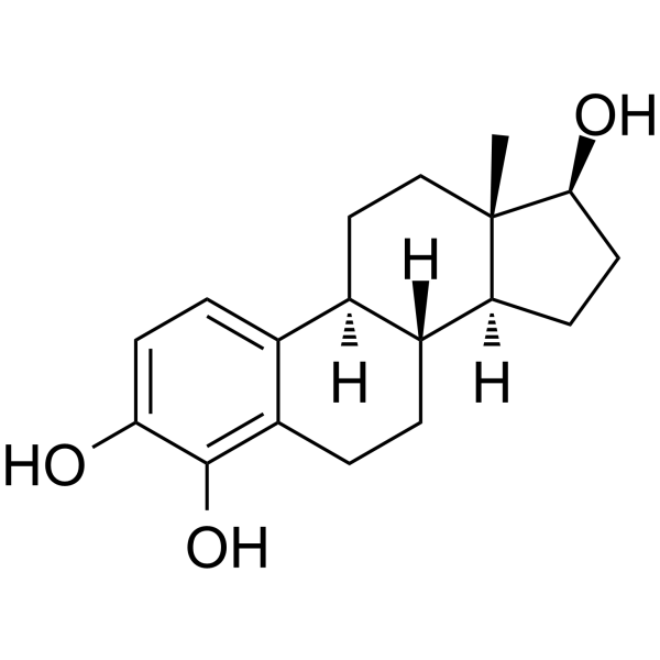
4-Hydroxyestradiol (4-Hydroxy-17β-estradiol; 4-OHE2) is an endogenous metabolite of Estradiol (HY-B0141). 4-Hydroxyestradiol binds to Estrogen Receptor (ER) with a Kd of 0.29 nM and competitively inhibits the binding of Estradiol to ER with a Ki of 0.48 nM. The reactive quinones generated from 4-Hydroxyestradiol via redox cycling, together with ROS, directly induce DNA damage while activating the ERK/Akt and downstream IKK-NF-κB-COX-2 signaling axis, thereby driving malignant cell transformation and conditional cell apoptosis. 4-Hydroxyestradiol can be used in breast cancer research.
商品は「研究用試薬」です。人や動物の医療用・臨床診断用・食品用の製品ではありません。
研究用途以外に使用した場合、当社は一切の責任を負いかねます。
- 純度 : 99.0%
- CAS 番号: 5976-61-4
- 分子式: C18H24O3
- 分子量:288.38
-
保管条件:Powder -20°C, 3 years ; In solvent -80°C, 6 months , -20°C, 1 month

To place orders, for customer services and technical support, please contact: MedChemExpress USA
Tel: 609-228-6898 E-mail: [email protected] [email protected]


-
 生物活性
-
化学情報
-
溶剤 & 溶解度
- プロトコル
- 純度とドキュメンテーション
- 参考文献
-
 Help & FAQs




生物活性
製品説明
IC50 & Target
[5]|
NF-κB |
COX-2 |
IKKβ |
IKKα |
Akt |
ERK |
体外実験
4-Hydroxyestradiol (50 μM; 6 h) induces intracellular ROS accumulation in MCF-10A cells, and this effect is attenuated by Trolox[1].
4-Hydroxyestradiol (20 μM; 6 h) increases oxidative DNA damage in MCF-10A cells by approximately 4-fold[1].
4-Hydroxyestradiol (20 μM; 1-6 h) increases intracellular ROS in MCF-10A cells, and this effect is reversed by NAC (HY-B0215)[5].
4-Hydroxyestradiol (20 μM; 2 h) increases NF-κB transcriptional activity in MCF-10A cells and induces nuclear translocation of phosphorylated p65[5].
4-Hydroxyestradiol (20 μM; 0.5-24 h) activates the phosphorylation of ERK, Akt, IKKα/β, and IκBα in MCF-10A cells[5].
4-Hydroxyestradiol induces COX-2 expression in MCF-10A cells[5].
4-Hydroxyestradiol (10-35 μM; 24 h) inhibits MCF-10A cell proliferation[1].
4-Hydroxyestradiol (20-50 μM; 24 h) induces cytotoxicity in MCF-10A cells, and this effect is attenuated by Trolox (HY-101445)[1].
4-Hydroxyestradiol (25 μM; 0.5-6 h) transiently activates NF-κB (but not AP-1) DNA-binding activity in MCF-10A cells[1].
4-Hydroxyestradiol (2 h) activates NF-κB-dependent transcription in MCF-10A cells[1].
4-Hydroxyestradiol (10-50 μM; 24 h) induces cytotoxicity in MCF-10A cells, and this effect is enhanced by co-exposure with TPCK (HY-124379)[1].
4-Hydroxyestradiol (25 μM; 15-300 min) rapidly activates ERK1/2 in MCF-10A cells and reaches a peak at 30 min[1].
4-Hydroxyestradiol (25 μM; 24 h) induces cytotoxicity in MCF-10A cells, and this effect is exacerbated by U0126 (HY-12031A)[1].
4-Hydroxyestradiol (200 nM; 18 h, multiple alternating treatments) has a weak mutagenic effect in BB rat2 embryonic cells, significantly increasing the cell mutation rate (especially A:T base pair mutations) under multiple low-dose exposures[2].
4-Hydroxyestradiol (0.007-70 nM; twice weekly for 2 weeks) induces microsatellite instability/loss of heterozygosity (LOH) in the 13q12.3 chromosomal region and a 5 bp deletion mutation in exon 4 of the p53 gene in MCF-10F, and this mutagenic effect cannot be prevented by the antiestrogen drug ICI182780 (HY-13636)[3].
4-Hydroxyestradiol (0.092-6 nM; 16-20 h) binds to estrogen receptors in human breast cancer cytoplasm with a Kd of 0.29 nM and a relative affinity of 151% that of estradiol-17β[4].
4-Hydroxyestradiol (16-20 h) inhibits the binding of [3H]estradiol-17β to ER with a Ki of 0.48 nM[4].
4-Hydroxyestradiol (2-20 μM; 3 weeks) induces anchorage-independent growth in MCF-10A cells[5].
4-Hydroxyestradiol (20 μM) increases PCNA expression and nuclear accumulation in MCF-10A cells[5].
4-Hydroxyestradiol (20 μM; 24 h-21 days) promotes the proliferation and migration of MCF-10A cells, and this effect is inhibited by Bay 11-7082 (HY-13453)[5].
MedChemExpress (MCE) has not independently confirmed the accuracy of these methods. They are for reference only.
-
Cell Line:MCF-10A
-
Concentration:10, 20, 30, 40, and 50 μM
-
Incubation Time:24 h
-
Result:Caused concentration-dependent inhibition of MCF-10A viability.
At 50 μM, caused more than 60% cell death.
-
Cell Line:MCF-10A
-
Concentration:10, 20, and 35 μM
-
Incubation Time:24 h
-
Result:Caused concentration-dependent inhibition of thymidine incorporation into MCF-10A cells.
-
Cell Line:MCF-10A
-
Concentration:25 μM
-
Incubation Time:2, 6, 12, and 24 h
-
Result:Caused progressive increases in the proportion of cleaved PARP, detected as the 85-kDa apoptotic fragment from the 116-kDa PARP protein.
-
Cell Line:MCF-10A
-
Concentration:25 μM
-
Incubation Time:15, 30, 60, 180, and 300 min
-
Result:Rapidly activated ERK via phosphorylation, which peaked at 30 min and declined thereafter.
Total ERK1/2 was also probed.
U0126 blocked ERK phosphorylation.
-
Cell Line:MCF-10A cells
-
Concentration:20 μM; 3 and 5 μM (Bay 11-7082)
-
Incubation Time:3 weeks
-
Result:At 20 μM, increased both the size and number of anchorage-independent foci.
Bay 11-7082 at 3 and 5 μM abrogated colony formation.
-
Cell Line:MCF-10A cells
-
Concentration:20 μM
-
Incubation Time:2 h
-
Result:Induced nuclear translocation of phosphorylated p65.
The nuclear translocation was abolished by excess complementary phosphorylated p65 peptide.
-
Cell Line:MCF-10A cells
-
Concentration:20 μM (time course); 20 μM (NAC co-incubation); 3 or 5 mM (NAC)
-
Incubation Time:0.5, 1, 3, 6, 12, 24 h (time course); 3 h (NAC co-incubation)
-
Result:Caused rapid phosphorylation of ERK and Akt over the time course.
Phosphorylation of IκBα and IKKα/β was increased at 3 h.
NAC decreased induced phosphorylation of ERK, Akt, IKKα/β, and IκBα.
-
Cell Line:MCF-10A cells
-
Concentration:20 μM; 3 and 5 μM (Bay 11-7082)
-
Incubation Time:24 h
-
Result:Promoted closure of the wound gap at 24 h.
Bay 11-7082 suppressed MCF-10A cell migration in a dose-dependent manner.
- Detection of Apoptosis
- TUNEL staining for apoptotic DNA fragmentation
- Annexin V plus membrane-impermeant dye apoptosis staining
- ROS/oxidative-stress fluorescent staining
- Research Protocol for Endocrine Diseases
- Apoptosis Solutions
- Research Protocol for Inflammation-related Diseases
- Breast Cancer Modeling
- Genotoxicity/Mutagenicity Study


Product Details
-
Structure Classification
-
Initial Source


化学情報
-
CAS 番号 5976-61-4
-
性状 Solid
-
分子量 288.38
-
分子式 C18H24O3
-
Color White to light yellow
-
SMILES
C[C@@]12[C@](CC[C@@H]2O)([H])[C@@](CCC3=C4O)([H])[C@@](CC1)([H])C3=CC=C4O
-
別名
4-ヒドロキシエストラジオール; 4-Hydroxy-17β-estradiol
-
輸送条件
Room temperature in continental US; may vary elsewhere.
-
保管条件
Powder -20°C 3 years In solvent -80°C 6 months -20°C 1 month


Publications (1)
-
Journal Impact Factor
-
Most Recent
-
Sci Rep
2025 Mar 11;15(1):8409. PMID: 40069295


溶剤 & 溶解度
体外:
DMSO : 25 mg/mL (86.69 mM; Need ultrasonic and warming; Hygroscopic DMSO has a significant impact on the solubility of product, please use newly opened DMSO)
Please refer to the solubility information to select the appropriate solvent. Once prepared, please aliquot and store the solution to prevent product inactivation from repeated freeze-thaw cycles.
Storage method and period of stock solution: -80°C, 6 months; -20°C, 1 month. When stored at -80°C, please use it within 6 months. When stored at -20°C, please use it within 1 month.
Please refer to the solubility information to select the appropriate solvent. Once prepared, please aliquot and store the solution to prevent product inactivation from repeated freeze-thaw cycles.
Storage method and period of stock solution: -80°C, 6 months; -20°C, 1 month. When stored at -80°C, please use it within 6 months. When stored at -20°C, please use it within 1 month.
濃度 (開始) × 体積 (開始) = 濃度 (終了) × 体積 (終了)
In Vivo Dissolution Calculator
Please enter the basic information of animal experiments:
-
-
-
-
Recommended: Prepare an additional quantity of animals to account for potential losses during experiments.
Working solution concentration: 0.22 mg/mL
純度とドキュメンテーション


参考文献
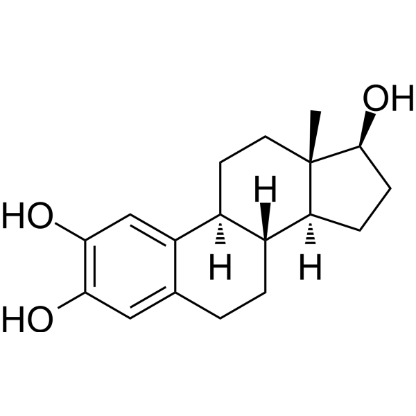
[3]. Fernandez SV, et al. Estradiol and its metabolites 4-hydroxyestradiol and 2-hydroxyestradiol induce mutations in human breast epithelial cells. International journal of cancer. 2006 Apr 15;118(8):1862-8. [Content Brief]
[4]. Van Aswegen CH, et al. Binding of 2-hydroxyestradiol and 4-hydroxyestradiol to estrogen receptors from human breast cancers. Journal of steroid biochemistry. 1989 Apr;32(4):485-92. [Content Brief]
[6]. Jin C, et al. Dynamic change of estrogen and progesterone metabolites in human urine during pregnancy. Scientific reports. 2025 Mar 11;15(1):8409. [Content Brief]

Complete Stock Solution Preparation Table
Please refer to the solubility information to select the appropriate solvent. Once prepared, please aliquot and store the solution to prevent product inactivation from repeated freeze-thaw cycles.
Storage method and period of stock solution: -80°C, 6 months; -20°C, 1 month. When stored at -80°C, please use it within 6 months. When stored at -20°C, please use it within 1 month.
| Optional Solvent | Concentration Solvent Mass | 1 mg | 5 mg | 10 mg | 25 mg |
|---|---|---|---|---|---|
| DMSO | 1 mM | 3.4676 mL | 17.3382 mL | 34.6765 mL | 86.6912 mL |
| 5 mM | 0.6935 mL | 3.4676 mL | 6.9353 mL | 17.3382 mL | |
| 10 mM | 0.3468 mL | 1.7338 mL | 3.4676 mL | 8.6691 mL | |
| 15 mM | 0.2312 mL | 1.1559 mL | 2.3118 mL | 5.7794 mL | |
| 20 mM | 0.1734 mL | 0.8669 mL | 1.7338 mL | 4.3346 mL | |
| 25 mM | 0.1387 mL | 0.6935 mL | 1.3871 mL | 3.4676 mL | |
| 30 mM | 0.1156 mL | 0.5779 mL | 1.1559 mL | 2.8897 mL | |
| 40 mM | 0.0867 mL | 0.4335 mL | 0.8669 mL | 2.1673 mL | |
| 50 mM | 0.0694 mL | 0.3468 mL | 0.6935 mL | 1.7338 mL | |
| 60 mM | 0.0578 mL | 0.2890 mL | 0.5779 mL | 1.4449 mL | |
| 80 mM | 0.0433 mL | 0.2167 mL | 0.4335 mL | 1.0836 mL |


4-Hydroxyestradiol 関連分類
HY-N10403 関連分類
 Powered by Bioz
Keywords
- 4-Hydroxyestradiol
- 5976-61-4
- 4-Hydroxy-17β-estradiol
- 4-OHE2
- Endogenous Metabolite
- Estrogen Receptor/ERR
- Reactive Oxygen Species (ROS)
- ERK
- Akt
- Apoptosis
- IKK
- NF-κB
- COX
- Extracellular signal regulated kinases
- PKB
- Protein kinase B
- IκB kinase
- I kappa B kinase
- Nuclear factor-κB
- Nuclear factor-kappaB
- Cyclooxygenase
- ERK1/2
- IKKα
- IKKβ
- IκBα
- RelA
- breast cancer
- estrogen receptor
- p53
- Inhibitor
- inhibitor
- inhibit