



Carveol
Based on 1 publication(s) in Google Scholar
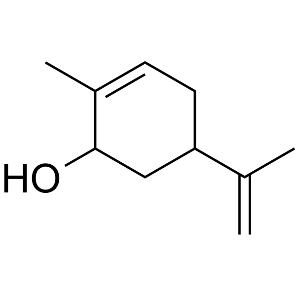
Carveol is an orally active monoterpenoid alcohol with neuroprotective, antioxidant, anti-inflammatory, anticonvulsant, antidiabetic, hypolipidemic and hepatoprotective activities. Carveol upregulates the expression of Bcl2, downregulates the expression of caspase-3, TNF-α, IL1β and IL6, activates the Nrf2/HO-1 antioxidant signaling pathway, inhibits the RAGE/NF-κB signaling pathway, and suppresses neuroinflammation and neuronal apoptosis. Carveol inhibits α-synuclein aggregation, improves cognitive ability, and inhibits α-amylase activity. Carveol exerts neuroprotective effects in a rat model of Parkinson's disease. Carveol exhibits antidiabetic effects in alloxan-induced diabetic rats. Carveol alleviates PTZ-induced seizures in rats. Carveol can be used in research related to Parkinson's disease, epilepsy, diabetes and ischemic stroke.
For research use only. We do not sell to patients.
- Purity : 98.32%
- CAS No.: 99-48-9
- Formula: C10H16O
- Molecular Weight:152.24
-
Storage:
-20°C, stored under nitrogen
* In solvent : -80°C, 6 months; -20°C, 1 month (stored under nitrogen)

To place orders, for customer services and technical support, please contact: MedChemExpress USA
Tel: 609-228-6898 E-mail: [email protected] [email protected]


-
 Biological Activity
-
Chemical Information
-
Solvent & Solubility
- Protocols
- Purity & Documentation
- References
-
 Help & FAQs



Publications Citing Use of MedChemExpress (MCE) Carveol
MoreAll Caspase Isoforms
MoreAll α-synuclein Isoforms
More

Biological Activity
Description
IC50 & Target
[1]|
IL-6 |
IL-1β |
Caspase 3 |
TNF-α |
In Vitro
Carveol (0.821-65.690 μM; 30 min) inhibits the activity of purified α-amylase in a concentration-dependent manner, with an inhibition rate of 73.01% at the concentration of 65.690 μM[3].
MedChemExpress (MCE) has not independently confirmed the accuracy of these methods. They are for reference only.
In Vivo
MedChemExpress (MCE) has not independently confirmed the accuracy of these methods. They are for reference only.
-
Animal Model:Rats (rotenone-induced Parkinson’s disease)[1]
-
Dosage:10 mg/kg; 20 mg/kg
-
Administration:i.p.; daily; 35 days
-
Result:Significantly reduced time spent in the open-field periphery, increased time spent in the center, and decreased grooming and rearing occurrences compared to rotenone-only rats.
Significantly increased striatal dopamine, serotonin, and GABA levels, and upregulated tyrosine hydroxylase immunoreactivity and brain-derived neurotrophic factor (BDNF) mRNA expression compared to rotenone-only rats; 20 mg/kg dose showed greater increases in these markers.
Significantly downregulated striatal α-synuclein protein expression compared to rotenone-only rats.
Significantly reduced striatal malondialdehyde and nitric oxide levels, and increased glutathione, superoxide dismutase, catalase, and glutathione peroxidase levels compared to rotenone-only rats.
Significantly upregulated nuclear factor erythroid 2-related factor 2 (Nrf2) and heme oxygenase 1 (HO-1) mRNA expression compared to rotenone-only rats, with the 20 mg/kg dose showing higher expression levels.
Significantly reduced striatal tumor necrosis factor-alpha, interleukin-1 beta, and interleukin-6 levels, and downregulated receptor for advanced glycation endproducts (RAGE) and nuclear factor kappa-B (NF-κB) mRNA and NF-κB protein expression compared to rotenone-only rats.
Significantly reduced glial fibrillary acidic protein (GFAP) immunoreactivity in the striatum and substantia nigra compared to rotenone-only rats, with the 20 mg/kg dose showing greater reduction.
Significantly increased Bcl-2 mRNA expression and reduced caspase-3 mRNA expression and immunoreactivity in the striatum and substantia nigra compared to rotenone-only rats.
Reduced the number of degenerated, shrunken neurons and pyknotic glial nuclei in hematoxylin and eosin-stained striatal and substantia nigra tissues, with the 20 mg/kg dose showing more preserved tissue architecture.
Reduced the number of darkly stained, shrunken neurons in Cresyl violet-stained striatal and substantia nigra tissues.
-
Animal Model:Sprague-Dawley (adult male, weight 250-300 g, PTZ-induced epilepsy kindling model)[2]
-
Dosage:10 mg/kg; 20 mg/kg
-
Administration:i.p.; 30 min before each PTZ injection; every 48 h; 15 days
-
Result:Reduced mean seizure intensity score, decreased seizure frequency, extended seizure latency, and improved survival to 85% at 10 mg/kg compared to PTZ-only rats.
Reduced mean seizure intensity score, decreased seizure frequency, extended seizure latency, and improved survival to 100% at 20 mg/kg compared to PTZ-only rats (71.4% survival).
Reduced escape latency in the Morris Water Maze hidden-platform test at both 10 mg/kg and 20 mg/kg compared to PTZ-only rats.
Increased time spent in the target quadrant at 10 mg/kg, and at 20 mg/kg compared to PTZ-only rats.
Significantly increased the number of intact neurons in the cortex, hippocampal CA1, CA3, and DG regions at 20 mg/kg, reversing PTZ-induced neuronal atrophy, pyknosis, and karyolysis.
Restored cortical and hippocampal levels of catalase, superoxide dismutase, glutathione-S-transferase, and reduced glutathione at both doses compared to PTZ-only rats.
Reduced thiobarbituric acid reactive substance in both brain regions at 20 mg/kg compared to PTZ-only rats.
Significantly upregulated cortical and hippocampal Nrf2 gene expression and protein localization, as well as downstream heme oxygenase-1 (HO-1) gene and protein expression at 20 mg/kg compared to PTZ-only rats.
Reduced cortical and hippocampal levels of tumor necrosis factor-alpha, phosphorylated nuclear factor kappa B, cyclooxygenase-2, and acetylcholinesterase at 20 mg/kg compared to PTZ-only rats.
Reduced cortical and hippocampal DG vascular endothelial growth factor (VEGF) expression at 20 mg/kg, reversing PTZ-induced BBB disruption.
Abolished all carveol-mediated protective effects when pretreated with the Nrf2 inhibitor ATRA.
-
Animal Model:Sprague-Dawley (SD) (adult, either sex, 7-11 weeks old, 250-280 g, alloxan-induced diabetic)[3]
-
Dosage:65.7 µM/Kg; 197 µM/Kg; 394.1 µM/Kg
-
Administration:12 days; 6 weeks (for biomarker analysis)
-
Result:Attenuated alloxan-induced hyperglycemia in a dose- and time-dependent manner across days 1, 3, 6, 9, and 12, with statistically significant reductions compared to the diabetic control group.
Improved oral glucose tolerance at 394.1 µM/Kg, with statistically significant reductions in blood glucose levels at 30, 60, 90, and 120 minutes after glucose load compared to the diabetic control group.
Reduced glycosylated hemoglobin (HbA1C) levels in a dose-dependent manner: 65.7 µM/Kg reduced HbA1C to 5.6%, 197 µM/Kg reduced it to 5.2%, and 394.1 µM/Kg reduced it to 4.9%.
Reduced triglycerides (TGs), LDL, and increased HDL in a dose-dependent manner: 65.7 µM/Kg reduced TGs to 131.3 mg/dL, LDL to 75.2 mg/dL, and increased HDL to 50.2 mg/dL; 197 µM/Kg reduced TGs to 122.3 mg/dL, LDL to 73.6 mg/dL, and increased HDL to 50.6 mg/dL; 394.1 µM/Kg reduced TGs to 120.4 mg/dL, LDL to 65 mg/dL, and increased HDL to 51 mg/dL.
Reduced hepatic biomarker levels in a dose-dependent manner: 65.7 µM/Kg reduced ALT to 38 u/L, AST to 27.7 u/L, ALP to 200.2 u/L, and total bilirubin (TB) to 0.67 mg/dL; 197 µM/Kg reduced ALT to 28.6 u/L, AST to 25 u/L, ALP to 185.3 u/L, and TB to 0.76 mg/dL; 394.1 µM/Kg reduced ALT to 25.6 u/L, AST to 23.8 u/L, ALP to 186.8 u/L, and TB to 0.7 mg/dL.
Reduced body weight across days 1, 3, 6, 9, and 12, with statistically significant differences compared to the diabetic control group.
-
Animal Model:Sprague-Dawley (male, 7-10 weeks old, 230-260 g, transient middle cerebral artery occlusion for 120 minutes followed by 72 hours of reperfusion)[4]
-
Dosage:10 mg/kg; 20 mg/kg
-
Administration:i.p.; administered at 30 min, 24, 48, and 72 h post-MCAO
-
Result:Significantly reversed MCAO-induced neurological deficits at 72 hours, with 10 mg/kg and 20 mg/kg showing dose-dependent efficacy.
Reduced MCAO-induced corrected infarct area from 33.14% to 26.5% and 19.2%.
Significantly increased the number of surviving neurons in the cortex and striatum and attenuated MCAO-induced neuronal morphological damage.
Restored cortical GSH levels to 41.4 μmoles/mg protein, GST activity to 27.9 μmoles CDNB conjugate/min/mg protein, and catalase activity to 17.1 μmoles H2O2/min/mg protein.
Reduced cortical LPO content to 63.8 TBARS-nM/min/mg protein.
Significantly reduced cortical TNF-α levels, p-JNK expression in the cortex and striatum, and p-NFκB expression.
Increased cortical Nrf2 protein expression and HO-1 protein expression.
Significantly reduced MCAO-induced brain water content.
Attenuated MCAO-induced caspase-3 expression and increased Bcl2 expression in the cortex and striatum.
- RNA extraction experimental
- Detection of Apoptosis
- TUNEL staining for apoptotic DNA fragmentation
- Annexin V plus membrane-impermeant dye apoptosis staining
- Human pluripotent stem cell midbrain dopaminergic neuron differentiation
- How to Select a Suitable Non-Mouse Animal Model
- Apoptosis Solutions
- Research Protocol for Inflammation-related Diseases
- Research Protocol for Neurological Diseases
- Research Protocol for Metabolic Diseases
- How to Choose the Right Model Animal


Chemical Information
-
CAS No. 99-48-9
-
Appearance Liquid (Density: 0.958 g/cm3)
-
Molecular Weight 152.24
-
Formula C10H16O
-
Color Colorless to light yellow
-
SMILES
OC1C(C)=CCC(C(C)=C)C1
-
Structure Classification
-
Initial Source
-
Shipping
Room temperature in continental US; may vary elsewhere.
-
Storage
-20°C, stored under nitrogen
* In solvent : -80°C, 6 months; -20°C, 1 month (stored under nitrogen)


Publications (1)
-
Journal Impact Factor
-
Most Recent
-
Chem Biol Interact
Carveol alleviates osteoarthritis progression by acting on synovial macrophage polarization transformation: An in vitro and in vivo study. [Abstract]2024 Jan 5:387:110781. PMID: 37967808


Solvent & Solubility
In Vitro:
DMSO : 100 mg/mL (656.86 mM; Need ultrasonic; Hygroscopic DMSO has a significant impact on the solubility of product, please use newly opened DMSO)
Please refer to the solubility information to select the appropriate solvent. Once prepared, please aliquot and store the solution to prevent product inactivation from repeated freeze-thaw cycles.
Storage method and period of stock solution: -80°C, 6 months; -20°C, 1 month (stored under nitrogen). When stored at -80°C, please use it within 6 months. When stored at -20°C, please use it within 1 month.
Please refer to the solubility information to select the appropriate solvent. Once prepared, please aliquot and store the solution to prevent product inactivation from repeated freeze-thaw cycles.
Storage method and period of stock solution: -80°C, 6 months; -20°C, 1 month (stored under nitrogen). When stored at -80°C, please use it within 6 months. When stored at -20°C, please use it within 1 month.
Concentration (start) × Volume (start) = Concentration (final) × Volume (final)
In Vivo:
Select the appropriate dissolution method based on your experimental animal and administration route.
- For the following dissolution methods, please ensure to first prepare a clear stock solution using an In Vitro approach and then sequentially add co-solvents:
- To ensure reliable experimental results, the clarified stock solution can be appropriately stored based on storage conditions. As for the working solution for In Vivo experiments, it is recommended to prepare freshly and use it on the same day.
- The percentages shown for the solvents indicate their volumetric ratio in the final prepared solution. If precipitation or phase separation occurs during preparation, heat and/or sonication can be used to aid dissolution.
Add each solvent one by one: 10% DMSO 40% PEG300 5% Tween-80 45% Saline
Solubility: ≥ 2.5 mg/mL (16.42 mM); Clear solution
This protocol yields a clear solution of ≥ 2.5 mg/mL (saturation unknown).
Taking 1 mL working solution as an example, add 100 μL DMSO stock solution (25.0 mg/mL) to 400 μL PEG300, and mix evenly; then add 50 μL Tween-80 and mix evenly; then add 450 μL Saline to adjust the volume to 1 mL.
Preparation of Saline: Dissolve 0.9 g sodium chloride in ddH₂O and dilute to 100 mL to obtain a clear Saline solution.
Add each solvent one by one: 10% DMSO 90% (20% SBE-β-CD in Saline)
Solubility: ≥ 2.5 mg/mL (16.42 mM); Clear solution
This protocol yields a clear solution of ≥ 2.5 mg/mL (saturation unknown).
Taking 1 mL working solution as an example, add 100 μL DMSO stock solution (25.0 mg/mL) to 900 μL 20% SBE-β-CD in Saline, and mix evenly.
Preparation of 20% SBE-β-CD in Saline (4°C, storage for one week): 2 g SBE-β-CD powder is dissolved in 10 mL Saline, completely dissolve until clear.
In Vivo Dissolution Calculator
Please enter the basic information of animal experiments:
-
-
-
-
Recommended: Prepare an additional quantity of animals to account for potential losses during experiments.
Please enter your animal formula composition:
-
%DMSO +
Recommended: Keep the proportion of DMSO in working solution below 2% if your animal is weak.
-
%+
-
+%Tween-80 + +
-
%Saline +
The co-solvents required include: DMSO, . All of co-solvents are available by MedChemExpress (MCE). , Tween 80. All of co-solvents are available by MedChemExpress (MCE).
Working solution concentration: 0.22 mg/mL
Method for preparing stock solution: mg drug dissolved in μL DMSO. Stock solution concentration: mg/mL. * In solvent : -80°C, 6 months; -20°C, 1 month (stored under nitrogen)
1. Take μL DMSO stock solution;
2. Add μL .
μL , mix evenly;
3. Then add μL Tween 80, mix evenly;
4. Then add μL
Please ensure that the stock solution in the first step is dissolved to a clear state, and add co-solvents in sequence. You can use ultrasonic heating (ultrasonic cleaner, recommended frequency 20-40 kHz), vortexing, etc. to assist dissolution.


Purity & Documentation
-
Data Sheet (295 KB)
-
SDS (394 KB)
- English - EN (394 KB)
- Français - FR (394 KB)
- Deutsch - DE (394 KB)
- Norwegian - NO (394 KB)
- Español - ES (394 KB)
- Swedish - SV (394 KB)
- Italian - IT (394 KB)
- Korean - KR (394 KB)
- Portuguese - PT (394 KB)
-
Handling Instructions (2659 KB)
References
[2]. Alvi AM, et al. Carveol Attenuates Seizure Severity and Neuroinflammation in Pentylenetetrazole-Kindled Epileptic Rats by Regulating the Signaling Pathway. Oxidative medicine and cellular longevity. 2021;2021:9966663. [Content Brief]
[3]. Ahmed MS, et al. Computational and Pharmacological Evaluation of Carveol for Antidiabetic Potential. Frontiers in pharmacology. 2020;11:919. [Content Brief]
[4]. Malik I, et al. Potent Natural Antioxidant Carveol Attenuates MCAO-Stress Induced Oxidative, Neurodegeneration by Regulating the Nrf-2 Pathway. Frontiers in neuroscience. 2020;14:659. [Content Brief]


Complete Stock Solution Preparation Table
Please refer to the solubility information to select the appropriate solvent. Once prepared, please aliquot and store the solution to prevent product inactivation from repeated freeze-thaw cycles.
Storage method and period of stock solution: -80°C, 6 months; -20°C, 1 month (stored under nitrogen). When stored at -80°C, please use it within 6 months. When stored at -20°C, please use it within 1 month.
| Optional Solvent | Concentration Solvent Mass | 1 mg | 5 mg | 10 mg | 25 mg |
|---|---|---|---|---|---|
| DMSO | 1 mM | 6.5686 mL | 32.8429 mL | 65.6858 mL | 164.2144 mL |
| 5 mM | 1.3137 mL | 6.5686 mL | 13.1372 mL | 32.8429 mL | |
| 10 mM | 0.6569 mL | 3.2843 mL | 6.5686 mL | 16.4214 mL | |
| 15 mM | 0.4379 mL | 2.1895 mL | 4.3791 mL | 10.9476 mL | |
| 20 mM | 0.3284 mL | 1.6421 mL | 3.2843 mL | 8.2107 mL | |
| 25 mM | 0.2627 mL | 1.3137 mL | 2.6274 mL | 6.5686 mL | |
| 30 mM | 0.2190 mL | 1.0948 mL | 2.1895 mL | 5.4738 mL | |
| 40 mM | 0.1642 mL | 0.8211 mL | 1.6421 mL | 4.1054 mL | |
| 50 mM | 0.1314 mL | 0.6569 mL | 1.3137 mL | 3.2843 mL | |
| 60 mM | 0.1095 mL | 0.5474 mL | 1.0948 mL | 2.7369 mL | |
| 80 mM | 0.0821 mL | 0.4105 mL | 0.8211 mL | 2.0527 mL | |
| 100 mM | 0.0657 mL | 0.3284 mL | 0.6569 mL | 1.6421 mL |


Carveol Related Classifications
HY-W017370 Related Classifications
 Powered by Bioz
Keywords
- Carveol
- 99-48-9
- Bcl-2 Family
- Caspase
- TNF Receptor
- Interleukin Related
- Keap1-Nrf2
- Heme Oxygenase (HO)
- NF-κB
- Apoptosis
- α-synuclein
- Amylases
- epilepsy
- Nrf2/HO-1 antioxidant signaling
- Parkinson’s disease
- blood-brain barrier
- diabetes mellitus
- RAGE/NF-κB signaling
- Keap1
- alpha-amylase
- Bcl2/caspase-3 apoptotic signaling
- Inhibitor
- inhibitor
- inhibit