



Satratoxin H

Based on 1 Customer Validation
Satratoxin H is a toxic metabolite of Stachybotrys atra. Satratoxin H induces caspase-3 and PARP cleavage via p38 MAPK and JNK pathways, stimulates JNK, ERK, and p38 MAPK phosphorylation, and activates JNK and p38 MAPK in a glutathione-sensitive manner. Satratoxin H induces DNA double-stranded breaks, apoptotic body formation, intracellular reactive oxygen species generation, and endoplasmic reticulum stress via ATF6, PERK, and IRE1 pathways. Satratoxin H can be used for the research of central nervous system disorders and melanoma.
For research use only. We do not sell to patients.
- Purity : 99.7%
- CAS No.: 53126-64-0
- Formula: C29H36O9
- Molecular Weight:528.59
-
Storage:Powder -20°C, 3 years , 4°C, 2 years ; In solvent -80°C, 6 months , -20°C, 1 month

To place orders, for customer services and technical support, please contact: MedChemExpress USA
Tel: 609-228-6898 E-mail: [email protected] [email protected]


-
 Biological Activity
-
Chemical Information
- Protocols
- Purity & Documentation
- References
-
 Help & FAQs


-
HY-130476
Satratoxin GLarge Cyclic Mucor Toxin. -
HY-N9599
Roridin APollen Development Inhibitor -

HY-125539
Roridin EG6Pase Inhibitor -
HY-W017227
Alloxan hydrateDiabetogenic Agent/Proteasome Inhibitor -
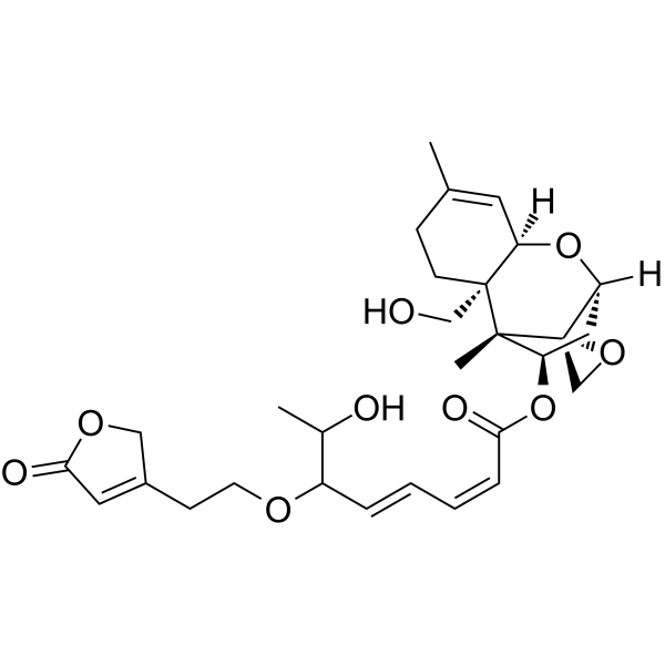
HY-N8400
Roridin L2Natural Product -

HY-N12908
Roridin HNatural Product -
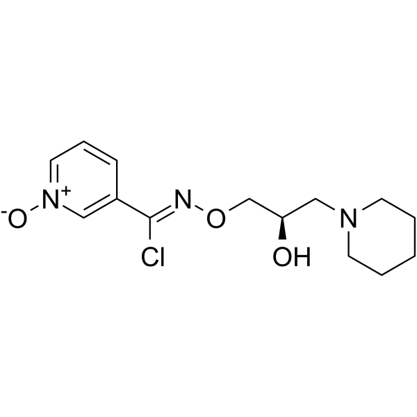
HY-106443
ArimoclomolHSP Co-Inducer -
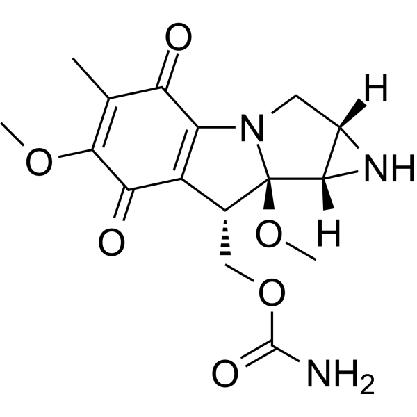
HY-130332
Mitomycin AAntitumor Agent -
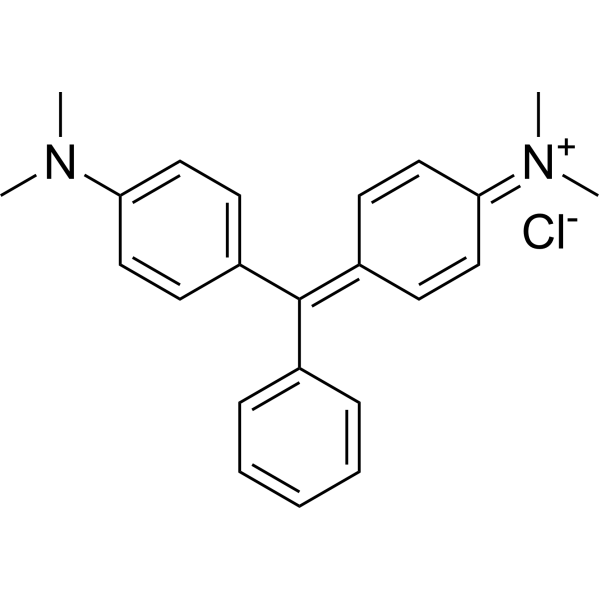
HY-D0203
Basic green 4Cationic Dye -
HY-152072S
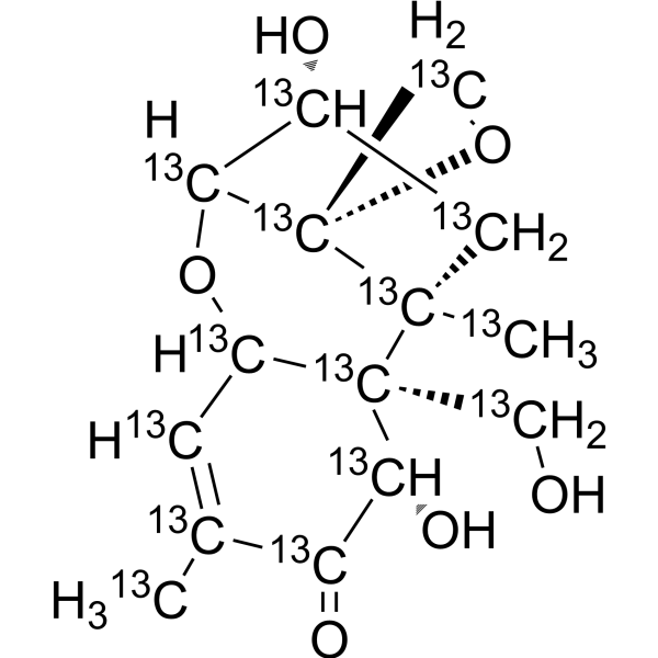
Deoxynivalenol-13C15 -

HY-N12261
Benastatin BGST Inhibitor


Biological Activity
Description
IC50 & Target
[1]|
Fungal Metabolite |
p38 MAPK |
ERK1 |
ERK2 |
Caspase-3 |
PARP |
In Vitro
Satratoxin H (50 nM; 0.5-24 hr) induces apoptosis in PC12 cells via p38 MAPK/JNK-mediated cleavage of caspase-3 and subsequent PARP cleavage[1].
Satratoxin H (5-20 nM; 24 hr) induces chromosome damage in PC12 cells[1].
Satratoxin H (5-50 nM; 12-24 hr) induces DNA double-stranded breaks in PC12 cells, as indicated by increased γH2AX staining at 50 nM (12 and 24 hours) and 5, 10, and 20 nM (24 hours)[1].
Satratoxin H (5-20 nM; 24 hr) does not induce DNA single-stranded breaks in PC12 cells at concentrations of 5, 10, and 20 nM after 24-hour exposure[1].
Satratoxin H (10-100 nM; 6-48 h) induces time- and concentration-dependent cytotoxicity in undifferentiated PC12 cells[2].
Satratoxin H (50 nM; 24 h) induces apoptotic morphological changes (condensed chromatin, nuclear fragmentation, apoptotic bodies) in undifferentiated PC12 cells after 24 h, with some concurrent necrosis[2].
Satratoxin H (50 nM; 18-24 h) increases the percentage of apoptotic undifferentiated PC12 cells to 4.85% after 18 h and 13.25% after 24 h, as measured by flow cytometric subdiploid DNA content analysis[2].
Satratoxin H (5-100 nM; 0.5-6 h) induces time- and concentration-dependent phosphorylation of p38 MAPK, ERK1/2, and JNK in undifferentiated PC12 cells, and co-treatment with antioxidants reduces satratoxin H-induced p38 MAPK phosphorylation[2].
Satratoxin H (1-100 nM; 6-48 h) reduces PC12 cell viability in a concentration- and time-dependent manner[3].
Satratoxin H (1 nM-50 nM; 24 h) inhibits B16F10 mouse melanoma cell proliferation with an IC50 of approximately 20 nM and completely suppresses colony formation at 50 nM[4].
Satratoxin H (1 nM-50 nM; 24 h) induces caspase-dependent apoptosis in B16F10 mouse melanoma cells by upregulating pro-apoptotic Bax and cleaved caspase-3, and downregulating anti-apoptotic Bcl-2, in a concentration-dependent manner[4].
Satratoxin H (1 nM-50 nM; 24 h) induces concentration-dependent activation of endoplasmic reticulum stress pathways (IRE1, PERK, ATF6) and upregulates pro-apoptotic Bim and CHOP mRNA levels in B16F10 mouse melanoma cells[4].
Satratoxin H (1 nM-50 nM; 2 h) induces concentration-dependent intracellular ROS production in B16F10 mouse melanoma cells, and this ROS generation is essential for its cytotoxic effects[4].
Satratoxin H (50 nM; 10 min) induces mitochondrial ROS production in B16F10 mouse melanoma cells, and this effect is dependent on endoplasmic reticulum stress[4].
MedChemExpress (MCE) has not independently confirmed the accuracy of these methods. They are for reference only.
-
Cell Line:PC12
-
Concentration:50 nM
-
Incubation Time:0.5 h; 1 h; 3 h; 6 h; 9 h; 12 h; 18 h; 24 h
-
Result:Induced cleavage of caspase-3, detected as active 19 kDa and 17 kDa fragments, from 3 to 24 hours.
Induced cleavage of PARP, with weak cleavage at 12 hours and strong cleavage at 24 hours.
Decreased the expression level of cleaved caspase-3 when cells were pre-incubated with 10 μM SB203580 or 10 μM SP600125.
Had no effect on the expression levels of pro-apoptotic Bax or anti-apoptotic Bcl-2 over 0.5 to 24 hours.
-
Cell Line:PC12
-
Concentration:5 nM; 10 nM; 20 nM; 50 nM
-
Incubation Time:12 h; 24 h
-
Result:Induced γH2AX (a marker of DNA double-stranded breaks) at 50 nM at 12 hours, with stronger staining observed at 24 hours.
Induced γH2AX at 5, 10, or 20 nM for 24 hours, with similar amounts detected across these concentrations.
-
Cell Line:undifferentiated rat pheochromocytoma PC12 cells
-
Concentration:50 nM
-
Incubation Time:24 h
-
Result:Caused nearly all PC12 cells to exhibit strong Hoechst 33342 staining and typical apoptotic morphological changes, including nuclear condensation, cleavage, and apoptotic bodies.
Induced necrosis in a subset of cells, as indicated by trypan blue positivity.
-
Cell Line:undifferentiated rat pheochromocytoma PC12 cells
-
Concentration:50 nM
-
Incubation Time:18 h; 24 h
-
Result:Increased the percentage of apoptotic PC12 cells (subdiploid DNA content) to 4.85% after 18 h and 13.25% after 24 h, compared to 0.54% in untreated control cells.
-
Cell Line:B16F10 mouse melanoma cells
-
Concentration:1 nM; 10 nM; 50 nM
-
Incubation Time:24 h
-
Result:Elevated protein levels of pro-apoptotic Bax in a concentration-dependent manner.
Decreased levels of anti-apoptotic Bcl-2 in a concentration-dependent manner.
Increased levels of cleaved caspase-3 in a concentration-dependent manner.


Chemical Information
-
CAS No. 53126-64-0
-
Appearance Solid
-
Molecular Weight 528.59
-
Formula C29H36O9
-
Color White to off-white
-
SMILES
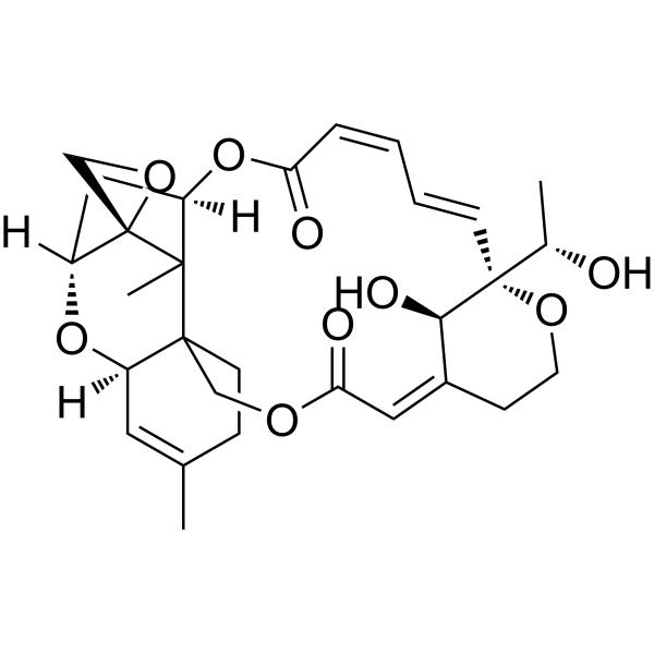
CC([C@]1([H])OC(/C=C\C=C\[C@@]2([C@@H](O)C)[C@H](O)/C(CCO2)=C\C3=O)=O)([C@@]4(OC4)[C@@]([H])(C1)O5)C6(CO3)CCC(C)=C[C@]65[H]
-
Structure Classification
-
Initial Source
Stachybotrys atra
-
Shipping
Room temperature in continental US; may vary elsewhere.
-
Storage
Powder -20°C 3 years 4°C 2 years In solvent -80°C 6 months -20°C 1 month


Purity & Documentation
-
Data Sheet (286 KB)
-
SDS (254 KB)
- English - EN (254 KB)
- Français - FR (254 KB)
- Deutsch - DE (254 KB)
- Norwegian - NO (254 KB)
- Español - ES (254 KB)
- Swedish - SV (254 KB)
- Italian - IT (254 KB)
- Korean - KR (254 KB)
- Portuguese - PT (254 KB)
-
Handling Instructions (2659 KB)
References
[1]. Nusuetrong P, et al. Apoptotic effects of satratoxin H is mediated through DNA double-stranded break in PC12 cells. J Toxicol Sci. 2012;37(4):803-812. [Content Brief]
[2]. Nusuetrong P, et al. Involvement of reactive oxygen species and stress-activated MAPKs in satratoxin H-induced apoptosis. Eur J Pharmacol. 2005;507(1-3):239-246. [Content Brief]
[3]. Nusuetrong P, et al. Satratoxin H generates reactive oxygen species and lipid peroxides in PC12 cells. Biol Pharm Bull. 2008;31(6):1115-1120. [Content Brief]
[4]. Lee J, et al. Roridin E and satratoxin H, macrocyclic trichothecene mycotoxins, induce endoplasmic reticulum stress-dependent apoptosis through ribosome interaction in B16 mouse melanoma cells. Bioorg Chem. 2025;164:108842. [Content Brief]

Calculators
Concentration (start) × Volume (start) = Concentration (final) × Volume (final)


Satratoxin H Related Classifications
HY-123647 Related Classifications
 Powered by Bioz