



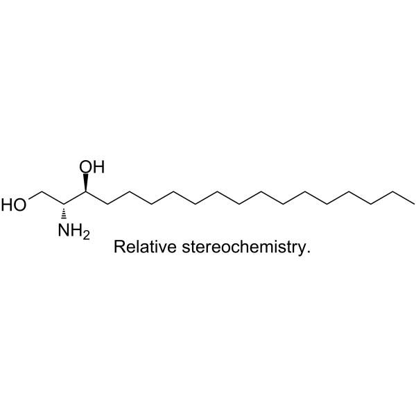
DL-erythro-Dihydrosphingosine

Based on 1 Customer Validation
DL-erythro-Dihydrosphingosine is a sphingosine-based PKC inhibitor. DL-erythro-Dihydrosphingosine directly inhibits the enzymatic activity of PLA2. DL-erythro-Dihydrosphingosine inhibits the activity of Bcl-2 protein involved in apoptosis. DL-erythro-Dihydrosphingosine inhibits the activity of matrix metalloproteinases (MMP). DL-erythro-Dihydrosphingosine is applicable for research on cancer and inflammatory diseases.
For research use only. We do not sell to patients.
- Purity : 98.0%
- CAS No.: 3102-56-5
- Formula: C18H39NO2
- Molecular Weight:301.52
-
Storage:Powder -20°C, 3 years ; In solvent -80°C, 6 months , -20°C, 1 month

To place orders, for customer services and technical support, please contact: MedChemExpress USA
Tel: 609-228-6898 E-mail: [email protected] [email protected]


-
 Biological Activity
-
Chemical Information
- Protocols
- Purity & Documentation
- References
-
 Help & FAQs


-

HY-W688565
(2S)-2-Amino-1-hydroxyoctadecan-3-oneCeramidase Substrate -
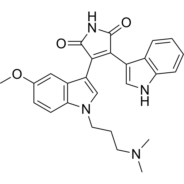
HY-13689
Go 6983PKC Inhibitor -
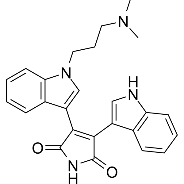
HY-13867
Bisindolylmaleimide IPKC Inhibitor -
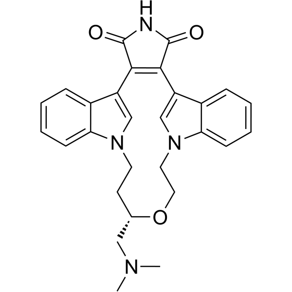
HY-10195
RuboxistaurinPKCβ Inhibitor -
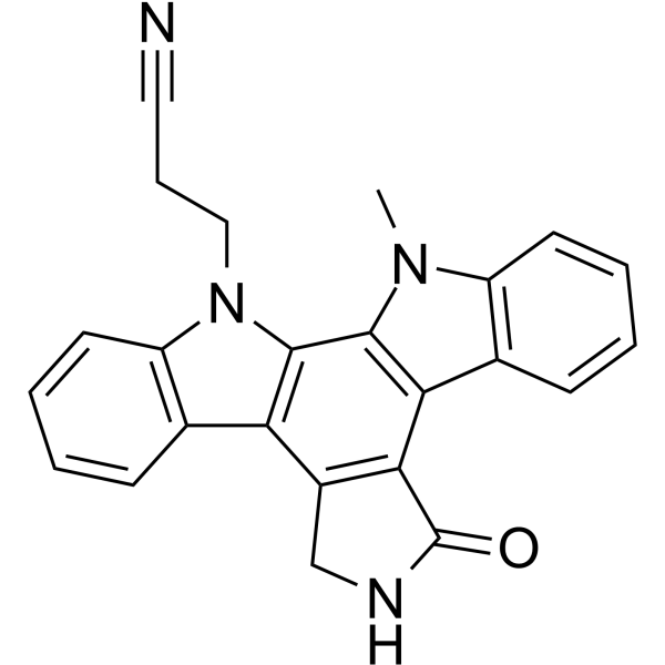
HY-10183
Go6976PKC Inhibitor -
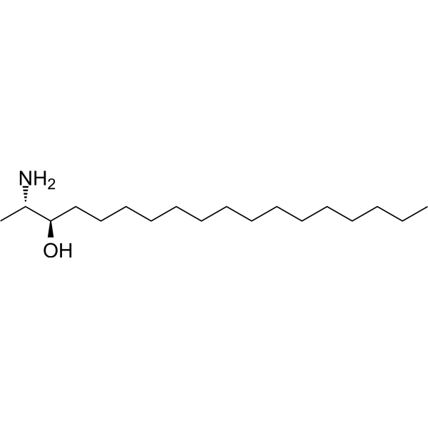
HY-13626
SpisulosineAnticancer Agent -
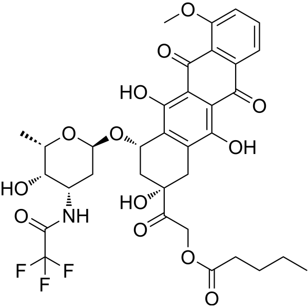
HY-13772
ValrubicinAntibiotic -
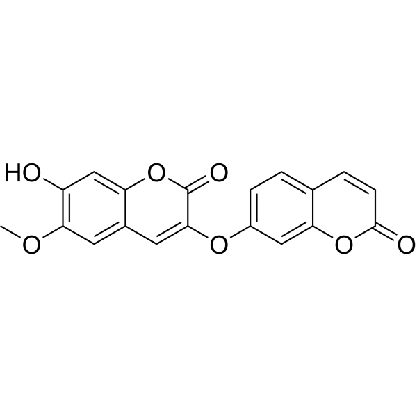
HY-N0699
DaphnoretinPKC Activator -
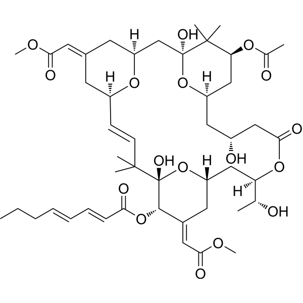
HY-105231
Bryostatin 1PKC Modulator -
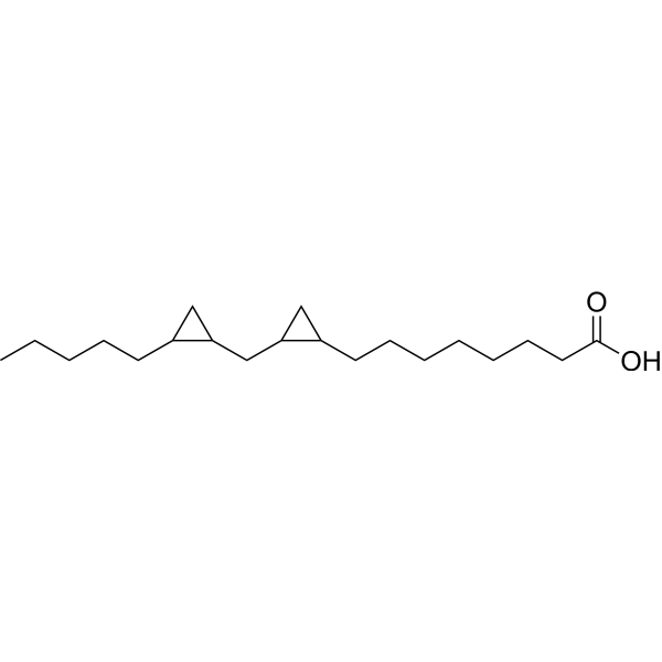
HY-108599
DCP-LAPKC Activator -
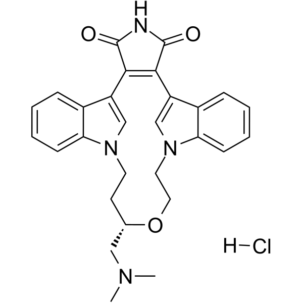
HY-10195B
Ruboxistaurin hydrochloridePKCβ Inhibitor -
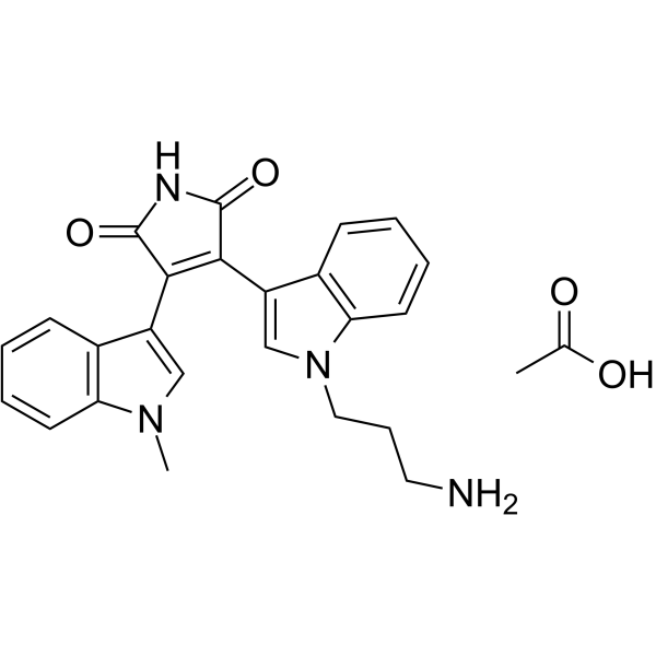
HY-129624A
Bisindolylmaleimide VIII acetatePKC Inhibitor
All Phospholipase Isoforms
More

Biological Activity
Description
Cellular Effect
|
Cell Line

|
Type | Value | Description | References |
|---|---|---|---|---|
| FL5.12 | IC50 |
3.5 μM
Compound: 7
|
Cytotoxicity against mouse FL5.12A cells after 48 hrs by DAPI staining-based flow cytometric analysis
Cytotoxicity against mouse FL5.12A cells after 48 hrs by DAPI staining-based flow cytometric analysis
|
[PMID: 27475534] |
| HBL-100 | GI50 |
25 μM
Compound: 10d
|
Antiproliferative activity against human HBL100 cells after 48 hrs incubation by SRB assay
Antiproliferative activity against human HBL100 cells after 48 hrs incubation by SRB assay
|
[PMID: 25899335] |
| HeLa | GI50 |
15 μM
Compound: 10d
|
Antiproliferative activity against human HeLa cells after 48 hrs incubation by SRB assay
Antiproliferative activity against human HeLa cells after 48 hrs incubation by SRB assay
|
[PMID: 25899335] |
| Jurkat | IC50 |
3.53 μM
Compound: Dihydrosphingosine
|
Inhibition of wild type human Jurkat cells after 18 hrs by MTT assay
Inhibition of wild type human Jurkat cells after 18 hrs by MTT assay
|
[PMID: 17400555] |
| Jurkat | IC50 |
3.68 μM
Compound: Dihydrosphingosine
|
Inhibition of 2-amino-4-(4-heptyloxyphenyl)-2-methylbutanol-resistant human Jurkat SBR2 mutant cells proliferation after 18 hrs by MTT assay
Inhibition of 2-amino-4-(4-heptyloxyphenyl)-2-methylbutanol-resistant human Jurkat SBR2 mutant cells proliferation after 18 hrs by MTT assay
|
[PMID: 17400555] |
| Jurkat | IC50 |
4.9 μM
Compound: Dihydrosphingosine
|
Inhibition of 2-amino-4-(4-heptyloxyphenyl)-2-methylbutanol-resistant human Jurkat SBR1 mutant cells proliferation after 18 hrs by MTT assay
Inhibition of 2-amino-4-(4-heptyloxyphenyl)-2-methylbutanol-resistant human Jurkat SBR1 mutant cells proliferation after 18 hrs by MTT assay
|
[PMID: 17400555] |
| Jurkat | IC50 |
6.31 μM
Compound: Dihydrosphingosine
|
Inhibition of human 2-amino-4-(4-heptyloxyphenyl)-2-methylbutanol-resistant human Jurkat SBR3 mutant cells proliferation after 18 hrs by MTT assay
Inhibition of human 2-amino-4-(4-heptyloxyphenyl)-2-methylbutanol-resistant human Jurkat SBR3 mutant cells proliferation after 18 hrs by MTT assay
|
[PMID: 17400555] |
| SW1573 | GI50 |
4.5 μM
Compound: 10d
|
Antiproliferative activity against human SW1573 cells after 48 hrs incubation by SRB assay
Antiproliferative activity against human SW1573 cells after 48 hrs incubation by SRB assay
|
[PMID: 25899335] |
| T47D | GI50 |
22 μM
Compound: 10d
|
Antiproliferative activity against human T47D cells after 48 hrs incubation by SRB assay
Antiproliferative activity against human T47D cells after 48 hrs incubation by SRB assay
|
[PMID: 25899335] |
| WiDr | GI50 |
19 μM
Compound: 10d
|
Antiproliferative activity against human WiDr cells after 48 hrs incubation by SRB assay
Antiproliferative activity against human WiDr cells after 48 hrs incubation by SRB assay
|
[PMID: 25899335] |
In Vitro
DL-erythro-Dihydrosphingosine inhibits the activity of Bcl-2 protein involved in apoptosis, inhibits the activity of matrix metalloproteinases (MMPs)[1][2].
MedChemExpress (MCE) has not independently confirmed the accuracy of these methods. They are for reference only. Further protocols information, click here.


Chemical Information
-
CAS No. 3102-56-5
-
Appearance Solid
-
Molecular Weight 301.52
-
Formula C18H39NO2
-
Color Off-white to light brown
-
SMILES
CCCCCCCCCCCCCCC[C@H](O)[C@H](N)CO
-
Shipping
Room temperature in continental US; may vary elsewhere.
-
Storage
Powder -20°C 3 years In solvent -80°C 6 months -20°C 1 month

Protocols
-
Apoptosis
Apoptosis, also called programmed cell death, is generally characterized by distinct morphological characteristics.
-
TUNEL staining for apoptotic DNA fragmentation
TUNEL staining detects DNA strand breaks by using terminal deoxynucleotidyl transferase to add labeled nucleotides to exposed 3′-OH DNA termini, generating either microscopic staining in fixed cells or tissue sections, or fluorescence/cytometric signal in cell suspensions. TUNEL positivity reflects DNA fragmentation but should not be interpreted alone as definitive apoptosis, because TUNEL can also label necrotic, autolytic, mechanically damaged, or DNA-repair-associated DNA breaks.
-
Mitochondrial membrane-potential fluorescent assay
Mitochondrial membrane potential fluorescent assays estimate ΔΨm in living cells using lipophilic cationic dyes such as TMRM, TMRE, rhodamine 123, and JC-1, which accumulate in mitochondria according to membrane polarization; loss of signal after FCCP or CCCP treatment is interpreted as mitochondrial depolarization. TMRM/TMRE and rhodamine 123 are commonly used for semi-quantitative live-cell microscopy or flow cytometry, while JC-1 can report a shift from red aggregate fluorescence to green monomer fluorescence during depolarization; interpretation requires controls because dye concentration, quenching mode, cell type, dye efflux, and mitochondrial mass can affect fluorescence independently of ΔΨm.
-
Annexin V plus membrane-impermeant dye apoptosis staining
Annexin V-based apoptosis assays rely on the detection of phosphatidylserine (PS) externalization from the inner leaflet of the plasma membrane to the outer leaflet, an early biochemical hallmark of apoptosis. Fluorescently labeled Annexin V binds PS in a calcium-dependent manner, enabling identification of early apoptotic cells by flow cytometry or fluorescence microscopy. When combined with a membrane-impermeant DNA-binding dye (e. g. , propidium iodide), this approach allows discrimination between viable (Annexin V−/dye−), early apoptotic (Annexin V+/dye−), and late apoptotic or necrotic (Annexin V+/dye+) cell populations by assessing membrane integrity and PS exposure.
-
Mitochondrial membrane-potential and mitochondrial mass staining
Mitochondrial membrane potential staining measures the electrochemical polarization across the mitochondrial inner membrane in live cells using lipophilic cationic fluorescent probes; early rhodamine-based work showed that selective mitochondrial dye accumulation is lost when the mitochondrial transmembrane potential is dissipated. JC-1 reports mitochondrial polarization by shifting from green monomer fluorescence to red J-aggregate fluorescence as dye concentration increases within energized mitochondria; therefore, the red/green fluorescence ratio is used as a relative readout of mitochondrial membrane potential. TMRE or TMRM staining provides a single-channel relative readout because these cationic rhodamine esters accumulate in polarized mitochondria, and lower fluorescence indicates reduced mitochondrial polarization when acquisition and dye-loading conditions are controlled. Mitochondrial mass staining is commonly performed with MitoTracker Green FM or related MitoTracker dyes as
-
Apoptosis Solutions
Apoptosis is a regulated, generally non-lytic cell-death pathway that removes unwanted, damaged, infected, or abnormal cells through coordinated morphological changes, caspase activation, DNA fragmentation, and membrane remodeling. The intrinsic apoptosis pathway is controlled mainly by mitochondrial outer membrane permeabilization, BCL-2 family proteins, cytochrome c release, apoptosome formation, caspase-9 activation, and downstream executioner caspase-3/7 activation. The extrinsic apoptosis pathway is initiated by death receptors such as Fas, TNFR, and TRAIL receptors, which recruit adaptor proteins and activate caspase-8 before engaging executioner caspases or mitochondrial amplification through BID cleavage. Apoptosis is linked to many phenotypes, including cancer cell killing, tissue homeostasis, immune regulation, neurodegeneration, infection response, and treatment-induced cytotoxicity; unresolved questions include how apoptosis interacts with necroptosis, pyroptosis, ferroptos


Purity & Documentation
-
Data Sheet (265 KB)
-
SDS (252 KB)
- English - EN (252 KB)
- Français - FR (252 KB)
- Deutsch - DE (252 KB)
- Norwegian - NO (252 KB)
- Español - ES (252 KB)
- Swedish - SV (252 KB)
- Italian - IT (252 KB)
- Korean - KR (252 KB)
- Portuguese - PT (252 KB)
-
Handling Instructions (2659 KB)
References
[1]. Chao R, et al. Retinoblastoma protein dephosphorylation induced by D-erythro-sphingosine. J Biol Chem. 1992;267(33):23459-23462. [Content Brief]
[2]. Nakamura H, et al. Inhibition of arachidonic acid release and cytosolic phospholipase A2 alpha activity by D-erythro-sphingosine. Eur J Pharmacol. 2004;484(1):9-17. [Content Brief]

Calculators
Concentration (start) × Volume (start) = Concentration (final) × Volume (final)


DL-erythro-Dihydrosphingosine Related Classifications
HY-101195 Related Classifications
 Powered by Bioz