



PIT-1

Based on 1 Customer Validation
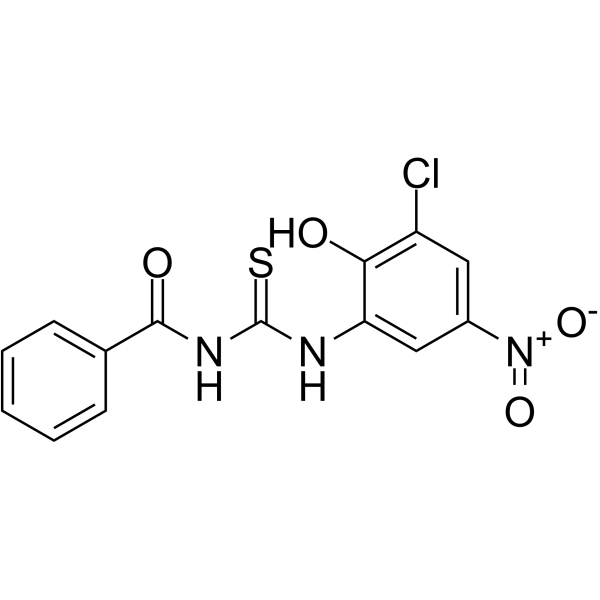
PIT-1 is a selective PIP3 (phosphatidylinositol-3,4,5-trisphosphate) antagonist targeting the PH domain, with a Kd of 43.2 μM for Akt-SH, and exhibits antitumor activity. PIT-1 blocks PIP3-mediated translocation of the PH domain to the plasma membrane, thereby specifically inhibiting the PI3K-PDK1-Akt signaling pathway and the ARF6 signaling pathway, consequently suppressing phosphorylation of downstream substrates, triggering AMPK/ACC energy metabolic stress and autophagy (autophagy), and inducing Bax/Bak- and caspase-dependent apoptosis (apoptosis). PIT-1 also binds to the TcAkt-PH domain of Trypanosoma cruzi, suggesting its antiparasitic potential. PIT-1 can be used for research on cancer and Chagas disease.
For research use only. We do not sell to patients.
- Purity : 98.08%
- CAS No.: 53501-41-0
- Formula: C14H10ClN3O4S
- Molecular Weight:351.76
-
Storage:
4°C, protect from light
* In solvent : -80°C, 6 months; -20°C, 1 month (protect from light)

To place orders, for customer services and technical support, please contact: MedChemExpress USA
Tel: 609-228-6898 E-mail: [email protected] [email protected]


-
 Biological Activity
-
Chemical Information
-
Solvent & Solubility
- Protocols
- Purity & Documentation
- References
-
 Help & FAQs


-
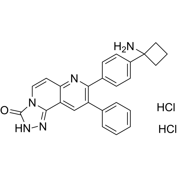
HY-10358
MK-2206 dihydrochlorideAllosteric Akt Inhibitor -
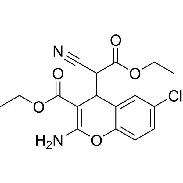
HY-18749
SC79Akt Activator -
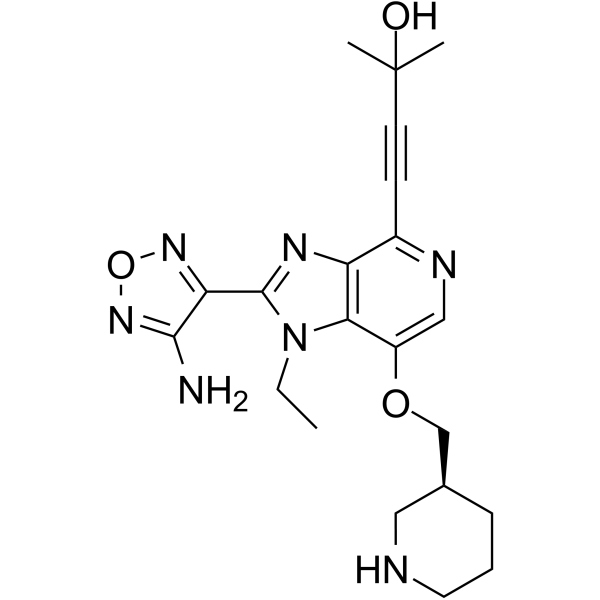
HY-10249
GSK-690693Akt Inhibitor -
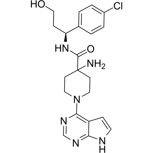
HY-15431
CapivasertibAkt Inhibitor -
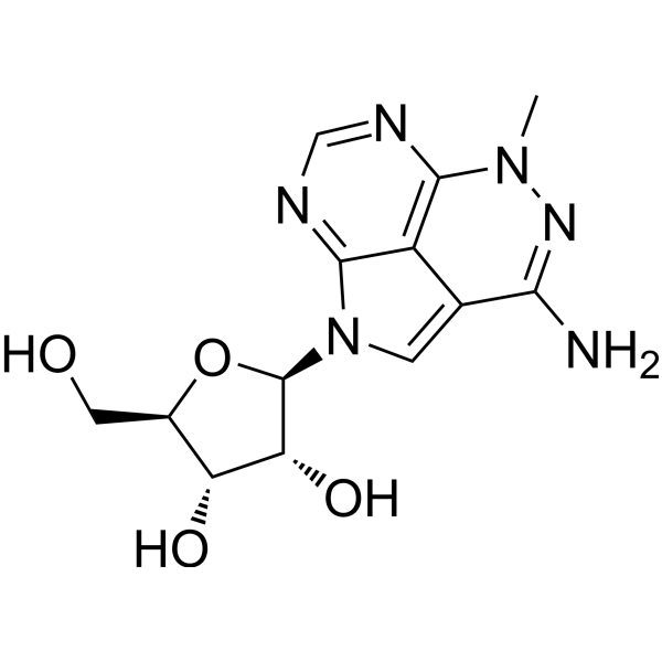
HY-15457
TriciribineHIV Inhibitor -
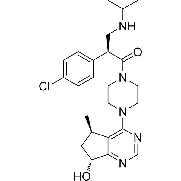
HY-15186
IpatasertibPan-Akt Inhibitor -
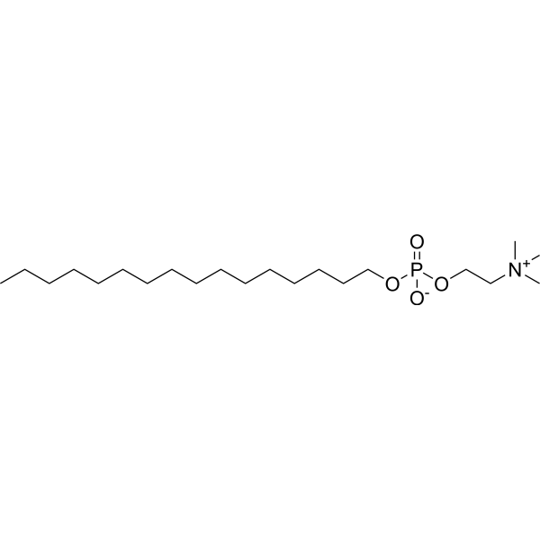
HY-13685
MiltefosinePI3K/Akt Inhibitor -

HY-103224R
PIT-1 (Standard)Reference Standard -
HY-15965
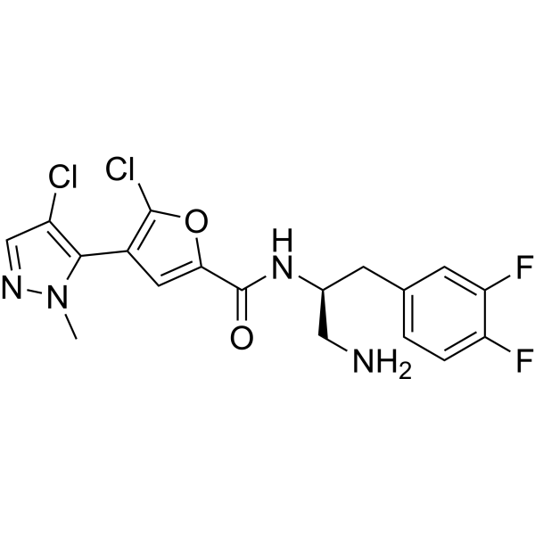
UprosertibAkt Inhibitor -
HY-50909
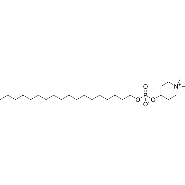
PerifosineAkt Inhibitor -
HY-10425
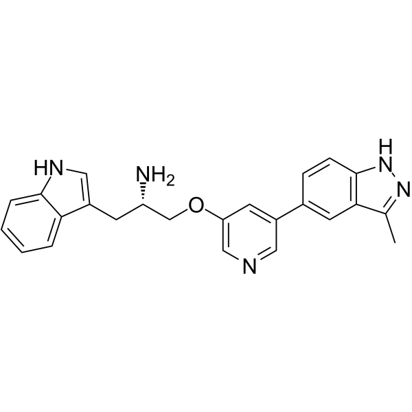
Rizavasertibpan-Akt Inhibitor


Biological Activity
Description
|
Akt-SH 43.2 μM (Kd) |
Caspase-3 |
ARF6 |
Cellular Effect
|
Cell Line

|
Type | Value | Description | References |
|---|---|---|---|---|
| U-87MG ATCC | IC50 |
37.4 μM
|
Cytotoxicity against human U87MG glioblastoma cells assessed as viability reduction by PIT-1.
Cytotoxicity against human U87MG glioblastoma cells assessed as viability reduction by PIT-1.
|
21041639 |
| U-87MG ATCC | IC50 |
103.6 μM
|
Cytotoxicity against human U87MG glioblastoma cells overexpressing PH-domain-deficient myristoylated Akt assessed as viability reduction by PIT-1.
Cytotoxicity against human U87MG glioblastoma cells overexpressing PH-domain-deficient myristoylated Akt assessed as viability reduction by PIT-1.
|
21041639 |
In Vitro
PIT-1 (12.5-100 μM; 48 h) dose-dependently reduces tumor cell viability in U87MG (IC50 = 39.9 μM), SUM159, A549, and MDA-MB-231 cells, and induces cell rounding, loss of adhesion, and death in U87MG cells[1].
PIT-1 (25-100 μM; 48 h) preferentially kills PTEN-inactivated cells in U87MG cells expressing wild-type PTEN or the inactivating mutant PTEN (R130M)[1].
PIT-1 (100 μM; 24 h) significantly enhances TRAIL-induced cell killing in TRAIL-resistant U87MG cells[1].
PIT-1 (100 μM; 48 h) significantly increases the phosphorylation levels of the energy metabolism stress markers P-AMPK and P-ACC in U87MG cells[1].
PIT-1 (50 μM; 2 h, followed by PDGF stimulation for 5 min) significantly inhibits PDGF-induced plasma membrane translocation of ARNO-PH (inhibition rate approximately 76%) and GRP1-PH in serum-starved SUM159 cells transfected with GFP-fused PH domain plasmids, but does not inhibit the plasma membrane localization and translocation of the PI (4,5) P2-specific EFA6-PH[2].
PIT-1 (50-100 μM; 2 h, followed by PDGF stimulation for 15 min) significantly inhibits PDGF-induced ARF6 activation in serum-starved SUM159 cells and shows a weaker inhibitory effect on ARF1 activation[2].
PIT-1 (25-100 μM; 2 h, followed by PDGF stimulation for 15 min) significantly inhibits PDGF-induced membrane ruffling/lamellipodia formation in serum-starved SUM159 cells, reduces F-actin at leading-edge lamellipodia and stress fibers, and induces polygonal radial morphological changes[2].
PIT-1 (2 h, followed by PDGF stimulation for 15 min) significantly inhibits lamellipodium formation and directional cell polarization at the scratch edge in serum-starved SUM159 cells[2].
PIT-1 (50-100 μM) inhibits ARF6-induced lamellipodia in SUM159 cells, but does not inhibit lamellipodia driven by constitutively active ARF6Q67L[2].
PIT-1 inhibits GRP1/ARNO-driven lamellipodia in SUM159 cells, whereas EFA6-driven lamellipodia are insensitive to it[2].
PIT-1 (6.25-100 μM; 8 h) inhibits the migration and invasion ability of SUM159[2].
PIT-1 (12.5 μM; 114 h, followed by washout for 96 h) reversibly inhibits the acquisition of invasive morphology by SUM159 cells in three-dimensional Matrigel culture[2].
PIT-1 (6.25-100 μM) inhibits scratch-wound migration of SUM159 cells overexpressing ARF6, GRP1, or ARNO, whereas constitutively active ARF6Q67L and EFA6 attenuate this inhibition[2].
PIT-1 (25-100 μM; 48 h) significantly increases the proportion of sub-G1 DNA cells in U87MG cells, induces dose-dependent cleavage and activation of Caspase-3 and PARP, and causes nuclear pyknosis and fragmentation[1].
PIT-1 (100 μM; pretreatment for 1 h, followed by PDGF stimulation for 5 min) significantly inhibits PDGF-induced plasma membrane translocation of Akt-PH and GRP1-PH, but not Btk-PH translocation, in SUM159 cells transfected with GFP-PH plasmid and serum-starved overnight[1].
PIT-1 (12.35-51.45 μM; association 60 s, dissociation 30 s) reversibly and directly binds the recombinant Akt-PH domain in SPR experiments, with Kd = 43.2 μM[1].
PIT-1 (100 μM; 24 h) exhibits significantly stronger viability inhibition against AKT1+/+ cells in mouse lung fibroblasts expressing Akt1 (AKT1+/+) and in fibroblasts with triple knockout of all Akt isoforms (AKT-/-)[1].
PIT-1 (25-100 µM) preferentially reduces the viability of PTEN-deficient U87MG cells compared with U87MG cells expressing wild-type PTEN[1].
PIT-1 (6.25-100 μM; 12 h) does not change intracellular PIP3 levels in U87MG cells[1].
PIT-1 (6.25-200 μM; 40 min at 15°C) inhibits the binding of PIP3 to the PH domains of recombinant Akt, GRP1, and ARNO in vitro, with IC50 values of 19.8, 26.3, and 30.2 μM, respectively[2].
PIT-1 (50 μM; 2 h, followed by fMLP stimulation for 3 min) significantly inhibits fMLP (HY-P0224)-induced cell polarization in actin fluorescence staining experiments of human neutrophil-like HL-60 cells[2].
PIT-1 (25-100 μM; 2 h, followed by bFGF stimulation for 15 min) inhibits bFGF-induced actin remodeling and lamellipodia in serum-starved HUVEC[2].
MedChemExpress (MCE) has not independently confirmed the accuracy of these methods. They are for reference only.
-
Cell Line:SUM159 human breast carcinoma cells
-
Concentration:25, 50, and 100 μM
-
Incubation Time:2 h (before PDGF stimulation); 15 min (PDGF stimulation)
-
Result:Reduced the percentage of PDGF-stimulated cells with lamellipodia at 25, 50, and 100 μM; representative images at 25 μM showed loss of lamellipodia.
-
Cell Line:Human cancer cell lines
-
Concentration:6.25, 12.5, 25, 50, and 100 μM
-
Incubation Time:8 h
-
Result:Inhibited transwell migration in a concentration-dependent manner; 6.25 and 12.5 μM suppressed migration at 8 h under non-cytotoxic conditions.
Inhibited wound closure in a concentration-dependent manner.
-
Cell Line:Human umbilical vein endothelial cells (HUVEC)
-
Concentration:3.13, 6.25, 12.5, 25, 50, and 100 μM
-
Incubation Time:8 h
-
Result:Inhibited HUVEC migration in both wound healing and transwell assays in a concentration-dependent manner; no loss of viability was observed under the assay conditions.
-
Cell Line:PTEN-deficient human glioblastoma U87MG cells
-
Concentration:25, 50, and 100 μM
-
Incubation Time:12 h
-
Result:Dose-dependently suppressed phosphorylation of Akt (Ser473/Thr308), GSK-3β (Ser9), p70S6K (Thr389), and S6 (Ser235/236).
In Vivo
MedChemExpress (MCE) has not independently confirmed the accuracy of these methods. They are for reference only.
- Kinase activity and phosphorylation assays
- Western Blot
- Detection of Apoptosis
- TUNEL staining for apoptotic DNA fragmentation
- Annexin V plus membrane-impermeant dye apoptosis staining
- Autophagy
- Apoptosis Solutions
- Protocol for Kinase activity and phosphorylation assays
- Lysosome and acidic-vesicle live-cell staining
- Subcutaneous Cell-Line-Derived Xenograft
- Macroautophagy Solutions


Chemical Information
-
CAS No. 53501-41-0
-
Appearance Solid
-
Molecular Weight 351.76
-
Formula C14H10ClN3O4S
-
Color Light yellow to green yellow
-
SMILES
O=C(NC(NC1=CC([N+]([O-])=O)=CC(Cl)=C1O)=S)C2=CC=CC=C2
-
Shipping
Room temperature in continental US; may vary elsewhere.
-
Storage
4°C, protect from light
* In solvent : -80°C, 6 months; -20°C, 1 month (protect from light)


Solvent & Solubility
In Vitro:
DMSO : 62.5 mg/mL (177.68 mM; ultrasonic and warming and heat to 60°C; Hygroscopic DMSO has a significant impact on the solubility of product, please use newly opened DMSO)
Please refer to the solubility information to select the appropriate solvent. Once prepared, please aliquot and store the solution to prevent product inactivation from repeated freeze-thaw cycles.
Storage method and period of stock solution: -80°C, 6 months; -20°C, 1 month (protect from light). When stored at -80°C, please use it within 6 months. When stored at -20°C, please use it within 1 month.
Please refer to the solubility information to select the appropriate solvent. Once prepared, please aliquot and store the solution to prevent product inactivation from repeated freeze-thaw cycles.
Storage method and period of stock solution: -80°C, 6 months; -20°C, 1 month (protect from light). When stored at -80°C, please use it within 6 months. When stored at -20°C, please use it within 1 month.
Concentration (start) × Volume (start) = Concentration (final) × Volume (final)
In Vivo Dissolution Calculator
Please enter the basic information of animal experiments:
-
-
-
-
Recommended: Prepare an additional quantity of animals to account for potential losses during experiments.
Working solution concentration: 0.22 mg/mL


Purity & Documentation


References

Complete Stock Solution Preparation Table
Please refer to the solubility information to select the appropriate solvent. Once prepared, please aliquot and store the solution to prevent product inactivation from repeated freeze-thaw cycles.
Storage method and period of stock solution: -80°C, 6 months; -20°C, 1 month (protect from light). When stored at -80°C, please use it within 6 months. When stored at -20°C, please use it within 1 month.
| Optional Solvent | Concentration Solvent Mass | 1 mg | 5 mg | 10 mg | 25 mg |
|---|---|---|---|---|---|
| DMSO | 1 mM | 2.8428 mL | 14.2142 mL | 28.4285 mL | 71.0712 mL |
| 5 mM | 0.5686 mL | 2.8428 mL | 5.6857 mL | 14.2142 mL | |
| 10 mM | 0.2843 mL | 1.4214 mL | 2.8428 mL | 7.1071 mL | |
| 15 mM | 0.1895 mL | 0.9476 mL | 1.8952 mL | 4.7381 mL | |
| 20 mM | 0.1421 mL | 0.7107 mL | 1.4214 mL | 3.5536 mL | |
| 25 mM | 0.1137 mL | 0.5686 mL | 1.1371 mL | 2.8428 mL | |
| 30 mM | 0.0948 mL | 0.4738 mL | 0.9476 mL | 2.3690 mL | |
| 40 mM | 0.0711 mL | 0.3554 mL | 0.7107 mL | 1.7768 mL | |
| 50 mM | 0.0569 mL | 0.2843 mL | 0.5686 mL | 1.4214 mL | |
| 60 mM | 0.0474 mL | 0.2369 mL | 0.4738 mL | 1.1845 mL | |
| 80 mM | 0.0355 mL | 0.1777 mL | 0.3554 mL | 0.8884 mL | |
| 100 mM | 0.0284 mL | 0.1421 mL | 0.2843 mL | 0.7107 mL |


PIT-1 Related Classifications
HY-103224 Related Classifications
 Powered by Bioz