



Epimedin B
Based on 5 publication(s) in Google Scholar
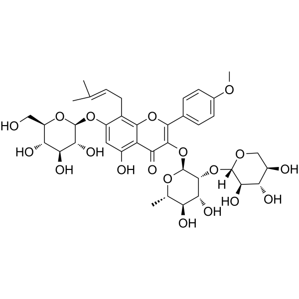
Epimedin B (Epmedin B) is a flavonoid active component found in Epimedium, with oral activity, and exhibits anti-osteoporotic and neuroprotective effects. Epimedin B inhibits RANKL-induced osteoclast differentiation, F-actin ring formation, and mature osteoclast bone resorption, inhibits the phosphorylation of JNK, p38 MAPK, PI3K, and AKT, activates the AMPK-Nrf2 antioxidant pathway, and reduces cellular and mitochondrial ROS. Epimedin B acts on ESR1 and exerts its effects via GPER. Epimedin B alleviates bone loss and improves bone microstructure in vivo, and in Parkinson's models protects dopaminergic neurons and maintains striatal dopamine levels through anti-apoptotic and anti-endoplasmic reticulum stress effects. Epimedin B can be used for research related to osteoporosis, diabetic osteoporosis, and Parkinson's disease.
For research use only. We do not sell to patients.
- Purity : 99.86%
- CAS No.: 110623-73-9
- Formula: C38H48O19
- Molecular Weight:808.78
-
Storage:
4°C, protect from light
* In solvent : -80°C, 6 months; -20°C, 1 month (protect from light)

To place orders, for customer services and technical support, please contact: MedChemExpress USA
Tel: 609-228-6898 E-mail: [email protected] [email protected]


-
 Biological Activity
-
Chemical Information
-
Solvent & Solubility
- Protocols
- Purity & Documentation
- References
-
 Help & FAQs


-
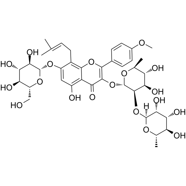
HY-N0260
Epmedin CAnti-inflammatory Agent -
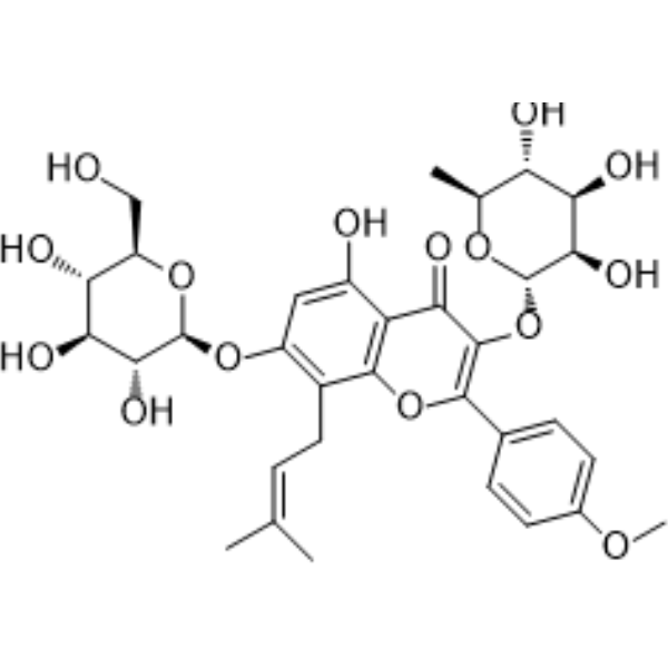
HY-N0014
IcariinPPAR Activator -
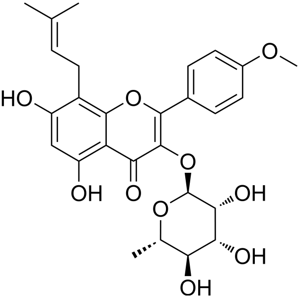
HY-N0011
Baohuoside ICXCR4 Inhibitor -
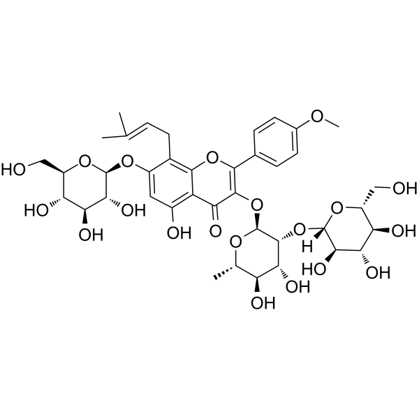
HY-N0257
Epimedin ANatural Product -
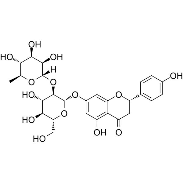
HY-N0153
NaringinCytochrome P450 Inhibitor -
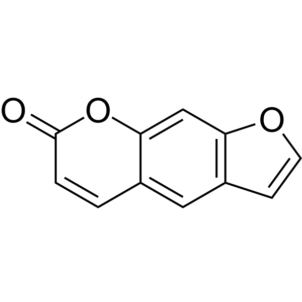
HY-N0053
PsoralenBioactive Natural Compound -
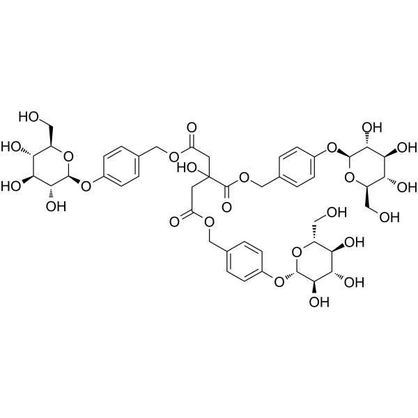
HY-N2031
ParishinSir2/Uth1/TOR Modulator -
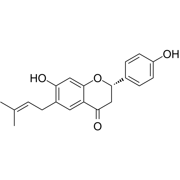
HY-N0233
BavachinPhytoestrogen -
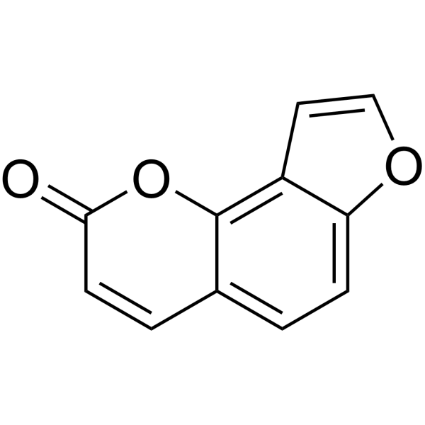
HY-N0763
AngelicinApoptosis Inducer
Publications Citing Use of MedChemExpress (MCE) Epimedin B
MoreAll AMPK Isoforms
More

Biological Activity
Description
In Vitro
Epimedin B (5-80 μM; 4 days) shows no cytotoxicity in BMMs[1].
Epimedin B (10-40 μM; 1-3 days) concentration-dependently inhibits RANKL-induced osteoclastogenesis in BMMs, with the strongest inhibitory effect observed in the early stage of differentiation[1].
Epimedin B (20-40 μM) concentration-dependently inhibits F-actin ring formation and nuclear translocation of NFATc1 in RANKL-induced BMMs[1].
Epimedin B (20-40 μM; 48 h) concentration-dependently reduces the bone resorption activity of mature osteoclasts derived from BMMs[1].
Epimedin B (20-40 μM; 1-5 days) downregulates RANKL-induced osteoclast-specific gene and protein expression in BMMs in a concentration-dependent manner[1].
Epimedin B (40 μM; 15-60 min) inhibits RANKL-induced phosphorylation of JNK, p38, PI3K, and AKT in BMMs, but does not affect ERK phosphorylation[1].
Epimedin B (40 μM; 15-60 min) activates the AMPK pathway in RANKL-induced BMMs, elevates p-AMPK levels, and its activating effect persists up to 60 min[1].
Epimedin B (40 μM; 48 h) alters the transcriptomic profile of RANKL-induced BMMs, with enrichment of pathways related to osteoclast differentiation, AMPK, PI3K-AKT, and MAPK signaling[1].
Epimedin B (2 h) directly binds to the ESR1 protein, stabilizing it during thermal degradation, with a predicted binding energy of −7.8 kcal/mol[1].
Epimedin B directly binds to the N-terminal hydrophobic core of mouse GPER in silico, with a binding free energy of −7.3 kcal/mol[2].
Epimedin B is a major component of ETF, with a concentration of 14.9 mg/g in the tested extract[3].
Epimedin B (20-40 μM; 48 h) concentration-dependently reduces intracellular and mitochondrial ROS levels in BMMs[1].
Epimedin B (20-40 μM; 1-5 days) activates the Nrf2-Keap1 antioxidant pathway and upregulates the expression of downstream antioxidant enzymes in BMMs[1].
MedChemExpress (MCE) has not independently confirmed the accuracy of these methods. They are for reference only.
-
Cell Line:bone marrow-derived macrophages (BMMs)
-
Concentration:5, 10, 20, 40, 80 μM
-
Incubation Time:4 days
-
Result:Exhibited no cytotoxic effects on BMM proliferation at concentrations up to 80 μM.
-
Cell Line:bone marrow-derived macrophages (BMMs)
-
Concentration:10, 20, 40 μM (concurrent with RANKL induction); 40 μM (stage-specific: days 1-3, days 3-5, or days 5-7)
-
Incubation Time:until differentiation assessment (concurrent RANKL induction); days 1-3, days 3-5, or days 5-7 (stage-specific)
-
Result:Suppressed RANKL-induced osteoclastogenesis in a concentration-dependent manner, as evidenced by reduced numbers of TRAP-positive multinucleated cells.
Suppressed osteoclast differentiation during the early stage (days 1-3) and mid-stage (days 3-5), with a more pronounced inhibitory effect observed in the early stage.
-
Cell Line:bone marrow-derived macrophages (BMMs)
-
Concentration:20 μM, 40 μM (gene expression analysis); 40 μM (protein expression analysis)
-
Incubation Time:upon significant osteoclast differentiation in control group (gene expression); 1, 3, and 5 days (protein expression)
-
Result:Suppressed the expression of osteoclast-specific genes-including Acp5, Nfatc1, Fos, Ctsk, Mmp9, and Atp6v0d2-which were upregulated upon RANKL stimulation, in a concentration-dependent manner.
Inhibited the expression of osteoclast differentiation-related proteins, such as NFATc1, MMP9, CTSK, and c-Fos, across the differentiation time course.
-
Cell Line:bone marrow-derived macrophages (BMMs)
-
Concentration:40 μM
-
Incubation Time:15, 30, 45, and 60 min
-
Result:Substantially inhibited phosphorylated JNK after 15 min of treatment.
Markedly suppressed phosphorylated p38 at 30 min.
Exhibited no significant effect on RANKL-mediated p-ERK levels over a 0-60 min period.
Downregulated phosphorylation levels of both PI3K and AKT after stimulation.
-
Cell Line:bone marrow-derived macrophages (BMMs)
-
Concentration:40 μM (Western blot); 40 μM (immunofluorescence)
-
Incubation Time:15, 30, 45, and 60 min (Western blot); 60 min (immunofluorescence)
-
Result:Significantly enhanced the phosphorylation level of AMPK at an early stage, leading to sustained activation of this pathway.
Molecular docking suggested a direct interaction between Epimedin B and the binding pocket of AMPK, with a binding affinity of −7.4 kcal/mol.
Upregulated p-AMPK expression, particularly in the cytoplasm, as demonstrated by immunofluorescence.
-
Cell Line:bone marrow-derived macrophages (BMMs)
-
Concentration:20 μM, 40 μM
-
Incubation Time:1, 3, 5 days
-
Result:Significantly upregulated Nrf2 expression and activated the Nrf2-Keap1 pathway, thereby enhancing the expression of downstream antioxidant enzymes including HO-1, SOD2, and Catalase.
Parmacokinetics
| Species | Dose | Route | AUC0-t | Cmax | Tmax | T1/2 | MRT0-t | CL/F | Vz/F |
|---|---|---|---|---|---|---|---|---|---|
| Rat[3] | 15 mg/kg | p.o. | 96.7 ng·h/mL | 33.6 ng/mL | 0.9 h | 2.3 h | 2.9 h | 163.9 L/h/kg | 553.6 L/kg |
In Vivo
Epimedin B (100 mg/kg; p.o.; single dose) alone does not induce significant liver injury in mice under normal physiological conditions[5].
Epimedin B (100 mg/kg; p.o.; single dose) does not cause significant liver injury in mice under TNF-α-mediated immune stress conditions[5].
Epimedin B (1-20 mg/kg; p.o.; once daily; for 8 consecutive days) exerts neuroprotective effects in MPTP (HY-W114750)-induced Parkinson's disease mice, increasing striatal dopamine levels and improving motor function[2].
Epimedin B (10 mg/kg; p.o.; once daily; for 8 consecutive days) enhances the survival of TH-immunoreactive neurons in MPTP-induced Parkinson's disease mice through anti-apoptotic and anti-endoplasmic reticulum stress effects, and this effect depends on GPER expression[2].
Epimedin B (10-40 mg/kg; intragastric administration; once daily; for 8 consecutive weeks) alleviates STZ (HY-13753)-induced diabetic osteoporosis in rats by promoting bone formation, inhibiting bone resorption and inflammatory responses, and regulating the OPG/RANKL axis[4].
MedChemExpress (MCE) has not independently confirmed the accuracy of these methods. They are for reference only.
-
Animal Model:C57BL/6J (female; 6 weeks old; bilateral ovariectomy-induced estrogen deficiency-associated bone loss)[1]
-
Dosage:10 mg/kg; 20 mg/kg
-
Administration:i.p.; every other day; 8 weeks
-
Result:Markedly attenuated ovariectomy-induced bone loss in tibiae, with improvements in trabecular number, trabecular separation, trabecular thickness, and bone volume fraction.
Showed analogous microstructural improvements in lumbar vertebrae.
Suppressed excessive osteoclast activity on tibial bone surfaces in histomorphometric analysis.
Significantly upregulated the expression of ESR1 and phosphorylated AMPK in bone tissue, predominantly localized near the growth plate.
Caused no mortality or significant adverse events, and no significant reduction in body weight.
-
Animal Model:C57BL/6 (male; 9-12 weeks old; MPTP-induced Parkinson's disease model)[2]
-
Dosage:1 mg/kg; 10 mg/kg; 20 mg/kg
-
Administration:p.o.; once daily; 8 days
-
Result:Increased the shortened latency to fall induced by MPTP in the rotarod test at 10 mg/kg and 20 mg/kg.
Attenuated MPTP-prolonged T-turn and T-total durations in the pole test at 10 mg/kg and 20 mg/kg.
Increased striatal DA content by 34.4% and DOPAC content by 19.2% at 10 mg/kg.
Increased striatal HVA content by 17.8% without statistical significance at 10 mg/kg.
Produced slight, non-significant increases in DA, DOPAC, and HVA compared with the MPTP group at 20 mg/kg.
Protected against MPTP-induced TH protein downregulation in the substantia nigra at 10 mg/kg and 20 mg/kg.
-
Animal Model:C57BL/6 (male; 9-12 weeks old; MPTP-induced Parkinson's disease model with GPER antagonist G15 (HY-103449) intracerebroventricular microinjection)[2]
-
Dosage:10 mg/kg
-
Administration:p.o.; once daily; 8 days
-
Result:Did not affect motor function compared with control when administered alone.
Increased TH-IR neuron survival to 91.6% of control and significantly antagonized MPTP-induced TH protein decrease in the substantia nigra.
Reversed MPTP-induced upregulation of Bax, GRP78, and CHOP and downregulation of Bcl-2 in the substantia nigra.
Did not alter Bax, Bcl-2, GRP78, or CHOP protein levels compared with control when administered alone.
-
Animal Model:C57BL/6 (male; 9-12 weeks old; GPER+/+ wild-type and GPER-/- knockout; MPTP-induced Parkinson's disease model)[2]
-
Dosage:10 mg/kg
-
Administration:p.o.; once daily; 8 days
-
Result:Alleviated MPTP-induced motor deficits in rotarod and pole tests in GPER+/+ mice.
Rescued MPTP-induced TH protein downregulation in the substantia nigra in GPER+/+ mice.
Significantly antagonized MPTP-induced changes in Bax, Bcl-2, GRP78, and CHOP protein levels in GPER+/+ mice.
Exhibited abolished protective effects on motor function, TH protein expression, and apoptosis/endoplasmic reticulum stress-related proteins in GPER-/- mice.
-
Animal Model:Sprague-Dawley (SD) (male; 180-230 g; diabetic osteoporosis model induced by high-fat diet combined with intraperitoneal streptozotocin injection)[4]
-
Dosage:10 mg/kg; 20 mg/kg; 40 mg/kg
-
Administration:i.g.; daily; 8 weeks
-
Result:Reversed STZ-induced weight loss and hyperglycemia in DOP rats.
Suppressed bone mass loss in femurs dose-dependently; at 10 and 20 mg/kg, markedly reversed DOP-induced decreases in bone mineral density (BMD), bone volume fraction (BV/TV), and trabecular number (Tb.N), as well as the increase in trabecular separation (Tb.Sp).
At 10 and 20 mg/kg, dose-dependently enhanced the maximum load borne by DOP rat femurs in three-point bending tests.
Improved trabecular structure, promoted new bone formation, and inhibited marrow adipogenesis in a dose-dependent manner, reducing bone marrow adipocyte density.
Reversed DOP-induced decreases in serum osteocalcin (OCN) and bone alkaline phosphatase (BAP) levels and the increase in serum tartrate-resistant acid phosphatase 5b (TRACP-5b) level.
Dose-dependently abolished STZ-induced elevation in serum levels of interleukin (IL)-6, tumor necrosis factor (TNF)-α, and monocyte chemoattractant protein-1 (MCP-1).
Increased osteoprotegerin (OPG) and decreased receptor activator of NF-κB ligand (RANKL) mRNA and protein levels in femoral tissue of DOP rats.
-
Animal Model:Balb/C (female; 6-8 weeks old; wild-type)[5]
-
Dosage:100 mg/kg
-
Administration:i.g.; single dose
-
Result:Showed no significant change in plasma ALT and AST levels compared with the normal group.
No significant pathohistologic changes were observed in liver tissue.\nNo significant pathohistologic changes were observed in liver tissue from the TNF-α + Epimedin B group.
- Kinase activity and phosphorylation assays
- Western Blot
- Detection of Apoptosis
- ROS/oxidative-stress fluorescent staining
- Human pluripotent stem cell midbrain dopaminergic neuron differentiation
- Apoptosis Solutions
- Protocol for Kinase activity and phosphorylation assays
- Osteoclast differentiation from monocyte/macrophage precursors
- Phalloidin F-actin cytoskeleton staining


Chemical Information
-
CAS No. 110623-73-9
-
Appearance Solid
-
Molecular Weight 808.78
-
Formula C38H48O19
-
Color Light yellow to yellow
-
SMILES
OC[C@@H]1[C@@H](O)[C@H](O)[C@@H](O)[C@H](OC2=CC(O)=C(C(C(O[C@@H]3O[C@@H](C)[C@H](O)[C@@H](O)[C@H]3O[C@@]4([H])[C@H](O)[C@@H](O)[C@H](O)CO4)=C(C5=CC=C(OC)C=C5)O6)=O)C6=C2C/C=C(C)/C)O1
-
Synonyms
Epmedin B
-
Structure Classification
-
Initial Source
-
Shipping
Room temperature in continental US; may vary elsewhere.
-
Storage
4°C, protect from light
* In solvent : -80°C, 6 months; -20°C, 1 month (protect from light)


Publications (5)
-
Journal Impact Factor
-
Most Recent
-
Acta Pharm Sin B
Chrysin serves as a novel inhibitor of DGK α/FAK interaction to suppress the malignancy of esophageal squamous cell carcinoma (ESCC). [Abstract]2021 Jan;11(1):143-155. PMID: 33532186 -
Food Chem
Effects of sun drying combined with baking processes on the flavor quality of Chongqing Tuocha raw tea. [Abstract]2025 Dec 30:497:146992. PMID: 41285060 -
Food Chem
Flavonoid-mediated metabolic underpinning quality variation in red bud-sport pear mutants. [Abstract]2025 Oct 15:489:144992. PMID: 40466530 -
Free Radic Biol Med
Epimedin B attenuates ovariectomy-induced bone loss by suppressing osteoclastogenesis through decreasing ROS production and targeting ESR1. [Abstract]2025 Aug 18:240:347-363. PMID: 40834910 -
J Pharm Pharmacol
Icariside I reduces breast cancer proliferation, apoptosis, invasion, and metastasis probably through inhibiting IL-6/STAT3 signaling pathway. [Abstract]2024 May 3;76(5):499-513. PMID: 37971302


Solvent & Solubility
In Vitro:
DMSO : 100 mg/mL (123.64 mM; Need ultrasonic; Hygroscopic DMSO has a significant impact on the solubility of product, please use newly opened DMSO)
Please refer to the solubility information to select the appropriate solvent. Once prepared, please aliquot and store the solution to prevent product inactivation from repeated freeze-thaw cycles.
Storage method and period of stock solution: -80°C, 6 months; -20°C, 1 month (protect from light). When stored at -80°C, please use it within 6 months. When stored at -20°C, please use it within 1 month.
Please refer to the solubility information to select the appropriate solvent. Once prepared, please aliquot and store the solution to prevent product inactivation from repeated freeze-thaw cycles.
Storage method and period of stock solution: -80°C, 6 months; -20°C, 1 month (protect from light). When stored at -80°C, please use it within 6 months. When stored at -20°C, please use it within 1 month.
Concentration (start) × Volume (start) = Concentration (final) × Volume (final)
In Vivo:
Select the appropriate dissolution method based on your experimental animal and administration route.
- For the following dissolution methods, please ensure to first prepare a clear stock solution using an In Vitro approach and then sequentially add co-solvents:
- To ensure reliable experimental results, the clarified stock solution can be appropriately stored based on storage conditions. As for the working solution for In Vivo experiments, it is recommended to prepare freshly and use it on the same day.
- The percentages shown for the solvents indicate their volumetric ratio in the final prepared solution. If precipitation or phase separation occurs during preparation, heat and/or sonication can be used to aid dissolution.
Add each solvent one by one: 10% DMSO 40% PEG300 5% Tween-80 45% Saline
Solubility: ≥ 0.83 mg/mL (1.03 mM); Clear solution
This protocol yields a clear solution of ≥ 0.83 mg/mL (saturation unknown).
Taking 1 mL working solution as an example, add 100 μL DMSO stock solution (8.3 mg/mL) to 400 μL PEG300, and mix evenly; then add 50 μL Tween-80 and mix evenly; then add 450 μL Saline to adjust the volume to 1 mL.
Preparation of Saline: Dissolve 0.9 g sodium chloride in ddH₂O and dilute to 100 mL to obtain a clear Saline solution.
Add each solvent one by one: 10% DMSO 90% (20% SBE-β-CD in Saline)
Solubility: ≥ 0.83 mg/mL (1.03 mM); Clear solution
This protocol yields a clear solution of ≥ 0.83 mg/mL (saturation unknown).
Taking 1 mL working solution as an example, add 100 μL DMSO stock solution (8.3 mg/mL) to 900 μL 20% SBE-β-CD in Saline, and mix evenly.
Preparation of 20% SBE-β-CD in Saline (4°C, storage for one week): 2 g SBE-β-CD powder is dissolved in 10 mL Saline, completely dissolve until clear.
In Vivo Dissolution Calculator
Please enter the basic information of animal experiments:
-
-
-
-
Recommended: Prepare an additional quantity of animals to account for potential losses during experiments.
Please enter your animal formula composition:
-
%DMSO +
Recommended: Keep the proportion of DMSO in working solution below 2% if your animal is weak.
-
%+
-
+%Tween-80 + +
-
%Saline +
The co-solvents required include: DMSO, . All of co-solvents are available by MedChemExpress (MCE). , Tween 80. All of co-solvents are available by MedChemExpress (MCE).
Working solution concentration: 0.22 mg/mL
Method for preparing stock solution: mg drug dissolved in μL DMSO. Stock solution concentration: mg/mL. * In solvent : -80°C, 6 months; -20°C, 1 month (protect from light)
1. Take μL DMSO stock solution;
2. Add μL .
μL , mix evenly;
3. Then add μL Tween 80, mix evenly;
4. Then add μL
Please ensure that the stock solution in the first step is dissolved to a clear state, and add co-solvents in sequence. You can use ultrasonic heating (ultrasonic cleaner, recommended frequency 20-40 kHz), vortexing, etc. to assist dissolution.


Purity & Documentation
-
Data Sheet (314 KB)
-
SDS (252 KB)
- English - EN (252 KB)
- Français - FR (252 KB)
- Deutsch - DE (252 KB)
- Norwegian - NO (252 KB)
- Español - ES (252 KB)
- Swedish - SV (252 KB)
- Italian - IT (252 KB)
- Korean - KR (252 KB)
- Portuguese - PT (252 KB)
-
Handling Instructions (2659 KB)
References
[1]. Lai Y, et al. Epimedin B attenuates ovariectomy-induced bone loss by suppressing osteoclastogenesis through decreasing ROS production and targeting ESR1. Free radical biology & medicine. 2025 Dec 01;240:347-363. [Content Brief]
[2]. Zhang M, et al. Epimedin B exerts neuroprotective effect against MPTP-induced mouse model of Parkinson's disease: GPER as a potential target. Biomedicine & pharmacotherapy = Biomedecine & pharmacotherapie. 2022 Dec;156:113955. [Content Brief]
[4]. Zhang X, et al. Epimedin B protects against bone loss and inflammation in diabetic osteoporosis rats by regulating OPG/RANKL pathway. Journal of orthopaedic surgery and research. 2025 Apr 22;20(1):403. [Content Brief]


Complete Stock Solution Preparation Table
Please refer to the solubility information to select the appropriate solvent. Once prepared, please aliquot and store the solution to prevent product inactivation from repeated freeze-thaw cycles.
Storage method and period of stock solution: -80°C, 6 months; -20°C, 1 month (protect from light). When stored at -80°C, please use it within 6 months. When stored at -20°C, please use it within 1 month.
| Optional Solvent | Concentration Solvent Mass | 1 mg | 5 mg | 10 mg | 25 mg |
|---|---|---|---|---|---|
| DMSO | 1 mM | 1.2364 mL | 6.1822 mL | 12.3643 mL | 30.9108 mL |
| 5 mM | 0.2473 mL | 1.2364 mL | 2.4729 mL | 6.1822 mL | |
| 10 mM | 0.1236 mL | 0.6182 mL | 1.2364 mL | 3.0911 mL | |
| 15 mM | 0.0824 mL | 0.4121 mL | 0.8243 mL | 2.0607 mL | |
| 20 mM | 0.0618 mL | 0.3091 mL | 0.6182 mL | 1.5455 mL | |
| 25 mM | 0.0495 mL | 0.2473 mL | 0.4946 mL | 1.2364 mL | |
| 30 mM | 0.0412 mL | 0.2061 mL | 0.4121 mL | 1.0304 mL | |
| 40 mM | 0.0309 mL | 0.1546 mL | 0.3091 mL | 0.7728 mL | |
| 50 mM | 0.0247 mL | 0.1236 mL | 0.2473 mL | 0.6182 mL | |
| 60 mM | 0.0206 mL | 0.1030 mL | 0.2061 mL | 0.5152 mL | |
| 80 mM | 0.0155 mL | 0.0773 mL | 0.1546 mL | 0.3864 mL | |
| 100 mM | 0.0124 mL | 0.0618 mL | 0.1236 mL | 0.3091 mL |


Epimedin B Related Classifications
HY-N0259 Related Classifications
 Powered by Bioz